Review

A Review of Northern Fur Seal (Callorhinus ursinus) Literature to Direct Future Health Monitoring Initiatives

1
College of Veterinary Medicine and Biomedical Sciences, Colorado State University, Fort Collins, CO 80526, USA
2
United States Department of Agriculture, Animal Plant and Health Inspection Service, Veterinary Services, Strategy and Policy, Center for Epidemiology and Animal Health, Fort Collins, CO 80526, USA
3
Sea Change Health, Sunnyvale, CA 94086, USA
4
College of Veterinary Medicine, Michigan State University, East Lansing, MI 48824, USA
5
Animal Emergency and Specialty Center, Reno, NV 89511, USA
6
National Marine Fisheries Service (Affiliate), Juneau, AK 99801, USA
*
Author to whom correspondence should be addressed.
Oceans 2022, 3(3), 303-318; https://doi.org/10.3390/oceans3030021
Submission received: 5 May 2022 / Revised: 23 June 2022 / Accepted: 5 July 2022 / Published: 7 July 2022
(This article belongs to the Special Issue Marine Mammal Health)

Abstract

:
Northern fur seals (Callorhinus ursinus, NFS) are a vulnerable species broadly distributed throughout the north Pacific. Although commercial hunting stopped in 1984, the population has continued to decline for unknown reasons. The goal of this scoping review was to synthesize and review 50 years of literature relevant to the health of NFS to inform the development of health surveillance recommendations. Search criteria were developed and applied to three databases, followed by title and abstract screening and full text review. Articles published between 1 January 1972 and 31 December 2021 were included. Articles were categorized by health determinant, and further as relating to ten subcategories of disease. Data were summarized descriptively. A total of 148 publications met the criteria for inclusion. Infectious disease reports were common, primarily relating to metazoan parasite presence. The presence of zoonotic pathogens such as Coxiella burnetii and Brucella spp. is of public health interest, although a failure to link disease research to individual animal or population health outcomes was consistent across the literature. A shift away from the single agent focus of disease programs toward more holistic, health-oriented perspectives will require broader interdisciplinary collaboration. These findings can inform stakeholders and help them to prioritize and strategize on future NFS health research efforts.

Graphical Abstract

1. Introduction

The northern fur seal (Callorhinus ursinus, further NFS) is an otariid (eared seal) that is broadly distributed throughout the north Pacific Ocean. NFSs are the largest of the fur seals who, similar to other members of the subfamily Arctocephalinae, exhibit significant sexual dimorphism; adult males can weigh up to 270 kg while adult females are typically ~50 kg [1]. They have relatively long lifespans, up to 18 and 27 years for males and females, respectively, and the generation length is estimated at ~14 years [2,3]. The species is highly pelagic, with animals typically only on land during the breeding (and parturition) season.
There are six different breeding populations, i.e., three in the United States and three in Russia: San Miguel Island, California; Bogoslof Island, eastern Bering Sea; Pribilof Islands, Alaska; Commander Islands; Kuril Islands; and Robben Island. The largest breeding population is on the Pribilof Islands, which supports about half the world’s NFS population [4]. After unregulated sealing ended in the 1950s, the population rose and was estimated at two million, but hunting of female NFS for their pelts from 1956 to 1968 significantly decreased the Pribilof Islands population [5]. Despite the cessation of commercial hunting in 1984, the population has continued to decline for reasons that are unknown, although a variety of contributory causes (e.g., entanglement in marine debris, disease and parasites, nutrition, toxins and pollutants, and predation) have all been proposed [1,2]. In 1988, the Pribilof Island population was listed as ‘depleted’ under The Marine Mammal Protection Act [6] and the species is currently ‘vulnerable’ according to the IUCN red list [1].
In general, studying the health of wildlife populations is challenged by the characteristics of the animals themselves as well as the environments in which they reside, therefore, making it difficult to access data or samples [7]. Such challenges are particularly apparent in the study of marine mammals where the complexities of investigations have great potential to introduce bias or limit the external validity of the study. For example, a review of infectious disease research in polar bears (Ursus maritimus) found that the most detailed health information was from captive animals housed in physical locations (e.g., zoos) and environments markedly different than their natural habitat, while information from wild populations was overwhelmingly disassociated from any clinical, pathological, or population health information [8]. Similarly, an extensive review of marine mammal disease literature from North America highlighted substantial publication biases and protracted lag times between disease events and information sharing [9]. How these biases have influenced NFS research, and how they may be addressed in the future, is less clear.
The definition and study of health in wildlife populations is a topic of increased attention. Health was once thought of as ‘the absence of disease’ but more modern concepts of health employ vulnerability, resilience, sustainability, and population stability [10]. In an effort to understand and apply a more dynamic concept of wildlife health, a determinants of health wildlife model has been proposed. These determinants of health include: needs for daily living, biologic endowment, physical and social environment, direct mortality pressures, and human expectations [11]. For a highly pelagic species such as the NFS, meaningfully assessing health, at least in part, by characteristics of their environment would be helpful given the logistical hurdles of observing or sampling the animals during all seasons and life stages.
Aggregation of historical and baseline health and disease information is an important step in the development of any wildlife surveillance program [7]. Scoping reviews are a particularly useful tool to assess the breadth and type of existing research on a topic, to identify knowledge gaps, to clarify concepts/definitions in the literature, and to investigate how research is conducted in a certain field [12]. The objective of this project was to synthesize and review literature relevant to the health of the NFS to inform the development of health surveillance recommendations.

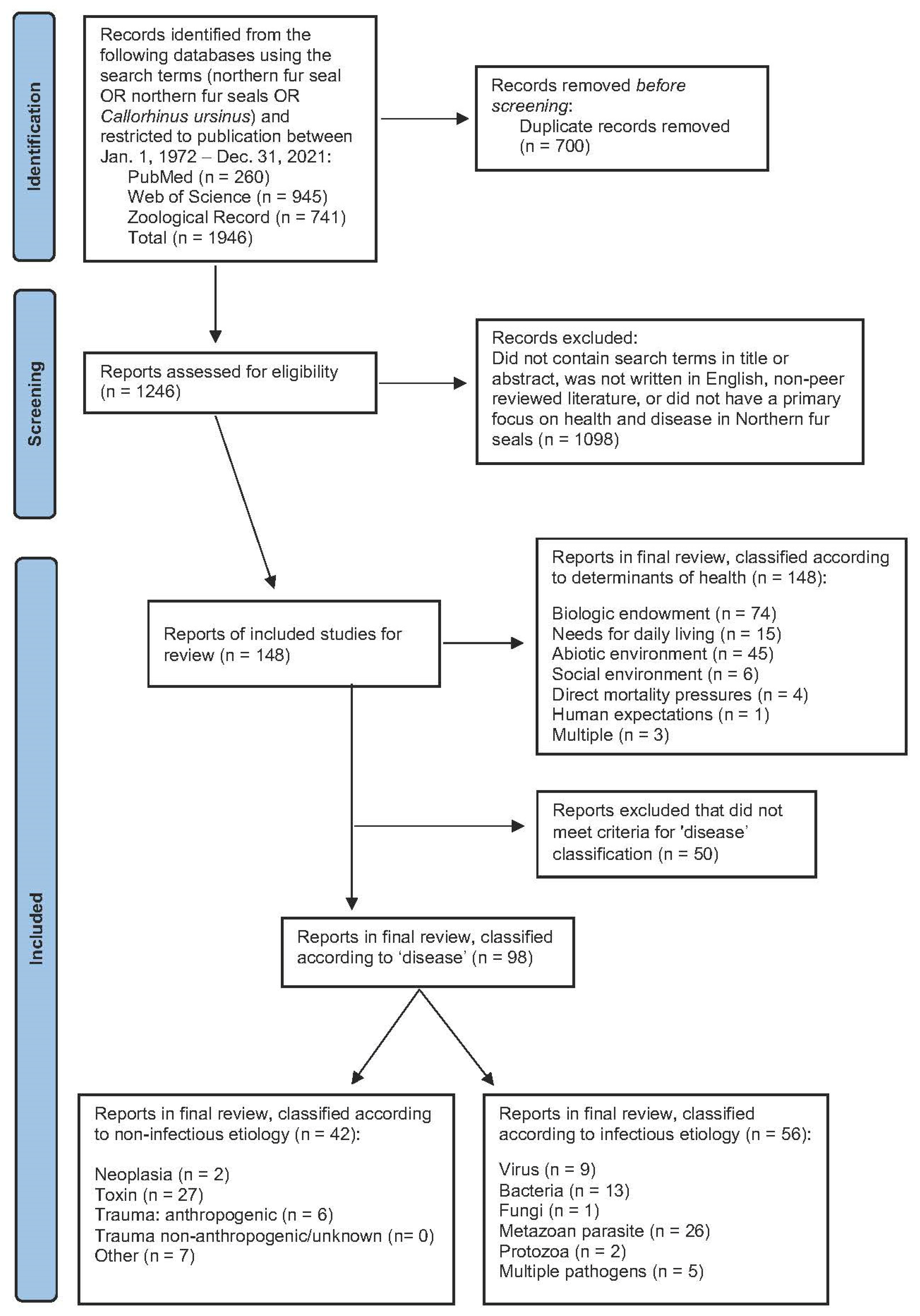
2. Materials and Methods

An overview of search criteria, inclusions and exclusions in accordance with PRISMA [13], is presented in Figure 1. We searched three electronic databases (PubMed, Web of Science, and Zoological Record) using the search terms “northern fur seal” OR “Callorhinus ursinus” OR “northern fur seals”. Specific inclusion criteria included publications between 1 January 1972, (in accordance with [9]) and 31 December 2021, and printed in English. Records were limited to articles published in peer-reviewed journals, with ‘grey literature’ such as government reports, books, and conference proceedings excluded. We read titles and abstracts and excluded publications that did not focus on NFS health [11] or disease [9] and publications which concentrated on tools and techniques (e.g., tagging, modeling, and methodologies).
Following the exclusion of duplicates and articles that failed to meet the inclusion criteria, titles and abstracts of all remaining records were reviewed and categorized independently by three authors (V.C., J.V., and C.D.) according to how they related to NFS health or disease. Health categories were based on the determinants of wildlife health model proposed by Wittrock et al., 2019: needs for daily living (e.g., nutrition and habitat quality); abiotic environment (e.g., physical surroundings, weather, and water quality); social environment (e.g., community dynamics, intra- and interspecies interactions); biologic endowment (e.g., physiological or pathological aspects of wildlife health); direct mortality pressures (e.g., factors that directly threaten survival); and human expectations (e.g., service-like entities involved in wildlife or ecosystem management) [11]. An article was classified as relating to disease or not if it covered one of the infectious or non-infectious disease conditions as described by Simeone et al., 2015 [9]. When classifications differed between authors, consensus was reached through discussion and complete article review as necessary.
All disease publications were reviewed in full and further divided into 10 subcategories; 5 infectious (viruses, bacteria, fungi, metazoan parasites, and protozoa) and 5 non-infectious (neoplasia, toxins and contaminants, anthropogenic trauma, non-anthropogenic/unknown source trauma and ‘other’), similar to classifications by Simeone et al., 2015 [9]. Within each of the subcategories, articles were evaluated according to the type of disease information available (i.e., indirect tests, direct tests, clinical disease/pathology, or other detectable health impact), the management of the NFS under study (wild or captive) and the location where the work was done, similar to Fagre et al., 2015 [8]. When articles included discussion of multiple disease categories that spanned multiple subcategories, they were assessed within the broader disease review section in addition to acknowledgement in relevant subcategories.

3. Results

A total of 148 publications met the criteria for inclusion in this study (Figure 1), all of which could be thematically classified according to the determinants of health. In contrast, 98 publications were classified as ‘disease’.

3.1. Disease Classification

The majority of the disease publications (n = 98) were focused on either a single agent, or a group of related infectious or non-infectious agents that fit within the specific categories that follow. A smaller number (n = 5) spanned multiple disparate categories. Most notable was an extensive case series of post-mortem findings from opportunistically collected NFSs on St. Paul Island, Alaska, between 1986 and 2006, by Spraker and Lander [14]. NFSs were also included in a large case series describing categories of disease in California stranded marine mammals from 1984 to 1990 [15]. The remaining multi-category publications were largely infectious disease-oriented and are referenced in relevant subsections below.

3.1.1. Infectious Disease

Viruses

Nine (9.2%) of the disease articles focused on viral diseases. Viral disease was also described in the necropsy study and referenced in several other of the multi-pathogen publications [14,16,17]. The majority of these articles, all published prior to 2000, were on caliciviruses isolated from wild NFS, most commonly in California but also Alaska [17,18,19,20,21,22]. Infection was typically devoid of pathology, although vesicular cutaneous lesions have also been described [23]. Proliferative cutaneous lesions associated with pox virus have been characterized by histopathology and electron microscopy, but were seen very infrequently relative to the number of animals examined post mortem [14,24].
The remaining reports of viral pathogens were those detected in the reproductive system. A novel polyomavirus was sequenced from a NFS placenta collected on St. Paul Island after viral inclusions were seen histologically [25]. The single affected placenta had been opportunistically collected from a rookery and no information was available regarding fetal or maternal health. There was a single report of Otarine herpes virus 4 detected by polymerase chain reaction (PCR) from vaginal swabs of free ranging NFSs in Alaska devoid of any associated pathology or description of associated clinical disease [26].

Bacteria

Thirteen (13%) of the disease articles focused on a single bacteria and several of the multi-pathogen articles also included information on bacterial species. The most frequent bacteria within this group was Coxiella burnetii. In 2010, 75% of the 146 opportunistically collected NFS placentas from St. Paul Island, Alaska, were PCR positive for C. burnetii and a subset (3%) of placentas had histologically identified intracytoplasmic bacteria confirmed as C. burnetii with immunohistochemistry (IHC) [27]. A similar molecular prevalence (77%) was reported in placentas collected from the same rookery the following year [28]. Infected placentas had decreased apoptosis of placental trophoblasts suggesting a functional change in the tissue, although no information on the associated NFS pup was available to support this claim [29]. PCR conducted on multiple tissue types from 50 subadult male NFSs harvested during subsistence hunting were tested for C. burnetii bacterial DNA by real-time PCR; there were no positive samples [30]. Similarly, archived vaginal swabs from adult female NFSs were all negative [31]. A serosurvey using archived samples collected from animals in the same location revealed high levels of exposure that varied by age class but appeared to increase between 1994 and 2009/2011 (49–69%) [31].
Brucella spp. have also been identified in the NFS population on St. Paul Island. From the same collection of placentas tested for C. burnetii above, a single case of necrotizing placentitis with intracytoplasmic bacteria was seen and attributed to Brucella spp. by IHC and PCR [32]. A total of 119 placentas collected during the 2011 pupping season were screened by IS711 PCR with 6 (5%) testing positive; and serology using archived samples suggested a similarly low level of exposure in the population (BMAT 2.5% positive, 30% borderline) [32]. Another serosurvey of 107 archived samples collected in the same area, but tested by enzyme-linked immunoassay (ELISA), were all negative [33]. Fifty subsistence harvested subadult male NFSs were tested by PCR using eight tissue types, but only a single spleen sample was positive and no disease was reportedly observed [30].
Pathogen-specific investigations have elucidated information on the presence, and in some cases pathogenicity, of different bacteria. A variety of Salmonella spp. have been isolated from the rectums of apparently healthy NFS pups in California and the authors concluded that the organism could cause opportunistic infections but did not usually cause disease in healthy animals [34]. That said, there was a single case report of a NFS pup with meningoencephalitis and septicemia where S. enteriditis was cultured from the brain and spinal cord [35]. Serologic screening and post-mortem examinations conducted on Pribilof Island NFSs in the 1970s revealed a rare leptospiral infection characterized by interstital nephritis in an adult and multisystem (renal, hepatic and placental) infection in neonates [17,36]. Erysipelothrix rhusiopathiae was isolated from the oral cavity of 2/12 otherwise presumed healthy, free-ranging subadult male NFSs on St. Paul Island as part of a multispecies investigation into the prevalence of the bacteria in the oral cavity of marine mammals and bite wounds from marine mammals [37]. A cross-sectional survey of oral Pasteurellaceae isolates from captive NFSs and other species concluded that the bacteria are part of normal marine mammal flora [38].
Multiple tissues have been cultured from subsistance-harvested subadult male NFSs in Alaska and a variety of mixed bacteria were identified; however, there was no association with disease and the high apparent prevalence in some normally sterile locations, suggested a high likelihood of contamination [39]. The remaining publications describing bacterial disease were typically case reports or case series investigations. While routine bacterial cultures were not conducted as part of the Alaska NFS necropsy program, beta-hemolytic Escherichia coli was isolated from 11 pups with pneumona [14]. Similarly, a captive subadult male NFS died following a period of anorexia and vomiting and a hemolytic E. coli isolated from the intestine was determined to be the causative agent [40].

Fungi

Only a single article was identified on fungal disease where Candida albicans was isolated from asymptomatic NFS in an aquarium setting where phocid seals exhibited clinical signs associated with infection [41].

Metazoan Parasites

The subcategory with the largest number of infectious disease articles (27%) was metazoan parasites. This body of research was well summarized in 2021 as an extensive literature review in addition to describing the intestinal helminth communities from hundreds of additional NFSs [42]. While this work explored both spatial and temporal patterns of infection as well as prevalence and abundance, samples were collected from presumably healthy animals and, as with the overwhelming majority of the literature on the topic, there was little association with pathology or population health impacts. Hookworm (Uncinaria lucasi) is an exception. The topic of hookworms made up more than half of the metazoan parasite publications and has been well reviewed by Lyons et al. (2011).The pattern of disease is variable by region, with hookworms recognized as a major cause of death in NFS pups in California but uncommon in other sites [43]. Gastric lesions associated with anisakid nematodes were identified in 21% of the stomachs from subadult males harvested on St. Paul Island, Alaska, 2011–2013; down from 92% as reported from the 1960s [44].

Protozoan Parasites

Two reports of protozoal infection were reviewed. Significant pathology was attributed to disseminated Toxoplasma gondii (diagnosed by IHC) infection in an adult female NFS that was stranded in California [45]. A brief description of histologically diagnosed sarcocystis in the muscle of a wild seal found on St. Paul Island was reported in the 1970s, but no additional information on the animal, or impact on the population, was included [46].

3.1.2. Non-Infectious Disease

Neoplasia

Two case reports focused on neoplastic disease; both reports were on neonates found dead on the Pribilof Islands, one of which had a renal fibrosarcoma [47] and the other diagnosed with multicentric lymphoma in which viral particles were suspected, but not confirmed, by electron microscopy [48]. A variety of neoplastic processes have been described by Spraker and Lander including a fetal ganglioneuroblastoma, an adrenal cortical carcinoma, ovarian dysgerminoma, fibromas, and a squamous papilloma [14].

Toxins and Contaminants

Twenty-eight percent of the disease publications focused on toxins or contaminants. Overwhelmingly, these publications report contaminant levels in a variety of tissue or secretory products, but are devoid of any association with individual or population health or disease information (Table 1). There were two reports on algal toxins, most notably a case series of stranded, multiple age class NFSs in California that characterized the clinical and post-mortem disease in NFS with domoic acid poisoning, including central nervous system signs and pathology of the nervous and cardiac systems consistent with disease in other species [49]. Lower domoic acid and saxitoxin exposures in NFSs relative to other marine mammals were attributed to differences in foraging behavior [50].

Trauma: Anthropogenic

Of the six articles focused on trauma, all reported traumas attributable to anthropogenic causes. The majority described the frequency and severity of NFS enganglement in different parts of their geographic range collectively highlighting the hazards of fishing materials and marine debris [76,77,78,79]. The energetics of entanglement were estimated in a study on captive animals which highlighted the difficulty that entangled animals can have both swimming and resting [80]. Enganglement as both a cause of death and cause of observed chronic pathology was also described in the Alaskan necropsy case series [14]. A single article described the pathology associated with acute head trauma sustained during NFS harvest activities [81].

Trauma Non-Anthropogenic/Unknown Source

As noted above, all of the disease articles that focused on trauma were classified as anthropogenic; however, non-anthropogenic or unknown trauma was a very common finding in the necropsy study in Alaska [14]. Trauma in adults was largely the result of fighting, while, in pups, crushing injuries and bite wounds were both commonly observed.

Other

Seven of the disease papers were classified as ‘other’ by default as they failed to align with the above criteria, but were broader than the congenital/metabolic category used by [9]. Two of these presumably uncommon disease conditions in captive animals were written up as case reports. These included an animal with gastric dilatation with volvulus and a gastric intramural hematoma with hemoperitoneum, both of unknown origin [82,83]. The remaining articles focused on the ocular or oral cavity. Cross-sectional surveys of opportunistically collected eyes from both wild and captive NFSs, contributed general information on gross and histologic changes in NFS eyes, but lacked associated information on individual or population health impacts [84,85,86]. Similarly, two articles described dental disease and temporomandibular joint pathology in museum collection NFS skulls, but nothing was reported about the relationship between observed lesions and other causes of morbidity or mortality [86,87].
An important condition of NFSs that was highlighted in the necropsy case series from Alaska and California, was emaciation [14,15]. Emaciation was not only the most common cause of death in NFS pups on St. Paul Island, but it has reportedly increased over time [14]. A variety of additional conditions such as congenital anomalies, predominantly musculoskeletal, were described in the longtudinal necropsy study in Alaska [14].

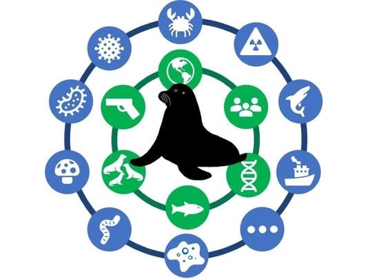
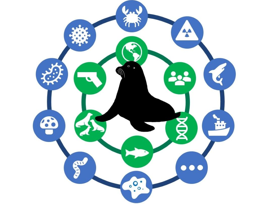
3.2. Health Classification

All of the 148 health and disease publications fit within one of the six determinants of health categories, although interestingly only 15 (10%) contained the words ‘health’ or ‘healthy’ in the title or abstract. The overwhelming majority (50%) of the articles were classified as biologic endowment. These were predominantly (89%) the infectious and non-infectious disease articles described above, with the remaining nine studies focused on fetal and pup growth and birth weight as related to survival or mortality, as well as several articles on reproductive indices. Thirty percent of the articles were classified as abiotic environment. These were largely about exposure to toxins or contaminants in the environment (n = 27, as included above) or anthropogenic trauma such as entanglements described above (n = 5). The remaining papers classified as abiotic environment were not included in any of the disease categories but described important topics such as the way adverse weather conditions and extreme water temperatures impact seal survival and dispersal.
All the remaining determinants of health categories were represented, but considerably less frequently. Needs for daily living (10%) included articles on how foraging behavior, food web dynamics, prey composition, and selection all affect NFS energetics and overall success. For example, an article by Short et al. (2021) tied prey availability to NFS pup survival by showing that commercial pollock fishing in close proximity to the Pribilof Islands thinned out schools of fish that were normally readily available to lactating female NFSs, which may have perpetuated low pup survival rates [88]. None of these articles was represented in the disease classification system.
Social environment (4%) also had no overlap with the disease classification system. These articles were largely investigations into inter- and intraspecific competition for prey, natal sites, and territoriality. None of these articles were represented in the disease category. Of particular interest were articles that linked this competition to population trends, such as a study by Kuhn et al. (2014) that showed increased densities of NFS may have negative effects on population growth, due to the increased energy output required to obtain prey [89]. Articles classified as direct mortality pressures largely addressed impacts from hunting and harvesting of NFSs with emphasis on how the sex of harvested seals could impact population growth. A single study in this category describing pathology associated with blunt head trauma in harvested seals [54], was also classified as traumatic, non-infectious disease. Only a single article that discussed the relationship between NFS management and population carrying capacity [90] was included in human expectations. Three articles spanned more than one category and were subsequently classified as ‘multiple’.

4. Discussion

Absence of evidence is not evidence of absence.
- Carl Sagan
The presence and absence of information from these 50 years of peer-reviewed NFS literature can both help to inform research and management programs specific to the species. Traditionally, such programs are disease focused and based on conditions that have been observed in the past, and there is a good foundation of NFS disease literature available to build upon. A particularly noteworthy contribution is the 20-year case series conducted on St. Paul Island, Alaska, by Spraker and Lander [14]. This work involved post-mortem examination of more than 3000 NFSs, creating a dataset that could be explored for trends, and could facilitate the collection of biologic samples, generate several hypothesis, and generally serve as a foundation for several other projects included in this review (e.g., [25,28,29,43,44]). Post-mortem examination has been cited as ‘the single most critical step in diagnosis for general wild animal disease surveillance’ [91]. There are several reasons for this including circumvention of hazards related to capture and handling of live animals, necropsy as a source of information on variations in ‘normal’ within a species, identification of several concurrent disease or physiologic changes, and as a sample source for more targeted disease investigations [92,93,94]. The long-term necropsy program on St. Paul Island is a unique opportunity that could serve as a foundation to which other NFS health and disease studies should be linked.
If necropsy data is to be used for these purposes however, it is important to understand both strengths and limitations of the work. The study of pinniped pathology is well known to be biased by issues of access; overrepresenting animals housed in captive facilities, species and age groups that strand more commonly, or populations that are easier to observe and sample [95]. Unique access to NFSs highlights this pathology bias. Necropsy work that has been conducted on St. Paul Island has been made possible by way of a ‘catwalk system’, i.e., raised walkways above some NFS rookeries from which researchers can safely observe a subset of NFS (those on the rookery) and collect deceased animals using hooked poles. Limitations to sample collection include size of the deceased animal (light enough to be picked up) and proximity to the catwalks. Animals included in the Spraker and Lander paper were overwhelmingly (90%) pups [14] which were largely representative of the number of pups in that location at that time and the lower survival probability of young animals, however, causes of death and disease are variable by life stage which limits the external validity of pup necropsy findings. Strategically aligning (e.g., examination, record keeping, sample collection, testing, and archiving) the necropsy program with other collection opportunities, such as young adult males harvested for consumption, could partially help to address this problem. Additionally, as the catwalk system serves as a sampling transect for the necropsy program, it will be important to ensure it is appropriately located relative to the rookery. Aerial photos clearly demonstrate changes in the distribution of NFSs on rookeries as the population declines, resulting in fewer animals adjacent to the catwalks [96,97]. Analysis of necropsy findings in conjunction with appropriate population (‘denominator’) information will help to keep findings in context.
The complete post-mortem examination process is typically overseen by pathologist(s) and informed by patient history, signalment, clinical disease, and gross necropsy findings. The nature and severity of changes seen within an organ system then drive the selection of additional diagnostic tests to confirm or exclude etiologic agents with the potential to cause the observed disease. However, because some etiologic agents of concern may not cause grossly identifiable pathology in any or all animals, routine screening of tissues for pathogens, toxins, or contaminants may be warranted. Despite numerous publications on the topic, as highlighted in this review, infectious disease was rarely (3%) implicated as a cause of pup mortality in the St. Paul Island necropsy study [14]. Unfortunately, systematic screening of tissues for pathogens, toxins, or contaminants, does not appear to have been conducted. Standardized protocols used to screen for infectious and non-infectious agents can ensure that the post-mortem examination is sensitive enough to identify any etiologic agents of concern.
As with biases associated with accessing animals for necropsy, our review highlighted biases associated with different sample types. The majority of the infectious disease articles reviewed in this study focused on intestinal parasites, however, with the exception of well described pathological and epidemiological investigations into hookworm infections [43], this work is largely devoid of associated health or disease information, making it challenging to tie much of the historical parasitological research into future monitoring programs. As noted in other reviews of Alaskan wildlife, the overrepresentation of parasites in disease research is likely, at least in part, to be a function of collection bias as fecal samples are relatively easy to collect [8].
Similarly, there were many publications on diseases of the NFS placenta (e.g., [25,27,29,32]). Publications on C. burnetii are undoubtably overrepresented in the literature because of the novelty of this pathogen in the species and geographic location, as well as its potential risk to humans. However, as with the collection of pups and material for parasitological investigations, sample availability undoubtably adds additional bias as the opportunity to systematically collect wild animal placentas is extremely uncommon, and therefore novel. While several of the pathogens within the NFS placentas have been demonstrated to cause disease in other species, including humans, use of this sample without any information on the associated maternal and pup outcomes makes it impossible to link these findings to the overall health of the population. Awareness of sample biases will be important to consider in future research efforts.
Failure to link disease research to individual or population outcomes was consistent across the NFS literature overall. This gap supports arguments for a paradigm shift away from the siloed, single agent focus of wildlife disease programs to the more holistic, health-oriented perspective that encompasses the broader social and environmental factors that are needed in resilient animal populations [10,98]. Doing this requires strong collaboration between those with broader (e.g., population, ecosystem) perspectives and experience, and also recognition that disciplines not classically thought of as ‘health’ domains are, in fact, most central to this work. Such disciplines may include, but are not limited to, wildlife (including fisheries) biologists and ecologists, oceanographers, climate scientists, immunologists, and toxicologists. Through these collaborations, we would be better positioned to transition the narrative away from looking at sick animals and screening for what is wrong (disease focus) to looking at healthy populations and identifying characteristics that help them be well (health focus).
As part of this review we included, and then subclassified, articles based on relevance to the six determinants of wildlife health proposed by Wittrock et al., 2019 [11]. The rank order of frequency of these in the NFS literature review (biologic endowment, abiotic environment, and needs for daily living being the top three) were similar to those of barren ground caribou (Rangifer tarandus groenlandicus) and Pacific salmon (Oncorhyncus spp.) [11]. It should be noted that our search methods (only including the common and scientific names for NFSs) differed from those of Wittrock et al. and the resulting list of publications is unlikely to fully represent the scope of research into factors influencing NFS health. For example, there is undoubtably a vast body of literature on abundance and distribution of NFS prey species that is relevant to NFS nutrition (‘needs for daily living’), but these publications are unlikely to have NFSs as a key word, and therefore, would not have been identified in our literature search. By restricting full-text review to articles with the common and/or scientific names for the NFS in the title or abstract we undoubtably excluded some relevant disease articles as well. That said, it is notable that our review included peer-reviewed publications spanning all six of the health determinants, suggesting that this framework may be appropriate for use in a NFS health program in the future. Interestingly, only 15 (all ‘biologic endowment’ or ‘abiotic environment’) of the 148 reports in the final review contained the words ‘health’ or ‘healthy’ in the title or abstract, the majority (n = 12) of which were also classified as disease publications. This highlights the fact that although the work is relevant to factors influencing the health of NFSs, numerous researchers and authors may not communicate it that way. Similarly, of the 148 reports included in our review, 50 publications were determined to be important to NFS health but were not classified as ‘disease’ articles and half of the health determinant categories such as (‘needs for daily living’, ‘social environment’, and ‘human expectations‘) contained no articles that were also captured in the ‘disease’ category. Collectively, this indicates how much important information could be missed using only a disease centric approach. Work is needed to engage with individuals and groups working in these more diverse branches of science that contribute to NFS population ‘health’, covering topics that include, but are not limited to, ecosystem dynamics, food availability, nutrition, genetic diversity, and stress.
To address concerns regarding bias and limitations of the historical work, and ways to better focus on health outcomes, it would be prudent to convene a group of NFS-invested individuals to develop a strategy for assessing health and disease of NFSs in the future. As resources (e.g., samples, time, and funding) are finite, it will be necessary to prioritize indices and conditions upon which to focus. Prioritization models developed and used successfully in public health livestock and wildlife domains (e.g., [99,100,101,102,103]) could help to inform similar efforts for the NFS. The general process involves compiling a list of conditions for prioritization, selecting appropriate measurement criteria, defining the range and weighting of levels for each criterion, aggregating scores for each condition, and ranking conditions by their total score for the final ordering [104]. While specific criteria may vary by species and location, those used in animal health typically include:
  • General characteristics of the condition in question (e.g., susceptible hosts, reservoirs, speed of spread, virulence, pathogenicity, and immune response);
  • Animal health impacts (e.g., morbidity, mortality, reproductive consequences, and welfare considerations);
  • Public health impacts (e.g., transmissibility to humans, severity of human disease, opportunities for human protection, food safety and security, bio/agroterrorism potential, spread amongst humans, and economic consequences);
  • Regulatory impacts (e.g., local, federal, or international trade consequences);
  • Mitigation (e.g., diagnosis, prevention, and treatment).
Our review has highlighted a body of literature that could aid in the prioritization process, particularly for diseases of the NFS. For example, several pathogens identified in this scoping review are zoonotic and some can have significant public health impacts. Individuals handling marine mammals, including researchers, have unique exposure opportunities to a variety of organisms they may not normally encounter [105,106]. Some of the zoonotic pathogens reported in NFSs, such as calicivirus or parapoxvirus, typically elicit only mild skin lesions, while others such as C. burnetii and Brucella spp., have the potential to cause more severe systemic disease [105,107]. Emphasis should be placed on conditions that are overrepresented in cohorts with increased opportunities for, or evidence of, exposure such as C. burnetii where the seroprevalence of Alaskan Native residents of the Pribilof Islands was almost four times the U.S. average [108]. Efforts should also be made to systematically survey for pathogens that may not have not been a topic of significant NFS research in the past, but are zoonotic, common in sympatric species, and have demonstrated ability to infect NFSs, such as Toxoplasma or Leptospira [45,95,109]. Engagement with public health personnel as part of the prioritization effort is scientifically justified and socially relevant, but may also be logistically and financially strategic as human and public health agencies can often access resources (e.g., laboratory expertise and funding sources) not typically utilized by wildlife professionals.
Similar to the benefits of collaborating with public health professionals, involving those who study conditions in sympatric species aids in the development of the initial disease list for use in the prioritization process. This is particularly important for novel animal health threats where little species-specific information is available; therfore, learning from others can help inform the development of a surveillance or testing program as necessary. Spatially clustered health risks (e.g., elevated mercury in Steller sea lions from the western part of their range [110]) could inform targeted data or sample collection. This collaborative effort may benefit multiple species. For example, infectious diseases such as leptospirosis or toxoplasmosis occur infrequently in NFSs [36,45], but can cause devastating disease in other marine species such as California sea lions and monk seals [111,112]. As the ecology of these pathogens is complex, synthesis of information from different species, geographic regions, and different environmental conditions may elucidate new information.
Finally, this prioritization process needs to be inclusive of all stakeholders and perspectives. Wild animals are a public resource. In addition to engaging scientists who may not already consider themselves a health professional as previously described, the NFS health prioritization should include members of the public. Involving the public, even those with little background on the topic, in zoonotic disease prioritization has been shown to yield meaningful results [113]. Northern fur seals are an important subsistence resource for indigenous people throughout their range, most notably Aleuts [114]. The benefit of incorporating local and traditional knowledge (LTK) is well recognized [115,116,117] and NFS population declines are a sign of changes within the Bering Sea ecosystem that is well recognized by the local community [118]. Frameworks to aid in the systematic and transparent approach to include LTK in wildlife assessments already exist [119]. Inclusivity of local hunters, fisherman, and indigenous populations with their unique LTK would make for a more holistic NFS health program.
Working as a team of NFS health and disease experts to prioritize and strategize on future research efforts would help to address several limitations present in this study. Our review included only English literature which undoubtably restricted the number of articles included. NFSs range throughout the North Pacific and research is conducted in many countries other than the United States. By convening an international team of NFS researchers and managers, publications identified in this review can be expanded to include results and perspectives from others that may not be captured here. Similarly, the smallest of our NFS health determinant categories was human expectations which was also likely a bias because of our exclusion of ‘grey literature’ in this review. According to [11], this category should include factors such as management policy, education programs, habitat funding and economics, and traditional knowledge. NFSs are federally managed, and therefore, inclusion of reports from governing bodies would markedly expand the available information on this topic. Similarly, the peer-reviewed scientific literature is not a channel though which LTK is typically shared, re-emphasizing the need to integrate this information using other established methods [119]. Collaboration would also facilitate the synthesis of ‘negative’ results which could be extremely important information for the prioritization process. The peer-reviewed literature is biased to focus on novel diseases and less likely to publish ‘negative’ findings [9]. Local people and managing agencies play a role in approving sample collection and animal handling; their records undoubtably contain considerably more information than is represented in the peer-reviewed literature.

5. Conclusions

This review of 50 years of scientific literature highlights the fact that NFS health is more than the presence or absence of innumerable disease-causing agents. Addressing the complexity of what makes an individual or population healthy requires a system approach to look at the many interacting factors (e.g., determinants of health and cumulative effects) from many different perspectives. Results of this work will be helpful in the next phase of an inclusive prioritization process that includes strategic planning and establishing mid- and long-range goals for consistent and targeted assessment and monitoring of NFS health.

Author Contributions

Conceptualization, C.D., K.P., C.S., K.S. and V.J.; methodology, C.D., K.P., J.V. and V.C.; formal analysis, J.V. and V.C.; investigation, V.C., K.P., C.S., J.V., V.J., K.S. and C.D.; data curation, V.C. and J.V.; writing—original draft preparation, C.D., J.V., V.C. and K.P.; writing—review and editing, V.C., K.P., C.S., J.V., V.J., K.S. and C.D.; visualization, V.C., K.P., J.V. and C.D.; supervision, C.D.; project administration, C.D.; funding acquisition, C.D., C.S., K.P. and V.J. All authors have read and agreed to the published version of the manuscript.

Funding

This research was funded by the NOAA Fisheries AK Region under award NA16NMF4390028 to Colorado State University.

Institutional Review Board Statement

Not applicable; retrospective literature review that did not involve live animals.

Informed Consent Statement

Not applicable.

Data Availability Statement

Not applicable.

Acknowledgments

This work was inspired by thoughtful discussions with our marine mammal colleagues, particularly Mike Williams, Tom Gelatt, and Rolf Ream. Special thanks also to Maddi Funk and Ah Young Kim for their assistance preparing our graphical abstract.

Conflicts of Interest

The sponsors had no role in the design, execution, interpretation, or writing of the study.

References

  1. Gelatt, T.; Ream, R.; Johnson, D. Callorhinus Ursinus. The IUCN Red List of Threatened Species 2015. Available online: https://www.iucnredlist.org/species/3590/45224953 (accessed on 5 May 2022).
  2. Fisheries, N. Northern Fur Seal. Available online: https://www.fisheries.noaa.gov/species/northern-fur-seal#:~:text=Male%20northern%20fur%20seals%20can,territories%20before%20the%20females%20arrive (accessed on 5 May 2022).
  3. Pacifici, M.; Santini, L.; Di Marco, M.; Baisero, D.; Francucci, L.; Grottolo Marasini, G.; Visconti, P.; Rondinini, C. Generation length for mammals. Nat. Conserv. 2013, 5, 5734. [Google Scholar] [CrossRef] [Green Version]
  4. Testa, J.W. Fur Seal Investigations, 2015–2016. 2018. Available online: https://doi.org/10.7289/V5/TM-AFSC-375 (accessed on 5 May 2022).
  5. Towell, R.G.; Ream, R.R.; York, A.E. Decline in northern fur seal (Callorhinus ursinus) pup production on the Pribilof Islands. Mar. Mammal Sci. 2006, 22, 486–491. [Google Scholar] [CrossRef]
  6. NMFS, Department of Commerce. North Pacific Fur Seal; Pribilof Island Population; Designation as Depleted. Fed. Reg. 1988, 53, 17888–17899. Available online: https://archives.federalregister.gov/issue_slice/1988/5/18/17881-17909.pdf#page=8 (accessed on 5 May 2022).
  7. Ryser-Degiorgis, M.-P. Wildlife health investigations: Needs, challenges and recommendations. BMC Vet. Res. 2013, 9, 223. [Google Scholar] [CrossRef] [PubMed] [Green Version]
  8. Fagre, A.C.; Patyk, K.A.; Nol, P.; Atwood, T.C.; Hueffer, K.; Duncan, C.G. A Review of Infectious Agents in Polar Bears (Ursus maritimus) and Their Long-Term Ecological Relevance. EcoHealth 2015, 12, 528–539. [Google Scholar] [CrossRef]
  9. Simeone, C.A.; Gulland, F.M.D.; Norris, T.; Rowles, T.K. A Systematic Review of Changes in Marine Mammal Health in North America, 1972–2012: The Need for a Novel Integrated Approach. PLoS ONE 2015, 10, e0142105. [Google Scholar] [CrossRef] [Green Version]
  10. Stephen, C. Toward a Modernized Definition of Wildlife Health. J. Wildl. Dis. 2014, 50, 427–430. [Google Scholar] [CrossRef]
  11. Wittrock, J.; Duncan, C.; Stephen, C. A Determinants of Health Conceptural Model for Fish and Wildlife Health. J. Wildl. Dis. 2019, 55, 285–297. [Google Scholar] [CrossRef]
  12. Munn, Z.; Peters, M.D.J.; Stern, C.; Tufanaru, C.; McArthur, A.; Aromataris, E. Systematic review or scoping review? Guidance for authors when choosing between a systematic or scoping review approach. BMC Med. Res. Methodol. 2018, 18, 143. [Google Scholar] [CrossRef]
  13. Page, M.J.; McKenzie, J.E.; Bossuyt, P.M.; Boutron, I.; Hoffmann, T.C.; Mulrow, C.D.; Shamseer, L.; Tetzlaff, J.M.; Akl, E.A.; Brennan, S.E.; et al. The PRISMA 2020 statement: An updated guideline for reporting systematic reviews. BMJ 2021, 372, n71. [Google Scholar] [CrossRef]
  14. Spraker, T.R.; Lander, M.E. Causes of Mortality in northern Fur Seals (Callorhinus ursinus), St. Paul Island, Pribilof Islands, Alaska, 1986–2006. J. Wildl. Dis. 2010, 46, 450–473. [Google Scholar] [CrossRef] [PubMed] [Green Version]
  15. Gerber, J.A.; Roletto, J.; Morgan, L.E.; Smith, D.M.; Gage, L.J. Findings in pinnipeds stranded along the central and northern California coast, 1984–1990. J. Wildl. Dis. 1993, 29, 423–433. [Google Scholar] [CrossRef] [PubMed] [Green Version]
  16. Smith, A.W.; Vedros, N.A.; Akers, T.G.; Gilmartin, W.G. Hazards of disease transfer from marine mammals to land mammals: Review and recent findings. J. Am. Vet. Med. Assoc. 1978, 173, 1131–1133. [Google Scholar] [PubMed]
  17. Smith, A.W.; Prato, C.M.; Gilmartin, W.G.; Brown, R.J.; Keyes, M.C. A Preliminary Report on Potentially Pathogenic Microbiological Agents Recently Isolated from Pinnipeds. J. Wildl. Dis. 1974, 10, 54–59. [Google Scholar] [CrossRef] [Green Version]
  18. Smith, A.W.; Skilling, D.E. Viruses and virus diseases of marine mammals. J. Am. Vet. Med. Assoc. 1979, 175, 918–920. [Google Scholar]
  19. Barlough, J.E.; Matson, D.O.; Skilling, D.E.; Berke, T.; Berry, E.S.; Brown, R.F.; Smith, A.W. Isolation of Reptilian Calicivirus Crotalus Type 1 From Feral Pinnipeds. J. Wildl. Dis. 1998, 34, 451–456. [Google Scholar] [CrossRef] [Green Version]
  20. Sawyer, J.C.; Madin, S.H.; Skilling, D.E. Isolation of San Miguel Sea Lion Virus from Samples of an animal food product produced from northern fur seal (Callorhinus ursinus) carcasses. Am. J. Vet. Res. 1978, 39, 137–139. [Google Scholar]
  21. Smith, A.W.; Prato, C.M.; Skilling, D.E. Characterization of two new serotypes of San Miguel sea lion virus. Intervirology 1977, 8, 30–36. [Google Scholar] [CrossRef]
  22. Smith, A.W.; Skilling, D.E.; Latham, A.B. Isolation and identification of five new serotypes of calicivirus from marine mammals. Am. J. Vet. Res. 1981, 42, 693–694. [Google Scholar]
  23. Prato, C.M.; Akers, T.G.; Smith, A.W. Serological evidence of calicivirus transmission between marine and terrestrial mammals. Nature 1974, 249, 255–256. [Google Scholar] [CrossRef]
  24. Hadlow, W.J.; Cheville, N.F.; Jellison, W.L. Occurrence of pox in a northern fur seal on the Pribilof Islands in 1951. J. Wildl. Dis. 1980, 16, 305–312. [Google Scholar] [CrossRef] [PubMed] [Green Version]
  25. Duncan, C.; Goldstein, T.; Hearne, C.; Gelatt, T.; Spraker, T. Novel polyomaviral infection in the placenta of a northern fur seal (Callorhinus ursinus) on the Pribilof Islands, Alaska, USA. J. Wildl. Dis. 2013, 49, 163–167. [Google Scholar] [CrossRef] [PubMed] [Green Version]
  26. Cortés-Hinojosa, G.; Gulland, F.M.D.; DeLong, R.; Gelatt, T.; Archer, L.; Wellehan, J.F.X. A Novel Gammaherpesvirus in Northern Fur Seals (Callorhinus ursinus) Is Closely Related to the California Sea Lion (Zalophus californianus) Carcinoma-Associated Otarine Herpesvirus-1. J. Wildl. Dis. 2016, 52, 88–95. [Google Scholar] [CrossRef] [PubMed]
  27. Duncan, C.; Kersh, G.; Spraker, T.; Patyk, K.; Fitzpatrick, K.; Massung, R.; Gelatt, T. Coxiella burnetii in Northern Fur Seal (Callorhinus ursinus) Placentas from St. Paul Island, Alaska. Vector-Borne Zoonotic Dis. 2012, 12, 192–195. [Google Scholar] [CrossRef] [PubMed] [Green Version]
  28. Duncan, C.; Savage, K.; Williams, M.; Dickerson, B.; Kondas, A.V.; Fitzpatrick, K.A.; Guerrero, J.L.; Spraker, T.; Kersh, G.J. Multiple strains of Coxiella burnetii are present in the environment of St. Paul Island, Alaska. Transbound. Emerg. Dis. 2013, 60, 345–350. [Google Scholar] [CrossRef] [PubMed] [Green Version]
  29. Myers, E.; Ehrhart, E.J.; Charles, B.; Spraker, T.; Gelatt, T.; Duncan, C. Apoptosis in normal and Coxiella burnetii-infected placentas from Alaskan northern fur seals (Callorhinus ursinus). Vet. Pathol 2013, 50, 622–625. [Google Scholar] [CrossRef] [PubMed] [Green Version]
  30. Duncan, C.; Dickerson, B.; Pabilonia, K.; Miller, A.; Gelatt, T. Prevalence of Coxiella burnetii and Brucella spp. in tissues from subsistence harvested northern fur seals (Callorhinus ursinus) of St. Paul Island, Alaska. Acta Vet. Scand. 2014, 56, 67. [Google Scholar] [CrossRef] [PubMed] [Green Version]
  31. Minor, C.; Kersh, G.J.; Gelatt, T.; Kondas, A.V.; Pabilonia, K.L.; Weller, C.B.; Dickerson, B.R.; Duncan, C.G. Coxiella burnetii in northern fur seals and Steller sea lions of Alaska. J. Wildl. Dis. 2013, 49, 441–446. [Google Scholar] [CrossRef] [Green Version]
  32. Duncan, C.G.; Tiller, R.; Mathis, D.; Stoddard, R.; Kersh, G.J.; Dickerson, B.; Gelatt, T. Brucella placentitis and seroprevalence in northern fur seals (Callorhinus ursinus) of the Pribilof Islands, Alaska. J. Vet. Diagn. Investig. 2014, 26, 507–512. [Google Scholar] [CrossRef] [Green Version]
  33. Nymo, I.H.; Rødven, R.; Beckmen, K.; Larsen, A.K.; Tryland, M.; Quakenbush, L.; Godfroid, J. Brucella Antibodies in Alaskan True Seals and Eared Seals—Two Different Stories. Front. Vet. Sci. 2018, 5, 8. [Google Scholar] [CrossRef] [Green Version]
  34. Gilmartin, W.G.; Vainik, P.M.; Neill, V.M. Salmonellae in feral pinnipeds off the Southern California coast. J. Wildl. Dis. 1979, 15, 511–514. [Google Scholar] [CrossRef] [PubMed]
  35. Stroud, R.K.; Roelke, M.E. Salmonella meningoencephalomyelitis in a northern fur seal (Callorhinus ursinus). J. Wildl. Dis. 1980, 16, 15–18. [Google Scholar] [CrossRef] [PubMed] [Green Version]
  36. Smith, A.W.; Brown, R.J.; Skilling, D.E.; Bray, H.L.; Keyes, M.C. Naturally-occurring leptospirosis in northern fur seals (Callorhinus ursinus). J. Wildl. Dis. 1977, 13, 144–148. [Google Scholar] [CrossRef] [PubMed] [Green Version]
  37. Suer, L.; Vedros, N.A. Erysipelothrix rhusiopathiae. I. Isolation and characterization from pinnipeds and bite/ abrasion wounds in humans. Dis. Aquat. Org. 1988, 5, 1–5. [Google Scholar] [CrossRef]
  38. Hansen, M.J.; Bertelsen, M.F.; Christensen, H.; Bisgaard, M.; Bojesen, A.M. Occurrence of Pasteurellaceae bacteria in the oral cavity of selected marine mammal species. J. Zoo Wildl. Med. 2012, 43, 828–835. [Google Scholar] [CrossRef]
  39. Vedros, N.A.; Quinlivan, J.; Cranford, R. Bacterial and fungal flora of wild northern fur seals (Callorhinus ursinus). J. Wildl. Dis. 1982, 18, 447–456. [Google Scholar] [CrossRef] [Green Version]
  40. Vanpelt, R.W.; Ohata, C.A. Hemolytic Escherichia coli as Cause of Acute Enterotoxemia in a Captive Northern Fur Seal. Vet. Med. Small Anim. Clin. 1974, 69, 1251–1254. [Google Scholar]
  41. Dunn, J.L.; Buck, J.D.; Spotte, S. Candidiasis in captive pinnipeds. J. Am. Vet. Med. Assoc. 1984, 185, 1328–1330. [Google Scholar]
  42. Kuzmina, T.A.; Kuzmin, Y.; Dzeverin, I.; Lisitsyna, O.I.; Spraker, T.R.; Korol, E.M.; Kuchta, R. Review of metazoan parasites of the northern fur seal (Callorhinus ursinus) and the analysis of the gastrointestinal helminth community of the population on St. Paul Island, Alaska. Parasitol. Res. 2021, 120, 117–132. [Google Scholar] [CrossRef]
  43. Lyons, E.T.; Spraker, T.R.; De Long, R.L.; Ionita, M.; Melin, S.R.; Nadler, S.A.; Tolliver, S.C. Review of research on hookworms (Uncinaria lucasi Stiles, 1901) in northern fur seals (Callorhinus ursinus Linnaeus, 1758). Parasitol. Res. 2011, 109, 257–265. [Google Scholar] [CrossRef]
  44. Kuzmina, T.A.; Lyons, E.T.; Spraker, T.R. Anisakids (Nematoda: Anisakidae) from stomachs of northern fur seals (Callorhinus ursinus) on St. Paul Island, Alaska: Parasitological and pathological analysis. Parasitol. Res. 2014, 113, 4463–4470. [Google Scholar] [CrossRef] [PubMed]
  45. Holshuh, H.J.; Sherrod, A.E.; Taylor, C.R.; Andrews, B.F.; Howard, E.B. Toxoplasmosis in a feral northern fur seal. J. Am. Vet. Med. Assoc. 1985, 187, 1229–1230. [Google Scholar] [PubMed]
  46. Brown, R.J.; Smith, A.W.; Keyes, M.C. Sarcocystis in the northern fur seal. J. Wildl. Dis. 1974, 10, 53. [Google Scholar] [CrossRef] [PubMed] [Green Version]
  47. Brown, R.J.; Smith, A.W.; Keyes, M.C. Renal fibrosarcoma in the northern fur seal. J. Wildl. Dis. 1975, 11, 23–25. [Google Scholar] [CrossRef]
  48. Stedham, M.A.; Casey, H.W.; Keyes, M.C. Lymphosarcoma in an infant northern fur seal (Callorhinus ursinus). J. Wildl. Dis. 1977, 13, 176–179. [Google Scholar] [CrossRef] [Green Version]
  49. Lefebvre, K.A.; Robertson, A.; Frame, E.R.; Colegrove, K.M.; Nance, S.; Baugh, K.A.; Wiedenhoft, H.; Gulland, F.M.D. Clinical signs and histopathology associated with domoic acid poisoning in northern fur seals (Callorhinus ursinus) and comparison of toxin detection methods. Harmful Algae 2010, 9, 374–383. [Google Scholar] [CrossRef]
  50. Lefebvre, K.A.; Quakenbush, L.; Frame, E.; Huntington, K.B.; Sheffield, G.; Stimmelmayr, R.; Bryan, A.; Kendrick, P.; Ziel, H.; Goldstein, T.; et al. Prevalence of algal toxins in Alaskan marine mammals foraging in a changing arctic and subarctic environment. Harmful Algae 2016, 55, 13–24. [Google Scholar] [CrossRef] [Green Version]
  51. Beckmen, K.B.; Duffy, L.K.; Zhang, X.; Pitcher, K.W. Mercury concentrations in the fur of Steller sea lions and northern fur seals from Alaska. Mar. Pollut. Bull. 2002, 44, 1130–1135. [Google Scholar] [CrossRef]
  52. Noda, K.; Ichihashi, H.; Loughlin, T.R.; Baba, N.; Kiyota, M.; Tatsukawa, R. Distribution of heavy metals in muscle, liver and kidney of northern fur seal (Callorhinus ursinus) caught off Sanriku, Japan and from the Pribilof Islands, Alaska. Environ. Pollut. 1995, 90, 51–59. [Google Scholar] [CrossRef]
  53. Goldblatt, C.J.; Anthony, R.G. Heavy Metals in Northern Fur Seals (Callorhinus ursinus) from the Pribilof Islands, Alaska. J. Environ. Qual. 1983, 12, 478–482. [Google Scholar] [CrossRef]
  54. Kim, K.C.; Chu, R.C.; Barron, G.P. Mercury in tissues and lice of Northern fur seals. Bull. Environ. Contam. Toxicol. 1974, 11, 281–284. [Google Scholar] [CrossRef] [PubMed]
  55. Anas, R.E. Heavy metals in the northern fur seal, Callorhinus ursinus, and harbor seal, Phoca vitulina richardi. Fish. Bull. 1974, 72, 133–137. [Google Scholar]
  56. Zeisler, R.; Demiralp, R.; Koster, B.J.; Becker, P.R.; Burow, M.; Ostapczuk, P.; Wise, S.A. Determination of inorganic constituents in marine mammal tissues. Sci. Total Environ. 1993, 139–140, 365–386. [Google Scholar] [CrossRef]
  57. Saeki, K.; Nakajima, M.; Noda, K.; Loughlin, T.R.; Baba, N.; Kiyota, M.; Tatsukawa, R.; Calkins, D.G. Vanadium Accumulation in Pinnipeds. Arch. Environ. Contam. Toxicol. 1999, 36, 81–86. [Google Scholar] [CrossRef]
  58. Arai, T.; Ikemoto, T.; Hokura, A.; Terada, Y.; Kunito, T.; Tanabe, S.; Nakai, I. Chemical Forms of Mercury and Cadmium Accumulated in Marine Mammals and Seabirds as Determined by XAFS Analysis. Environ. Sci. Technol. 2004, 38, 6468–6474. [Google Scholar] [CrossRef]
  59. Fujihara, J.; Kunito, T.; Kubota, R.; Tanabe, S. Arsenic accumulation in livers of pinnipeds, seabirds and sea turtles: Subcellular distribution and interaction between arsenobetaine and glycine betaine. Comp. Biochem. Physiol. Part C Toxicol. Pharmacol. 2003, 136, 287–296. [Google Scholar] [CrossRef]
  60. Saeki, K.; Nakajima, M.; Loughlin, T.R.; Calkins, D.C.; Baba, N.; Kiyota, M.; Tatsukawa, R. Accumulation of silver in the liver of three species of pinnipeds. Environ. Pollut. 2001, 112, 19–25. [Google Scholar] [CrossRef]
  61. Donohue, M.J.; Masura, J.; Gelatt, T.; Ream, R.; Baker, J.D.; Faulhaber, K.; Lerner, D.T. Evaluating exposure of northern fur seals, Callorhinus ursinus, to microplastic pollution through fecal analysis. Mar. Pollut. Bull. 2019, 138, 213–221. [Google Scholar] [CrossRef]
  62. Wang, D.; Shelver, W.L.; Atkinson, S.; Mellish, J.A.; Li, Q.X. Tissue distribution of polychlorinated biphenyls and organochlorine pesticides and potential toxicity to Alaskan northern fur seals assessed using PCBs congener specific mode of action schemes. Arch. Environ. Contam. Toxicol. 2010, 58, 478–488. [Google Scholar] [CrossRef]
  63. Mössner, S.; Barudio, I.; Spraker, T.S.; Antonelis, G.; Early, G.; Geraci, J.R.; Becker, P.R.; Ballschmiter, K. Determination of HCHs, PCBs, and DDTs in brain tissues of marine mammals off different age. Fresenius J. Anal. Chem. 1994, 349, 708–716. [Google Scholar] [CrossRef]
  64. Norstrom, R.J.; Muir, D.C. Chlorinated hydrocarbon contaminants in arctic marine mammals. Sci. Total Environ. 1994, 154, 107–128. [Google Scholar] [CrossRef]
  65. Schantz, M.M.; Koster, B.J.; Wise, S.A.; Becker, P.R. Determination of PCBs and chlorinated hydrocarbons in marine mammal tissues. Sci. Total Environ. 1993, 139–140, 323–345. [Google Scholar] [CrossRef]
  66. Beckmen, K.B.; Blake, J.E.; Ylitalo, G.M.; Stott, J.L.; O’Hara, T.M. Organochlorine contaminant exposure and associations with hematological and humoral immune functional assays with dam age as a factor in free-ranging northern fur seal pups (Callorhinus ursinus). Mar. Pollut. Bull. 2003, 46, 594–606. [Google Scholar] [CrossRef]
  67. Loughlin, T.R.; Castellini, M.A.; Ylitalo, G. Spatial aspects of organochlorine contamination in northern fur seal tissues. Mar. Pollut. Bull. 2002, 44, 1024–1034. [Google Scholar] [CrossRef]
  68. Beckmen, K.B.; Ylitalo, G.M.; Towell, R.G.; Krahn, M.M.; O’Hara, T.M.; Blake, J.E. Factors affecting organochlorine contaminant concentrations in milk and blood of northern fur seal (Callorhinus ursinus) dams and pups from St. George Island, Alaska. Sci. Total Environ. 1999, 231, 183–200. [Google Scholar] [CrossRef]
  69. Mössner, S.; Spraker, T.R.; Becker, P.R.; Ballschmiter, K. Ratios of enantiomers of alpha-HCH and determination of alpha-, beta-, and gamma-HCH isomers in brain and other tissues of neonatal Northern fur seals (Callorhinus ursinus). Chemosphere 1992, 24, 1171–1180. [Google Scholar] [CrossRef]
  70. Reiner, J.L.; Becker, P.R.; Gribble, M.O.; Lynch, J.M.; Moors, A.J.; Ness, J.; Peterson, D.; Pugh, R.S.; Ragland, T.; Rimmer, C.; et al. Organohalogen Contaminants and Vitamins in Northern Fur Seals (Callorhinus ursinus) Collected During Subsistence Hunts in Alaska. Arch. Environ. Contam. Toxicol. 2016, 70, 96–105. [Google Scholar] [CrossRef] [Green Version]
  71. Nomiyama, K.; Kanbara, C.; Ochiai, M.; Eguchi, A.; Mizukawa, H.; Isobe, T.; Matsuishi, T.; Yamada, T.K.; Tanabe, S. Halogenated phenolic contaminants in the blood of marine mammals from Japanese coastal waters. Mar. Environ. Res. 2014, 93, 15–22. [Google Scholar] [CrossRef]
  72. Tanabe, S.; Sung, J.-K.; Choi, D.-Y.; Baba, N.; Kiyota, M.; Yoshida, K.; Tatsukawa, R. Persistent organochlorine residues in northern fur seal from the Pacific coast of Japan since 1971. Environ. Pollut. 1994, 85, 305–314. [Google Scholar] [CrossRef]
  73. Kajiwara, N.; Ueno, D.; Takahashi, A.; Baba, N.; Tanabe, S. Polybrominated diphenyl ethers and organochlorines in archived northern fur seal samples from the Pacific coast of Japan, 1972–1998. Environ. Sci. Technol. 2004, 38, 3804–3809. [Google Scholar] [CrossRef]
  74. Iwata, H.; Tanabe, S.; Iida, T.; Baba, N.; Ludwig, J.P.; Tatsukawa, R. Enantioselective Accumulation of α-Hexachlorocyclohexane in Northern Fur Seals and Double-Crested Cormorants:  Effects of Biological and Ecological Factors in the Higher Trophic Levels. Environ. Sci. Technol. 1998, 32, 2244–2249. [Google Scholar] [CrossRef]
  75. Ruedig, E.; Duncan, C.; Dickerson, B.; Williams, M.; Gelatt, T.; Bell, J.; Johnson, T.E. Fukushima derived radiocesium in subsistence-consumed northern fur seal and wild celery. J. Environ. Radioact. 2016, 152, 1–7. [Google Scholar] [CrossRef] [PubMed]
  76. Kuzin, A.E.; Trukhin, A.M. Entanglement of northern fur seals (Callorhinus ursinus) in marine debris on Tyuleniy Island (Sea of Okhotsk) in 1998–2013. Mar. Pollut. Bull. 2019, 143, 187–192. [Google Scholar] [CrossRef] [PubMed]
  77. Hanni, K.D.; Pyle, P. Entanglement of Pinnipeds in Synthetic Materials at South-east Farallon Island, California, 1976–1998. Mar. Pollut. Bull. 2000, 40, 1076–1081. [Google Scholar] [CrossRef]
  78. Kiyota, M.; Baba, N. Entanglement in marine debris among adult female northern fur seals at St. Paul Island, Alaska in 1991–1999. Bull.-Natl. Res. Inst. Far Seas Fish. 2001, 38, 13–20. [Google Scholar]
  79. Stewart, B.S.; Yochem, P.K. Entanglement of pinnipeds in synthetic debris and fishing net and line fragments at San Nicolas and San Miguel Islands, California, 1978–1986. Mar. Pollut. Bull. 1987, 18, 336–339. [Google Scholar] [CrossRef]
  80. Feldkamp, D.M.; Costa, D.P.; Dekrey, G.K. Energetic and Behavioral Effects of Net Entanglement on Juvenile Northern Fur Seals, Callorhinus ursinus. Fish. Bull. 1989, 87, 85–94. [Google Scholar]
  81. Jortner, B.S. Neuropathologic Observations of Head Trauma in the Northern Fur Seal. J. Wildl. Dis. 1974, 10, 121–129. [Google Scholar] [CrossRef]
  82. Frasca, S., Jr.; Van Kruiningen, H.J.; Dunn, J.L.; St Aubin, D.J. Gastric intramural hematoma and hemoperitoneum in a captive northern fur seal. J. Wildl. Dis. 2000, 36, 565–569. [Google Scholar] [CrossRef] [Green Version]
  83. Frasca, S., Jr.; Dunn, J.L.; Van Kruiningen, H.J. Acute gastric dilatation with volvulus in a northern fur seal (Callorhinus ursinus). J. Wildl. Dis. 1996, 32, 548–551. [Google Scholar] [CrossRef] [Green Version]
  84. Stoskopf, M.K.; Zimmerman, S.; Hirst, L.W.; Green, R. Ocular anterior segment disease in northern fur seals. J. Am. Vet. Med. Assoc. 1985, 187, 1141–1144. [Google Scholar] [PubMed]
  85. Miller, S.; Colitz, C.M.H.; St. Leger, J.; Dubielzig, R. A retrospective survey of the ocular histopathology of the pinniped eye with emphasis on corneal disease. Vet. Ophthalmol. 2013, 16, 119–129. [Google Scholar] [CrossRef] [PubMed]
  86. Colitz, C.M.H.; Renner, M.S.; Manire, C.A.; Doescher, B.; Schmitt, T.L.; Osborn, S.D.; Croft, L.; Olds, J.; Gehring, E.; Mergl, J.; et al. Characterization of progressive keratitis in Otariids. Vet. Ophthalmol. 2010, 13, 47–53. [Google Scholar] [CrossRef] [PubMed]
  87. Aalderink, M.T.; Nguyen, H.P.; Kass, P.H.; Arzi, B.; Verstraete, F.J. Dental and Temporomandibular Joint Pathology of the Northern Fur Seal (Callorhinus ursinus). J. Comp. Pathol. 2015, 152, 325–334. [Google Scholar] [CrossRef] [PubMed]
  88. Short, J.W.; Geiger, H.J.; Fritz, L.W.; Warrenchuk, J.J. First-Year Survival of Northern Fur Seals (Callorhinus ursinus) Can Be Explained by Pollock (Gadus chalcogrammus) Catches in the Eastern Bering Sea. J. Mar. Sci. Eng. 2021, 9, 975. [Google Scholar] [CrossRef]
  89. Kuhn, C.E.; Baker, J.D.; Towell, R.G.; Ream, R.R. Evidence of localized resource depletion following a natural colonization event by a large marine predator. J. Anim. Ecol. 2014, 83, 1169–1177. [Google Scholar] [CrossRef]
  90. Eberhardt, L.L.; Siniff, D.B. Population Dynamics and Marine Mammal Management Policies. J. Fish. Res. Board Can. 1977, 34, 183–190. [Google Scholar] [CrossRef]
  91. Leighton, F.A. Surveillance of wild animal diseases in Europe. Rev. Sci. Et Tech. Int. Off. Epizoot. 1995, 14, 819–830. [Google Scholar]
  92. Cooper, J.E. Diagnostic pathology of selected diseases in wildlife. Rev. Sci. Tech. 2002, 21, 77–89. [Google Scholar] [CrossRef]
  93. Küker, S.; Faverjon, C.; Furrer, L.; Berezowski, J.; Posthaus, H.; Rinaldi, F.; Vial, F. The value of necropsy reports for animal health surveillance. BMC Vet. Res. 2018, 14, 191. [Google Scholar] [CrossRef] [Green Version]
  94. McAloose, D.; Colegrove, K.M.; Newton, A.L. Chapter 1—Wildlife Necropsy. In Pathology of Wildlife and Zoo Animals; Terio, K.A., McAloose, D., Leger, J.S., Eds.; Academic Press: Cambridge, MA, USA, 2018; pp. 1–20. [Google Scholar]
  95. Colegrove, K.M.; Burek-Huntington, K.A.; Roe, W.; Siebert, U. Pinnipediae. Pathol. Wildl. Zoo Anim. 2018, 4, 569–592. [Google Scholar] [CrossRef]
  96. Yano, K.; Fowler, C. Population Changes in Northern Fur Seal Rookeries at Reef Rookery on St. Paul Island of the Pribilof Islands, Alaska. Available online: https://apps-afsc.fisheries.noaa.gov/pubs/posters/pdfs/pYano02_n-fur-seal-aerial.pdf (accessed on 15 April 2022).
  97. Yano, K.M.; Tingg, J.; Fowler, C. Northern Fur Seal Rookery Photo Archive: Aerial and Ground-Level Photos, Pribilof Islands, Alaska, 1895–2006; Alaska Fisheries Science Center, NOAA, National Marine Fisheries Service: Seattle, WA, USA, 2009; p. 62.
  98. Patyk, K.A.; Duncan, C.; Nol, P.; Sonne, C.; Laidre, K.; Obbard, M.; Wiig, Ø.; Aars, J.; Regehr, E.; Gustafson, L.L.; et al. Establishing a definition of polar bear (Ursus maritimus) health: A guide to research and management activities. Sci. Total Environ. 2015, 514, 371–378. [Google Scholar] [CrossRef] [PubMed] [Green Version]
  99. Krause, G. How can infectious diseases be prioritized in public health? A standardized prioritization scheme for discussion. EMBO Rep. 2008, 9 (Suppl. S1), S22–S27. [Google Scholar] [CrossRef] [PubMed] [Green Version]
  100. O’Brien, D.; Scudamore, J.; Charlier, J.; Delavergne, M. DISCONTOOLS: A database to identify research gaps on vaccines, pharmaceuticals and diagnostics for the control of infectious diseases of animals. BMC Vet. Res. 2016, 13, 1. [Google Scholar] [CrossRef] [Green Version]
  101. Humblet, M.-F.; Vandeputte, S.; Albert, A.; Gosset, C.; Kirschvink, N.; Haubruge, E.; Fecher-Bourgeois, F.; Pastoret, P.-P.; Saegerman, C. Multidisciplinary and evidence-based method for prioritizing diseases of food-producing animals and zoonoses. Emerg. Infect. Dis. 2012, 18, e1. [Google Scholar] [CrossRef]
  102. Cardoen, S.; Van Huffel, X.; Berkvens, D.; Quoilin, S.; Ducoffre, G.; Saegerman, C.; Speybroeck, N.; Imberechts, H.; Herman, L.; Ducatelle, R.; et al. Evidence-based semiquantitative methodology for prioritization of foodborne zoonoses. Foodborne Pathog. Dis. 2009, 6, 1083–1096. [Google Scholar] [CrossRef] [Green Version]
  103. McKenzie, J.; Simpson, H.; Langstaff, I. Development of methodology to prioritise wildlife pathogens for surveillance. Prev. Vet. Med. 2007, 81, 194–210. [Google Scholar] [CrossRef]
  104. Ng, V.; Sargeant, J.M. A stakeholder-informed approach to the identification of criteria for the prioritization of zoonoses in Canada. PLoS ONE 2012, 7, e29752. [Google Scholar] [CrossRef]
  105. Waltzek, T.B.; Cortés-Hinojosa, G.; Wellehan, J.F.X., Jr.; Gray, G.C. Marine mammal zoonoses: A review of disease manifestations. Zoonoses Public Health 2012, 59, 521–535. [Google Scholar] [CrossRef]
  106. Hunt, T.D.; Ziccardi, M.H.; Gulland, F.M.D.; Yochem, P.K.; Hird, D.W.; Rowles, T.; Mazet, J.A.K. Health risks for marine mammal workers. Dis. Aquat. Org. 2008, 81, 81–92. [Google Scholar] [CrossRef]
  107. Parker, N.R.; Barralet, J.H.; Bell, A.M. Q fever. Lancet 2006, 367, 679–688. [Google Scholar] [CrossRef]
  108. Kersh, G.J.; Fitzpatrick, K.; Pletnikoff, K.; Brubaker, M.; Bruce, M.; Parkinson, A. Prevalence of serum antibodies to Coxiella burnetii in Alaska Native Persons from the Pribilof Islands. Zoonoses Public Health 2020, 67, 89–92. [Google Scholar] [CrossRef] [PubMed]
  109. Dubey, J.P.; Murata, F.H.A.; Cerqueira-Cézar, C.K.; Kwok, O.C.H.; Grigg, M.E. Recent epidemiologic and clinical importance of Toxoplasma gondii infections in marine mammals: 2009–2020. Vet. Parasitol. 2020, 288, 109296. [Google Scholar] [CrossRef] [PubMed]
  110. Rea, L.D.; Castellini, J.M.; Avery, J.P.; Fadely, B.S.; Burkanov, V.N.; Rehberg, M.J.; O’Hara, T.M. Regional variations and drivers of mercury and selenium concentrations in Steller sea lions. Sci. Total Environ. 2020, 744, 140787. [Google Scholar] [CrossRef]
  111. Gulland, F.M.; Koski, M.; Lowenstine, L.J.; Colagross, A.; Morgan, L.; Spraker, T. Leptospirosis in California sea lions (Zalophus californianus) stranded along the central California coast, 1981–1994. J. Wildl. Dis. 1996, 32, 572–580. [Google Scholar] [CrossRef] [Green Version]
  112. Barbieri, M.M.; Kashinsky, L.; Rotstein, D.S.; Colegrove, K.M.; Haman, K.H.; Magargal, S.L.; Sweeny, A.R.; Kaufman, A.C.; Grigg, M.E.; Littnan, C.L. Protozoal-related mortalities in endangered Hawaiian monk seals Neomonachus schauinslandi. Dis. Aquat. Org. 2016, 121, 85–95. [Google Scholar] [CrossRef]
  113. Ng, V.; Sargeant, J.M. A quantitative and novel approach to the prioritization of zoonotic diseases in North America: A public perspective. PLoS ONE 2012, 7, e48519. [Google Scholar] [CrossRef]
  114. Veltre, D.W.; Veltre, M.J. The Northern Fur Seal: A Subsistence and Commercial Resource for Aleuts of the Aleutian and Pribilof Islands, Alaska. Études Inuit Stud. 1987, 11, 51–72. [Google Scholar]
  115. Ostertag, S.K.; Loseto, L.L.; Snow, K.; Lam, J.; Hynes, K.; Gillman, D.V. “That’s how we know they’re healthy”: The inclusion of traditional ecological knowledge in beluga health monitoring in the Inuvialuit Settlement Region. Arct. Sci. 2018, 4, 292–320. [Google Scholar] [CrossRef] [Green Version]
  116. Breton-Honeyman, K.; Furgal, C.M.; Hammill, M.O. Systematic Review and Critique of the Contributions of Traditional Ecological Knowledge of Beluga Whales in the Marine Mammal Literature. Arctic 2016, 69, 37–46. [Google Scholar] [CrossRef]
  117. Wyllie de Echeverria, V.R.; Thornton, T.F. Using traditional ecological knowledge to understand and adapt to climate and biodiversity change on the Pacific coast of North America. Ambio 2019, 48, 1447–1469. [Google Scholar] [CrossRef] [PubMed] [Green Version]
  118. Huntington, H.P.; Braem, N.M.; Brown, C.L.; Hunn, E.; Krieg, T.M.; Lestenkof, P.; Noongwook, G.; Sepez, J.; Sigler, M.F.; Wiese, F.K.; et al. Local and traditional knowledge regarding the Bering Sea ecosystem: Selected results from five indigenous communities. Deep. Sea Res. Part II Top. Stud. Oceanogr. 2013, 94, 323–332. [Google Scholar] [CrossRef]
  119. Peacock, S.J.; Mavrot, F.; Tomaselli, M.; Hanke, A.; Fenton, H.; Nathoo, R.; Aleuy, O.A.; Di Francesco, J.; Aguilar, X.F.; Jutha, N.; et al. Linking co-monitoring to co-management: Bringing together local, traditional, and scientific knowledge in a wildlife status assessment framework. Arct. Sci. 2020, 6, 247–266. [Google Scholar] [CrossRef]
Figure 1. Schematic representation of the scoping literature process including original searches through to classification of articles by determinants of health, as well as subcategories of disease.
Oceans 03 00021 g001
Table 1. A summary of the identified literature on contaminants in NFS.
ContaminantUSJapan
Heavy metals (e.g., mercury, cadmium, arsenic, silver, vanadium)[51,52,53,54,55,56,57][52,57,58,59,60]
Microplastics[61]
Persistent organic pollutants (e.g., PCB, DDT, PBDEs)[62,63,64,65,66,67,68,69,70][71,72,73,74]
Radiocesium[75]
Publisher’s Note: MDPI stays neutral with regard to jurisdictional claims in published maps and institutional affiliations.

