Communication

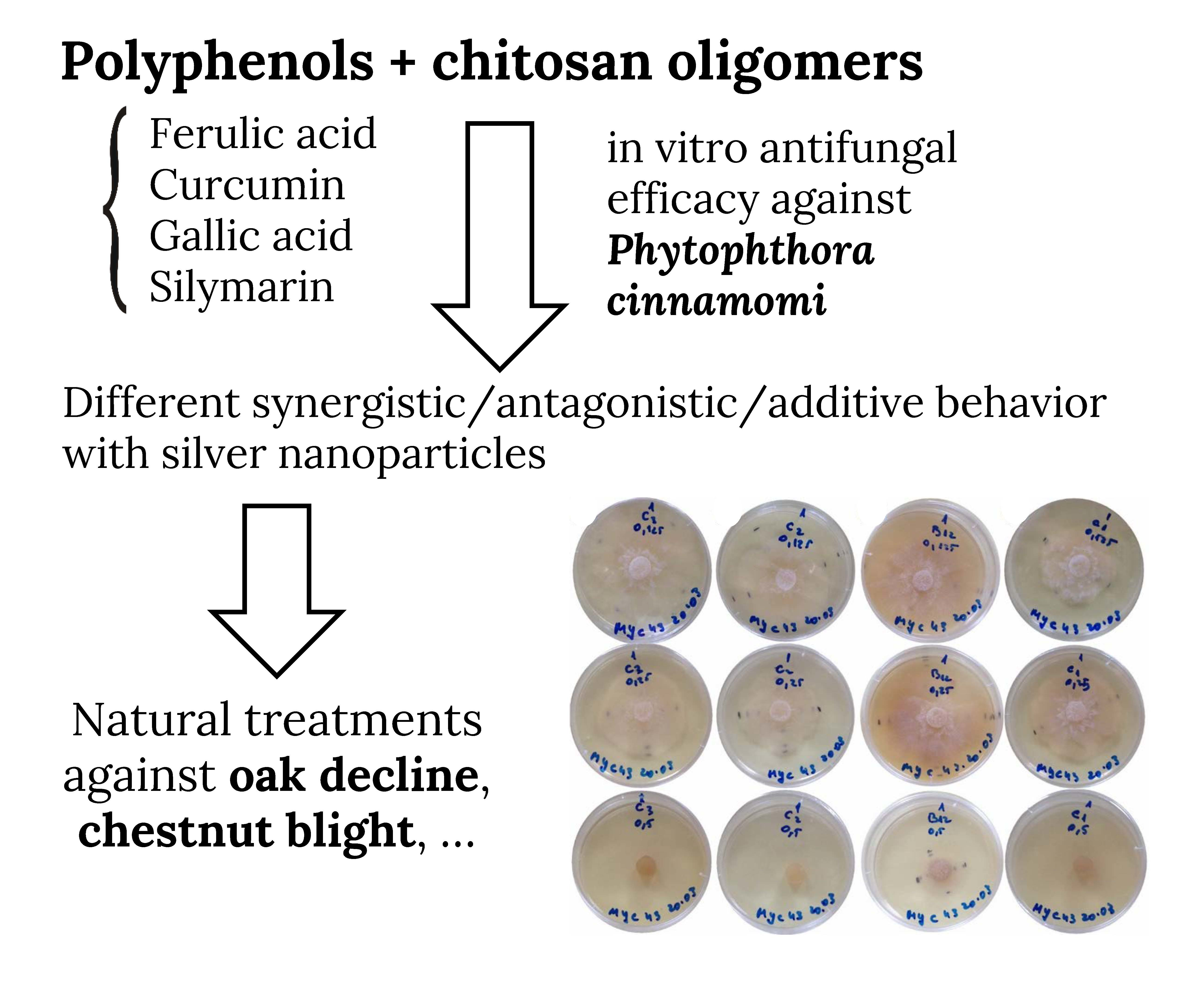
In Vitro Antifungal Activity of Chitosan-Polyphenol Conjugates against Phytophthora cinnamomi

1
Department of Bioengineering of Horticultural and Viticultural Systems, University of Agricultural Sciences and Veterinary Medicine of Bucharest, Bulevardul Mărăști 59, 011464 București, Romania
2
Departamento de Ingeniería Agroforestal, ETSIIAA, Universidad de Valladolid, Avenida de Madrid 44, 34004 Palencia, Spain
3
Servicio de Microbiología y Parasitología, Hospital Universitario Rio Hortega, SACYL, Calle Dulzaina 2, 47012 Valladolid, Spain
4
Instituto Universitario de Investigación en Ciencias Ambientales de Aragón (IUCA), EPS, Universidad de Zaragoza, Carretera de Cuarte, s/n, 22071 Huesca, Spain
*
Author to whom correspondence should be addressed.
AgriEngineering 2020, 2(1), 72-77; https://doi.org/10.3390/agriengineering2010005
Submission received: 31 December 2019 / Revised: 22 January 2020 / Accepted: 22 January 2020 / Published: 24 January 2020
(This article belongs to the Special Issue Selected Papers from 10th Iberian Agroengineering Congress)

Abstract

:
Phytophthora cinnamomi is responsible for radical rot in a wide range of hosts, resulting in large economic and ecological losses worldwide. In Spain, it is responsible for diseases such as the oak decline or the chestnut blight. In this study, different polyphenol-stevioside inclusion compounds dispersed in a hydroalcoholic solution of chitosan oligomers have been investigated, with a view to their application as natural bioactive complexes to replace conventional systemic fungicides against this fungus. The polyphenols tested in vitro were curcumin, ferulic acid, gallic acid and silymarin. Three concentrations (125, 250 and 500 µg·mL−1) were assayed, with and without silver nanoparticles (AgNPs), and notable differences were found in the inhibition of mycelium growth, with EC50 and EC90 values ranging from 171 to 373.6 µg·mL−1, and from 446.2 to 963.7 µg·mL−1, respectively. The results obtained showed that the addition of AgNPs, despite their antimicrobial activity, did not always lead to synergies. In the case of P. cinnamomi, an unexpected antagonistic behavior was found for two of the polyphenols (curcumin and silymarin), while an additive behavior for ferulic acid and a synergistic behavior for gallic acid were attained. In view of their inhibitory power, the preparations based on ferulic acid with AgNPs and on silymarin without AgNPs are proposed for applications in crop and forests protection against P. cinnamomi.

Graphical Abstract

1. Introduction

Phytophthora cinnamomi is an oomycete that lives on the ground nourishing itself thanks to decomposing matter. This pathogen, responsible for “root rot” or “regressive death”, is one of the most invasive species worldwide. It infects close to 5000 species of plants [1], affecting a variety of plant families: conifers, grasses, ferns, ornamental plants and food crops such as pineapple or avocado [2]. Its expansion has been attributed to the phenomenon of climate change, given that the potential disease range is influenced by winter temperature (disease development is strongly hampered by cold winters) and other climatic variables, such as summer temperatures and hydrologic variables. For instance, in Mediterranean forests, the initiation of the disease at the root level occurs during temporal waterlogging, caused by the higher frequency of extreme rain events [3]. Symptoms of P. cinnamomi infection include: wilting, reduced fruit size, death of young shoots, chlorosis of leaves and stem cankers [4].
In the Iberian Peninsula, P. cinnamomi is responsible for diseases such as the oak decline, with an enormous impact on the dehesa or montado ecosystem (a “man-made” ecosystem characterized by a savannah-like physiognomy that occupies extensive areas in Southern Spain and Portugal), or of chestnut blight [5].
At present there is no treatment able to eradicate regressive death by P. cinnamomi, although the stem injection of phosphites (which are elicitors that indirectly behave as fungicides) [6] and foliar spraying with metalaxyl + mancozeb (fungicides with contact and systemic activity) have been tested.
As an alternative to these conventional treatment agents, it is possible to use natural bioactive complexes extracted from plants, such as phenolic acids, flavonoids, tannins or lignans. In particular, polyphenols have been shown to feature a remarkable antimicrobial activity [7,8,9,10]. However, their applicability is limited by their low solubility and bioavailability [11], which may be improved through the formation of inclusion compounds with, for instance, terpene glycosides [12]. In this study, polyphenol-stevioside inclusion compounds dispersed in a hydroalcoholic solution of chitosan oligomers have been assayed. The polyphenols selected for the in vitro tests were curcumin, ferulic acid, gallic acid and silymarin, at different concentrations, with and without silver nanoparticles (AgNPs). The aim of the research has been to investigate the activity of the different chitosan-polyphenol conjugates against P. cinnamomi and to study synergistic effects upon addition of AgNPs to the conjugates.

2. Materials and Methods

2.1. Reagents and Preparation of the Bioactive Conjugates

Curcumin (CAS No. 458-37-7 ), ferulic acid (CAS No. 1135-24-6), gallic acid (CAS No. 149-91-7) and silymarin (MDL number MFCD01776359) were supplied by Sigma-Aldrich (Merck, Darmstadt, Germany); stevioside (CAS No. 57817-89-7) was supplied by Wako (Osaka, Japan); and medium molecular weight chitosan (MMWC) was purchased from Hangzhou Simit Chemical Technology Co. Ltd. (Hangzhou, China).
The preparation of chitosan oligomers from MMWC was carried out according to the methodology reported by Sun et al. [13]. Microwave-assisted aqueous biphasic system separation was used to prepare the polyphenol inclusion compounds. The conjugates with chitosan oligomers in hydroalcoholic solution medium were prepared according to the procedure previously reported in Matei et al. [14], and were characterized by infrared spectroscopy (FTIR), SEM and TEM microscopy techniques to ensure the reproducibility of the results presented in patent P201731489 [15].
It should be clarified that the antifungal efficacy assays reported herein and those reported in Matei et al. [14] (focused on the comparison between two dispersion media and on differences between polyphenols, but with AgNPs in all cases) were run in parallel. Since they were conducted at the same time and in the same conditions, the impact of physiological changes of the fungus or differences in abiotic conditions may be excluded.

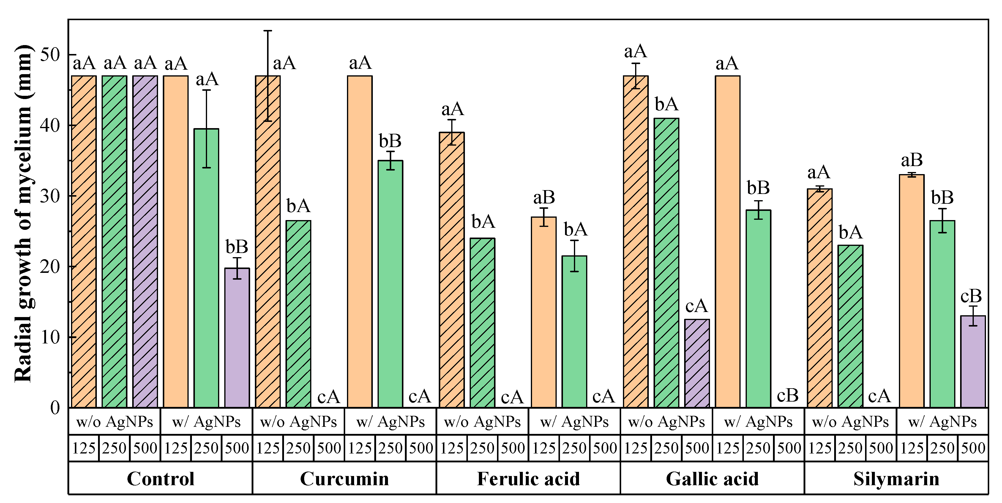
2.2. Fungal Isolate, Growth Conditions and In Vitro Tests of Mycelial Growth Inhibition

The fungal isolate of Phytophthora cinnamomi used for the in vitro sensitivity assays (MYC43) was provided by ICMC-IPROCOR (CICYTEX, Mérida, Spain) and was maintained in potato-dextrose-agar (PDA) culture medium at 4 °C. Fresh subcultures to obtain the inoculum for the tests were obtained by transferring hyphae plugs to Petri dishes containing PDA medium, which were incubated at 25 °C in the dark for 7 days.
To test the antifungal activity of the conjugates, the agar dilution method was used. Final concentrations of 125, 250 and 500 µg·mL−1 were obtained by adding aliquots of the stock solutions discussed in previous subsection to the PDA medium. Eight-mm mycelial disks from the margins of these fresh subcultures were then placed in these PDA plates. Pure PDA culture medium was used as the control. Three experiments, with three replicates per experiment, were carried out.
After 7 days of incubation at 25 °C in the dark, mycelial growth inhibition for each treatment and concentration was determined according to the formula: ((dcdt)/dc)×100, where dc and dt are the average diameters of the control and treated fungal colonies, respectively [16].
The concentrations that reduced mycelial growth by 50% and 90% (EC50 and EC90, respectively) were determined by regressing the values of radial growth inhibition (%) against the log10 values of the concentration of the conjugates.
To assess the joint action of the bioactive products in mixtures, Wadley’s method was used. In this method, if the synergy factor (SF) is 1, the hypothesis of similar joint action (additivity) can be accepted; if SF > 1, there is synergistic action; and if SF < 1 there is antagonistic action between the bioactive products [17].

2.3. Statistical Analyses

Data from the results obtained herein and from those reported in a previous study with AgNPs [14] were subjected to analysis of variance (ANOVA), followed by post hoc comparison of means through Tukey’s HSD (honest significant difference) test at p < 0.05. SPSS Statistics v.25 software (IBM; Armonk, NY, USA) was used.

3. Results and Discussion

The radial growth of the mycelium was monitored to study the in vitro activity of the different treatments. The results of the sensitivity assays are shown Figure 1 and Figure S1, in which one may observe that a reduction in the radial growth of the mycelium was attained in all cases when the concentration of the conjugates was increased from 125 to 500 µg·mL−1.
It is worth noting that the addition of AgNPs only resulted in a noticeable enhancement of activity for the conjugates based on gallic acid. For the conjugates based on ferulic acid the improvement was statistically significant only at the lowest dose; and for the conjugates based on curcumin or silymarin, the addition of AgNPs led to a lower performance (particularly evident for the conjugate with silymarin at 500 µg·mL−1).
The results from the factorial ANOVA (Table S1) indicated statistically significant one-way, two-way and three-way interaction effects. A classification of the treatments, both with and without AgNPs, according to Tukey’s HSD test is shown in Table S2.
The effective concentrations EC50 and EC90 for each treatment are summarized in Table 1. The presence/absence of AgNPs in the conjugates and the phenolic compound had a noticeable influence on the sensitivity of the isolate. While the presence of AgNPs barely modified the EC90 values for curcumin and ferulic acid, it significantly improved those of gallic acid conjugates (by 34% and 112% for EC50 and EC90, respectively).
In relation to the influence of the polyphenol, for the treatments without AgNPs, both of on the basis of the least square means and the EC50 values, the efficacy would follow the sequence: silymarin > ferulic acid > curcumin > gallic acid. On the basis of the EC90 values, the sensitivities of P. cinnamomi to curcumin, ferulic acid and silymarin would be similar, and gallic acid would be the least preferred choice.
According to Wadley’s method [17] for quantification of the level of interaction, a synergy factor FS = 1.7 was obtained for gallic acid, indicative of a synergistic interaction with AgNPs; for ferulic acid, FS = 1.0 was found, indicative of an additive behavior between the two antifungal products; and for silymarin and curcumin, FS values of 0.5 and 0.9 were calculated, respectively, indicative of an antagonistic behavior with the AgNPs.
Although rare in the literature, it should be clarified that, for example, cases of antagonistic behavior of AgNPs with amoxicillin versus methicillin-resistant Staphylococcus aureus (MRSA) have been reported [18]. These authors, in trials with 7 organisms and 19 antibiotics, completed 96 tests, finding 5 combinations with synergistic behavior, 89 with additive behavior and 2 with antagonistic behavior. This work would provide evidence of the existence of analogous complex interactions with AgNPs in the case of phenolic complexes.

4. Conclusions

The in vitro tests led to notable differences in the inhibition of mycelium growth of P. cinnamomi, observing a superior performance of the preparations based on ferulic acid with AgNPs and silymarin without AgNPs (thus evidencing that the addition of AgNPs, in spite of their antimicrobial activity, does not always result in synergies). In fact, an unexpected antagonistic behavior was detected for two of the polyphenols (curcumin and silymarin), an additive behavior was observed for ferulic acid and a synergistic behavior was found for gallic acid. In any case, in view of the EC50 and EC90 concentrations, these preparations can be promising bioactive products for protection applications of crops and forests against P. cinnamomi.

Supplementary Materials

The following are available online at https://www.mdpi.com/2624-7402/2/1/5/s1, Figure S1: Example of sensitivity test. Radial growth of mycelium for the control and treatments with curcumin, ferulic acid, gallic acid and silymarin; Table S1: Test of between-subjects effects; Table S2: Categories as a function of radial growth values for each polyphenol*dose*AgNPs combination, with a confidence interval of 95%, by Tukey’s HSD test.

Author Contributions

Conceptualization, E.P.-L., J.M.-G., M.C.R.-S. and P.M.-R.; Formal analysis, L.B.-D. and P.M.-R.; Funding acquisition, J.M.-G.; Investigation, P.M.M. and B.M.I.; Methodology, E.P.-L., J.M.-G. and M.C.R.-S.; Resources, J.M.-G.; Supervision, L.B.-D. and M.C.R.-S.; Validation, L.B.-D., M.C.R.-S. and P.M.-R.; Visualization, P.M.-R.; Writing – original draft, L.B.-D., E.P.-L., J.M.-G., M.C.R.-S. and P.M.-R.; Writing – review & editing, L.B.-D., J.M.-G. and P.M.-R. All authors have read and agreed to the published version of the manuscript.

Funding

This work was funded by JUNTA DE CASTILLA Y LEÓN under project VA258P18, with FEDER co-funding. P.M.-R. acknowledges the support of Universidad de Zaragoza under project UZ2019-TEC-07.

Conflicts of Interest

The authors declare no conflict of interest. The founding sponsors had no role in the design of the study; in the collection, analyses, or interpretation of data; in the writing of the manuscript, and in the decision to publish the results.

References

  1. Hardham, A.R.; Blackman, L.M.  Phytophthora cinnamomi. Mol. Plant Pathol. 2018, 19, 260–285. [Google Scholar] [CrossRef] [Green Version]
  2. Ramírez Gil, J.G. Incidencia, diagnóstico, comportamiento y alternativas de manejo de la marchitez del aguacate con énfasis en Phytophthora cinnamomi Rands; Universidad Nacional de Colombia: Medellín, Colombia, 2013. [Google Scholar]
  3. Homet, P.; González, M.; Matías, L.; Godoy, O.; Pérez-Ramos, I.M.; García, L.V.; Gómez-Aparicio, L. Exploring interactive effects of climate change and exotic pathogens on Quercus suber performance: Damage caused by Phytophthora cinnamomi varies across contrasting scenarios of soil moisture. Agricultural and Forest Meteorology 2019, 276, 107605. [Google Scholar] [CrossRef]
  4. Lowe, S.; Browne, M.; Boudjelas, S.; De Poorter, M. 100 of the World’s Worst Invasive Alien Species: A Selection from the Global Invasive Species Database; Invasive Species Specialist Group Auckland: Auckland, NZ, USA, 2004; p. 12. [Google Scholar]
  5. Sánchez, M.; Caetano, P.; Romero, M.; Navarro, R.; Trapero, A. Phytophthora root rot as the main factor of oak decline in southern Spain. In Progress in Research on Phytophthora Diseases of Forest Trees; Brasier, C., Jung, T., Oßwald, W., Eds.; Forest Research: Farnham, Surrey, UK, 2004; pp. 149–154. [Google Scholar]
  6. Jackson, T.J.; Burgess, T.; Colquhoun, I.; Hardy, G.E.S. Action of the fungicide phosphite on Eucalyptus marginata inoculated with Phytophthora cinnamomi. Plant Pathol. 2000, 49, 147–154. [Google Scholar] [CrossRef]
  7. Fialova, S.; Rendekova, K.; Mucaji, P.; Slobodnikova, L. Plant natural agents: Polyphenols, alkaloids and essential oils as perspective solution of microbial resistance. Curr. Org. Chem. 2017, 21. [Google Scholar] [CrossRef]
  8. Zacchino, S.A.; Butassi, E.; Liberto, M.D.; Raimondi, M.; Postigo, A.; Sortino, M. Plant phenolics and terpenoids as adjuvants of antibacterial and antifungal drugs. Phytomedicine 2017, 37, 27–48. [Google Scholar] [CrossRef]
  9. Teplova, V.V.; Isakova, E.P.; Klein, O.I.; Dergachova, D.I.; Gessler, N.N.; Deryabina, Y.I. Natural polyphenols: Biological activity, pharmacological potential, means of metabolic engineering (review). Appl. Biochem. Microbiol. 2018, 54, 221–237. [Google Scholar] [CrossRef]
  10. Gupta, M.; Chandra, A.; Aggarwal, G. Curcumin: potential therapeutic moiety for fungal infections. Current Traditional Medicine 2019, 4, 249–262. [Google Scholar] [CrossRef]
  11. Hussain, Z.; Thu, H.E.; Amjad, M.W.; Hussain, F.; Ahmed, T.A.; Khan, S. Exploring recent developments to improve antioxidant, anti-inflammatory and antimicrobial efficacy of curcumin: A review of new trends and future perspectives. Mater. Sci. Eng. C 2017, 77, 1316–1326. [Google Scholar] [CrossRef] [PubMed]
  12. Nguyen, T.T.H.; Si, J.; Kang, C.; Chung, B.; Chung, D.; Kim, D. Facile preparation of water soluble curcuminoids extracted from turmeric (Curcuma longa L.) powder by using steviol glucosides. Food Chem. 2017, 214, 366–373. [Google Scholar] [CrossRef] [Green Version]
  13. Sun, T.; Zhou, D.; Xie, J.; Mao, F. Preparation of chitosan oligomers and their antioxidant activity. Eur. Food Res. Technol. 2007, 225, 451–456. [Google Scholar] [CrossRef]
  14. Matei, P.; Martín-Gil, J.; Michaela Iacomi, B.; Pérez-Lebeña, E.; Barrio-Arredondo, M.; Martín-Ramos, P. Silver nanoparticles and polyphenol inclusion compounds composites for Phytophthora cinnamomi mycelial growth inhibition. Antibiotics 2018, 7, 76. [Google Scholar] [CrossRef] [PubMed] [Green Version]
  15. Martín-Gil, J.; Matei Petruta, M.; Pérez Lebeña, E. Complejo de Inclusión para Mejorar la Biodisponibilidad de Compuestos BiolóGicamente Activos no Hidrosolubles. US patent p201731489, 28 December 2017. [Google Scholar]
  16. Şesan, T.E.; Enache, E.; Iacomi, B.M.; Oprea, M.; Oancea, F.; Iacomi, C. In vitro antifungal activity of some plant extracts against Fusarium oxysporum in blackcurrant (Ribes nigrum L.). Acta Sci. Pol. Hortorum 2017, 16, 167–176. [Google Scholar] [CrossRef]
  17. Wadley, F.M. The Evidence Required to Show Synergistic Action of Insecticides and a Short Cut in Analysis; U.S. Government Printing Office: Washington DC, USA, 1945.
  18. De Souza, A.; Mehta, D.; Leavitt, R. Bactericidal activity of combinations of silver-water dispersion with 19 antibiotics against seven microbial strains. Curr. Sci. 2006, 91, 1825–1851. [Google Scholar]
Figure 1. Radial growth values of P. cinnamomi mycelium in the presence of the conjugates, composed of various polyphenol inclusion compounds with (w/) or without (w/o) silver nanoparticles (AgNPs) [14] at different concentrations (in µg·mL−1). For each treatment, concentrations labelled with the different lowercase letters are significantly different at p < 0.05 by Tukey’s test. For treatments with the same polyphenol and at the same dose, different uppercase letters indicate that the absence/presence of AgNPs resulted in significant differences at p < 0.05 by Tukey’s test. Mean values of three experiments with three replicates are presented, with error bars representing the standard deviation.
Agriengineering 02 00005 g001
Table 1. Effective concentrations of the conjugates that inhibited mycelial growth by 50% and 90% (EC50 and EC90, respectively).
TreatmentEC50 (µg·mL−1)EC90 (µg·mL−1)
w/o AgNPsw/AgNPsw/o AgNPsw/AgNPs
Control--458.4--1192.8
Curcumin257.5279.9448.3487.4
Ferulic acid228.7171.6446.2450.4
Gallic acid373.6261.3795.3455.6
Silymarin195.5261.8453.1963.7

