Case Report

Gastro-Splenic Fistula Related to Large B Cell Lymphoma

1
Haematology Department, Royal Blackburn Hospital, Blackburn BB2 3HH, UK
2
Gastroenterology Department, Royal Blackburn Hospital, Blackburn BB2 3HH, UK
3
Respiratory Department, Royal Blackburn Hospital, Blackburn BB2 3HH, UK
4
Radiology Department, Royal Blackburn Hospital, Blackburn BB2 3HH, UK
5
Department of Pathology, Royal Blackburn Hospital, Blackburn BB2 3HH, UK
6
Department of Surgery, Royal Blackburn Hospital, Blackburn BB2 3HH, UK
7
Haematology Department, Blackpool Victoria Hospital, Blackpool FY3 8NR, UK
8
Haematopathology Department, Leeds Teaching Hospitals, Leeds LS1 3EX, UK
*
Author to whom correspondence should be addressed.
Reports 2020, 3(2), 17; https://doi.org/10.3390/reports3020017
Submission received: 1 May 2020 / Revised: 13 June 2020 / Accepted: 13 June 2020 / Published: 17 June 2020

Abstract

:
We report a case of spontaneous gastrosplenic fistula in a 57 year old female who presented to the emergency department with abdominal pain and weight loss. From the physical examination, she had a palpable abdominal mass. A CT scan was performed and showed a mass involving the proximal greater curve of the stomach, infiltrating the spleen and pancreas. There was a 12 mm defect in the cardia of the stomach with gas entering the large mass but there was no free gas in the abdomen. The defect was a gastrosplenic fistula. A gastroscopic biopsy confirmed the diagnosis of diffuse large B cell lymphoma. Surgical removal of the mass was not feasible; therefore she was treated with RCHOP chemotherapy, achieving complete remission.

1. Introduction

Gastrosplenic fistula (GSF) is a rare disease entity resulting from gastric, splenic malignancies and rarely benign conditions. In the literature, there are 28 cases reported since 1962, first case report was from Belgium [1]. With chemotherapy there is a risk of fistula formation as a result of tumor necrosis and gastric wall invasion. The majority of the reported cases were treated with splenectomy and partial gastrectomy followed by chemotherapy [2,3]. When surgical options are deemed risky due to poor general conditions, the outcome is generally poor [3]. However our patient’s condition was complicated by gastro intestinal bleed and pulmonary embolism. Surgical intervention was not feasible and therefore she was treated with chemotherapy. This is the third case in the literature where chemotherapy was administered to a lymphoma patient with GSF, prior any surgical intervention. In the majority of the cases, GSF occurred during chemotherapy or after the completion of treatment. If it was diagnosed prior the administration of chemotherapy, surgery was performed. In our case, chemotherapy was administered concomitantly with GSF.

2. Case Presentation

A 57 year old female presented to the emergency department with 6 weeks history of right upper abdominal pain. She had lost 6 kg in weight and was experiencing night sweats. On admission she was febrile and hypotensive. Her temperature was 40.0 °C and her oxygen saturation was 95% on room air with a blood pressure of 90/59 mmHg.
Written informed consent was obtained from the patient for publication of this case report and any accompanying images.
The physical examination revealed a palpable right upper quadrant mass and abdominal tenderness. Bowel sounds were normal and there was no generalized lymphadenopathy. Spleen tip was palpable.
Laboratory results were as follows:
WBC: 20.0 × 10⁹/L (normal:4.0–11.0), NE: 16.4 × 10⁹/L (normal 2.0–7.5), LY: 1.4 × 10⁹/L (normal 1.5–4.0), MO: 1.8 × 10⁹/L (normal 0.2–0.8), HB: 82 g/L (normal 115–165), MCV: 77.6 fL (normal 76–100), RDW: 15.9% (normal 10.0–15.7), PLTS: 710 × 109/L (normal 150-450), crea: 56 umol/L (normal 46–92), urea: 5.2 mmol/L (normal 2.5–7.8), bili: 9 umol/L (normal 0–21), ALT: 31 IU/L (normal 3–53), ALP: 137 IU/L (normal 30–130), alb: 32 g/L (normal 35–50), amy: <30 IU/L (normal 30–110), CRP: 342 mg/L (normal 0–10), LDH: 1303 IU/L (normal 313–618), iron: 3.0 umol/L (normal 7.0–30.0), B2-microglobulin: 2.2 mg/L (normal: 1.2–2.4).
The CT scan revealed a 13.3 × 10.7 cm lesion of the large distal body and tail of pancreas. This communicated with the cardia of the stomach and contained multiple specks of gas. The lesion infiltrated the spleen which was difficult to identify separately. There was also a smaller 1.4 × 1.2 cm low attenuation lesion anterior to the larger lesion.
Additionally, a non-obstructing 2.6 × 2.5 cm soft tissue density mass involved the ileocecal valve (Figure 1 and Figure 2).
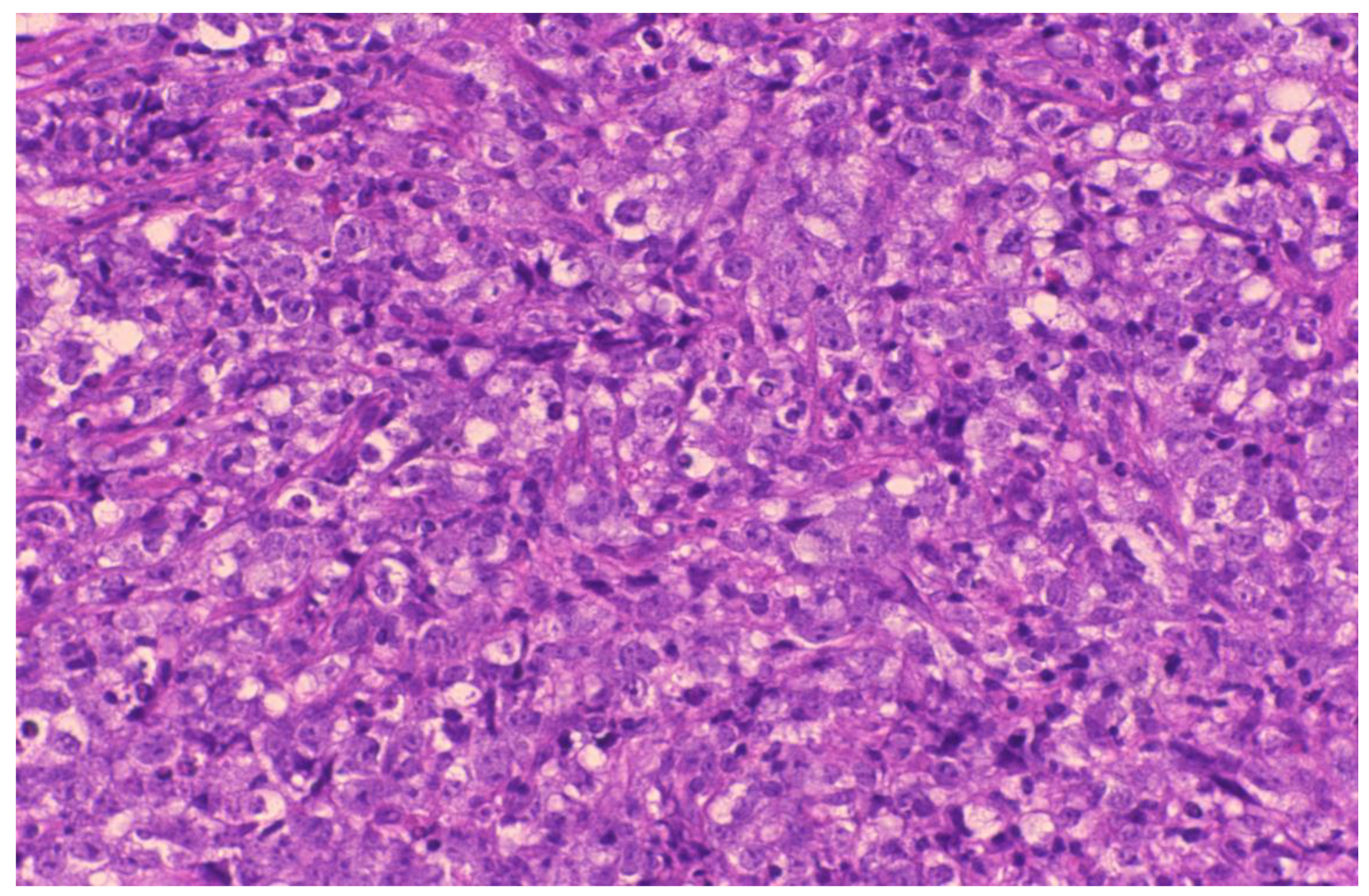
A gastroscopy performed showed a malignant looking ulcer on the gastric fundus. Figure 3 shows the heterotypic population of lymphoma cells from the gastric biopsy.
Figure 4 shows gastric mucosa with infiltration by large lymphoid cells expressing a post germinal center phenotype and a high proliferation index. There was co-expression of CD30. Immunohistochemistry testing showed BCL-2+ BCL-6+ CD10 CD20+ CD23 CD3 CD30+ CD5 CD79+ CyclinD1 IRF4+ Ki6790% LMP-1 expression.
FISH was carried out on thin paraffin sections. (Dako probe sets) and the lymphoid cells had no evidence of an MYC gene rearrangement.
Her stage was IVB, with a revised IPI of 3 and CNS IPI risk score suggested a 10% risk of CNS relapse within 2 years.
The patient was started on intravenous antibiotics and there were multiple discussions regarding the management of her case.
Surgical treatment of the gastro splenic fistula was not considered feasible. The collection resulting from the fistula was contained but surrounded by an inflammatory phlegmon. The fistula itself could not be repaired and surgical treatment would have involved at least a total gastrectomy, splenectomy and distal pancreatectomy. This would not remove the whole mass and would have been very high risk in her clinical condition. It would have been non curative and delayed potentially curative chemotherapy.
Following lengthy MDT discussions it was decided to administer six cycles of RCHOP with three cycles of intrathecal methotrexate and high dose methotrexate at the end.
During the patient’s hospital stay, she had two events of gastric bleeding with an approximate blood loss of 600 mL. Prophylactic tinzaparin was stopped. Four days after the gastric bleed she was considered stable so she received her first cycle of RCHOP. The team appreciated that there were major risks of complications from the administration of RCHOP, however it was deemed to be the most appropriate treatment option for our patient.
On day +9 post RCHOP and whilst being on prophylactic Tinzaparin for five days, the patient became significantly hemodynamically compromised.
She collapsed whilst returning from the bathroom. Oxygen saturation was 88% and patient became hypotensive with a blood pressure of 75/60 mmHg.
We decided to proceed with CT scans to exclude cerebral bleeding, to review the lymphoma and to rule out a pulmonary embolism.
Regarding the lymphoma, the large mass in the upper abdomen had increasing locules of gas and clear communication was seen with the stomach.
Unfortunately, the CT scan also revealed multiple pulmonary emboli involving the right main pulmonary, right upper and lower lobe and main left lower lobe arteries. The main pulmonary artery was not dilated. The patient had a small peripheral wedge shaped area of opacification in the left lower lobe, ground glass changes in the upper lobes bilaterally, which was concerning for pulmonary infarction. She also had a right internal iliac vein thrombus and thrombus in the left femoral vein. Following discussion with the Acute Care Team it was decided that thrombolysis with alteplase was considered a reasonable option even though the risk for a lethal bleed was apparent. This was discussed with the patient who agreed to proceed.
Thrombolysis was beneficial in this case and there were no bleeding complications afterwards.
The patient completed six cycles of RCHOP, three cycles of intrathecal methotrexate (cytospin at diagnosis was clear) and high dose methotrexate in the end.
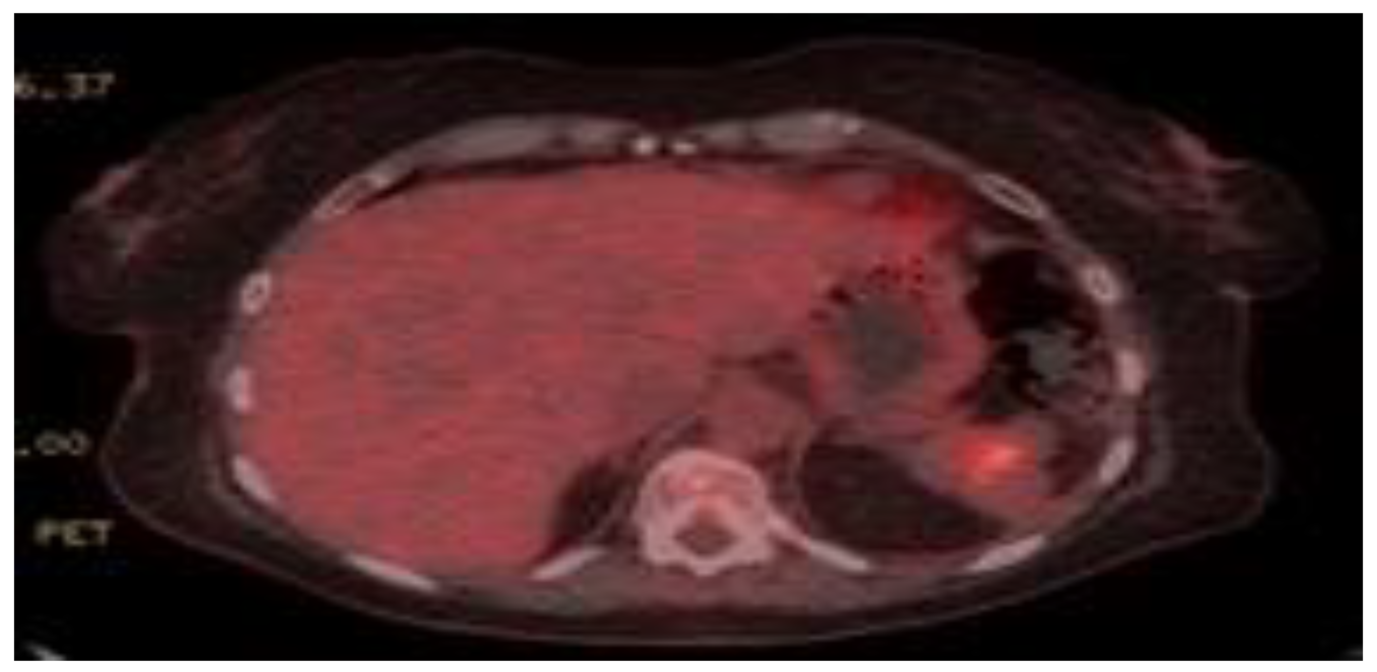
The end of treatment PET CT scan showed complete remission. The patient is still in remission 2 years post completion of treatment (Figure 5).

3. Discussion

Gastrosplenic fistula is a rare complication seen mainly in gastric and splenic lymphomas [4,5,6,7,8,9,10,11,12,13,14,15,16,17,18,19,20,21,22,23]. The treatment is usually surgical, followed by chemotherapy. Al-Ashgar et al. recommend avoiding extensive open laparoscopic procedures and suggest simple laparoscopic fistulectomy with partial gastric resection [22].
One of the most feared complications of GSF is massive hematemesis, which can be lethal.
By reviewing the literature, massive hematemesis has been reported in 2 lymphoma cases with erosion of gastric contents into the splenic circulation [15,17]. In the event of GSF massive bleeding, Moghazy KM suggests splenic artery embolization, followed by splenectomy and gastric resection [10].
Diffuse Large B cell Lymphoma seems to have a tendency to invade the gastric wall and cause widespread necrosis causing fistulation with the adjacent organs [19]. Also, Crohn’s disease [23] and benign gastric ulcers [24,25,26,27] are known causes.
In our case, the patient received no surgical treatment and underwent chemotherapy. This is the first case in the literature where chemotherapy has been given first. The fistula resolved with continued chemotherapy. There are also other reports in the literature where fistulas resolved post chemotherapy [4]. Early diagnosis of GSF may be life-saving but can be challenging and should be considered in every DLBCL patient undergoing routine abdominal imaging. Abdominal CT seems superior to other radiological tests for diagnosing GSF [13].
We thought that this case was worth mentioning, as GSF is recognized as a rare complication of gastric or splenic lymphoma.

Author Contributions

Conceptualization, D.T.; Methodology, J.A.; Software, M.S.A., S.A.-I., R.T.; Validation, H.C.; Formal Analysis, A.B.A.; Investigation, I.C.; Resources, M.M.; Data Curation, D.T., R.T., N.S.; Writing-Original Draft Preparation, D.T.; Writing-Review & Editing, J.A., M.P.; Visualization, I.G.; Supervision, A.K.; Project Administration, L.F. All authors have read and agreed to the published version of the manuscript.

Funding

This research received no external funding.

Acknowledgments

We would like to thank the Haematological Malignancy Diagnostic Service–Leeds (HMDS) for their continuous support.

Conflicts of Interest

The authors declare no conflict of interests.

References

  1. De Scoville, A.; Bovy, P.; Demeester, P. Radiological splenomegaly by lymphosarcoma splenic necrosing with double fistulation in the digestive tract. Acta Gastroenterol. Belg. 1967, 30, 840–846. [Google Scholar]
  2. Ding, Y.-L.; Wang, S.-Y. Gastrosplenic fistula due to splenic large B-cell lymphoma. J. Res. Med. Sci. Off. J. Isfahan Univ. Med. Sci. 2012, 17, 805–807. [Google Scholar]
  3. Kang, D.H.; Huh, J.; Lee, J.H.; Jeong, Y.K.; Cha, H.J. Gastrosplenic fistula occurring in lymphoma patients: Systematic review with a new case of extranodal NK/T-cell lymphoma. World J. Gastroenterol. 2017, 23, 6491–6499. [Google Scholar] [CrossRef] [PubMed]
  4. A Carolin, K.; Prakash, S.H.; Silva, Y.J. Gastrosplenic fistulas: a case report and review of the literature. Am. Surg. 1997, 63, 1007–1010. [Google Scholar] [PubMed]
  5. Aribaş, B.K.; Başkan, E.; Altinyollar, H.; Ungül, U.; Cengiz, A.; Erdil, H.F. Gastrosplenic fistula due to splenic large cell lymphoma diagnosed by percutaneous drainage before surgical treatment. Turk. J. Gastroenterol. 2008, 19, 69–70. [Google Scholar]
  6. Jain, V.; Pauli, E.; Sharzehi, K.; Moyer, M. Spontaneous gastrosplenic fistula secondary to diffuse large B-cell lymphoma. Gastrointest. Endosc. 2011, 73, 608–609. [Google Scholar] [CrossRef] [PubMed]
  7. Seib, C.D.; Rocha, F.G.; Hwang, D.G.; Shoji, B.T. Gastrosplenic Fistula from Hodgkin’s Lymphoma. J. Clin. Oncol. 2009, 27, e15–e17. [Google Scholar] [CrossRef]
  8. Marín, A.G.; García, L.B.; Rodríguez, A.V.; L, M.V.; Fuentes, F.T. Spontaneous gastrosplenic fistula secondary to primary gastric lymphoma. Rev. Esp. Enferm. Dig. 2009, 101, 76–78. [Google Scholar]
  9. Yang, S.E.; Jin, J.-Y.; Song, C.W.; Park, J.C.; Lee, J.I.; Kim, W.; Kim, J.; Lee, H.G. Gastrosplenic Fistula Complicated in a Patient with Non- Hodgkin’s Lymphoma. Cancer Res. Treat. 2002, 34, 153–156. [Google Scholar] [CrossRef]
  10. Moghazy, K. Gastrosplenic fistula following chemotherapy for lymphoma. Gulf J. Oncol. 2008, 3, 64–67. [Google Scholar]
  11. Puppala, S.; Williams, R.; Harvey, J.; Crane, M. Spontaneous gastrosplenic fistula in primary gastric lymphoma: case report and review of literature. Clin. Radiol. Extra 2005, 60, 20–22. [Google Scholar] [CrossRef] [Green Version]
  12. Maillo, C.; Bau, J. [Gastrosplenic and thoracosplenic fistula due to primary untreated splenic lymphoma]. Rev. Esp. Enferm. Dig. 2009, 101, 222–223. [Google Scholar] [PubMed]
  13. Palmowski, M.; Zechmann, C.M.; Satzl, S.; Bartling, S.; Hallscheidt, P. Large gastrosplenic fistula after effective treatment of abdominal diffuse large-B-cell lymphoma. Ann. Hematol. 2007, 87, 337–338. [Google Scholar] [CrossRef]
  14. Rothermel, L.D.; Chadwick, C.L.; Thambi-Pillai, T. Gastrosplenic fistula: etiologies, diagnostic studies, and surgical management. Int. Surg. 2010, 95, 270–272. [Google Scholar] [PubMed]
  15. A Bird, M.; Amjadi, D.; Behrns, K.E. Primary splenic lymphoma complicated by hematemesis and gastric erosion. South. Med. J. 2002, 95, 941–942. [Google Scholar] [CrossRef]
  16. Harris, N.L.; Aisenberg, A.C.; Meyer, J.E.; Ellman, L.; Elman, A. Diffuse large cell (Histiocytic) lymphoma of the spleen: Clinical and pathologic characteristics of ten cases. Cancer 1984, 54, 2460–2467. [Google Scholar] [CrossRef]
  17. Hiltunen, K.-M.; Airo, I.; Mattila, J.; Helve, O. Massively Bleeding Gastrosplenic Fistula Following Cytostatic Chemotherapy of a Malignant Lymphoma. J. Clin. Gastroenterol. 1991, 13, 478–480. [Google Scholar] [CrossRef]
  18. Kerem, M.; Sakrak, O.; Yilmaz, T.U.; Gultekin, F.A.; Bedirli, A.; Dursun, A. Spontaneous Gastrosplenic Fistula in Primary Gastric Lymphoma: Surgical Management. Asian J. Surg. 2006, 29, 287–290. [Google Scholar] [CrossRef] [Green Version]
  19. Choi, J.E.; Chung, H.J.; Lee, H.G. Spontaneous gastrosplenic fistula: a rare complication of splenic diffuse large cell lymphoma. Abdom. Imaging 2002, 27, 728–730. [Google Scholar] [CrossRef] [PubMed]
  20. Bubenik, O.; Lopez, M.J.; Greco, A.O.; Kraybill, W.G.; Cherwitz, D.L. Gastrosplenic fistula following successful chemotherapy for disseminated histiocytic lymphoma. Cancer 1983, 52, 994–996. [Google Scholar] [CrossRef]
  21. Blanchi, A.; Bour, B.; Alami, O. Spontaneous gastrosplenic fistula revealing high-grade centroblastic lymphoma: Endoscopic findings. Gastrointest. Endosc. 1995, 42, 587–589. [Google Scholar] [CrossRef]
  22. Al-Ashgar, H.I.; Khan, M.Q.; Ghamdi, A.M.; Bamehriz, F.Y.; Maghfoor, I. Gastrosplenic fistula in Hodgkin’s lymphoma treated successfully by laparoscopic surgery and chemotherapy. Saudi Med. J. 2007, 28, 1898–1900. [Google Scholar] [PubMed]
  23. Cary, E.R.; Tremaine, W.J.; Banks, P.M.; Nagorney, D.M. Isolated Crohn’s disease of the stomach. Mayo Clin. Proc. 1989, 64, 776–779. [Google Scholar] [CrossRef] [Green Version]
  24. Ching, J.L.C.; Owen, M.J.; Heller, C.A. Radiological gastric filling defect due to penetration into the spleen by a large gastric ulcer. Br. J. Radiol. 1983, 56, 488–489. [Google Scholar] [CrossRef] [PubMed]
  25. Glick, S.N.; Levine, M.S.; Teplick, S.K.; Gasparaitis, A. Splenic penetration by benign gastric ulcer: Preoperative recognition with CT. Radiology 1987, 163, 637–639. [Google Scholar] [CrossRef]
  26. Joffe, N.; Antonioli, D.A. Penetration into spleen by benign gastric ulcer. Clin. Radiol. 1981, 32, 177–181. [Google Scholar] [CrossRef]
  27. Frenkel, A.; Bichovsky, Y.; Perry, Z.H.; Peiser, J.; Roy-Shapira, A.; Brotfain, E.; Koyfman, L.; Binyamin, Y.; Nalbandyan, K.; Klein, M. Management of gastrosplenic fistula in the emergency setting—A case report and review of the literature. Ann. Med. Surg. 2018, 29, 26–29. [Google Scholar] [CrossRef] [PubMed]
Figure 1. Early CT showing loss of stomach (*) wall integrity and fistulation into spleen (+). The fistulous tract is shown by the arrow.
Reports 03 00017 g001
Figure 2. Image from a later CT showing complete breakdown of the stomach (*) wall continuity into a large collection replacing the spleen (+). Clear communication seen between the stomach and collection defined by the arrow.
Reports 03 00017 g002
Figure 3. Biopsy revealed lymphoma cells with round, oval, nuclei, multiple punctate nucleoli and scant cytoplasm (hematoxylin and eosin ×20).
Reports 03 00017 g003
Figure 4. Hematoxylin and eosin stained section (top left) shows gastric mucosa with infiltration of the lamina propria by sheets of large lymphoid cells. On immunohistochemistry, they are positive for CD20, bcl2 and bcl6, express a post germinal center phenotype (IRF4 positive, CD10 negative) and show a high proliferation index (Ki67—90%). There is co-expression of CD30. The images were taken on an a Nikon Eclipse 80i microscope with Plan Apo lens ×20 magnification, using a Nikon DSFi1 camera and DS-L2 Nikon digital sight system.
Reports 03 00017 g004
Figure 5. End of treatment Axial PET image showing excellent response with minimal non-specific residual FDG activity in spleen considered likely post inflammatory.
Reports 03 00017 g005