Management and Prevention of Oral Self-Injuries in Lesch–Nyhan Syndrome


CIR Dental School, Section of Paediatric Dentistry, University of Turin, 10124 Torino, Italy
*
Author to whom correspondence should be addressed.
Reports 2018, 1(1), 8; https://doi.org/10.3390/reports1010008
Submission received: 19 March 2018
/
Revised: 28 March 2018
/
Accepted: 6 April 2018
/
Published: 9 April 2018
Abstract
:Lesch–Nyhan syndrome (LNS) is a rare X-linked recessive disorder with an incidence of 1/100,000–380,000 live births. It is characterized by neurological manifestations, including symptoms of compulsive self-mutilation, which result in the destruction of oral and perioral tissues. This report describes a case of a four-year-old boy diagnosed with LNS, who was referred for evaluation and treatment of self-injury behaviour (SIB). The parents requested the prevention of self-mutilation of the lower lip and tongue by the child’s own teeth. After a thorough discussion with the parents, it was agreed that a conservative approach, avoiding extraction, should be followed initially. A removable dental appliance was fabricated. The parents were instructed and trained about insertion, removal, and cleaning of the appliance. The child was re-examined after one week: biting of the lips and tongue improved immediately after the insertion of the appliances. Initial healing of the lesion was observed. After two and four weeks, positive results were seen. The lesion had resolved completely. In conclusion, appropriate preventive methods have to be developed for each individual patient on the basis of the observation of each single case. Oral appliances represent a conservative solution for SIB and an alternative to more invasive approaches. They can be the initial solution for the management of oral self-injury in LNS patients.
1. Introduction
Lesch–Nyhan syndrome (LNS) is a rare X-linked recessive disorder [1] that was first described in 1964. This disorder primarily affects males and has an incidence of 1/100,000–380,000 live births [2] LNS is characterized by a biochemical abnormality of the enzyme hypoxanthine-guanine phosphoribosyltransferase (HPRT) [3]. The excessive uric acid production, due to the deficiency of HPRT, forms urate crystals, which are deposited in peripheral organs and tissues and produce neurological, renal, and musculoskeletal manifestations, such as developmental delay, growth and intellectual disability, nephrolithiasis, obstructive nephropathy, and acute gouty arthritis [1,4]. The compulsive self-injury behaviour (SIB) is the most distinctive symptom in LNS. Hypertonicity and involuntary movements appear around the first year of age [5] In children with SIB, persistent biting of the lips, tongue, fingers, and shoulders [6] and partial or total destruction of perioral tissues, especially of the lower lip, have been observed. Children can have partial or complete amputation of fingers, toes, and tongue [7]. Usually, physical restrains are necessary, since they are sensitive to pain but cannot control SIB [8]. Allopurinol is used for the pharmacological treatment of LNS; it reduces uric acid and prevents the development of renal and musculoskeletal injury, but it has no effect on the neurological manifestations and does not reduce the self-injuries behaviour [5,9]. Thanks to the delay of renal failure, which is the common cause of death in early childhood, a significant increase in life expectancy has been observed [10,11,12]. Benzodiazepines, neuroleptics, antidepressants, chloralhydrates, and anticonvulsive drugs have also been proposed to control SIB [13,14]. Recent therapeutic trials also include gabapentin, which is free from side effects [15], botulinum toxin A (BTX-A), which acts on the central and peripheral nervous system [16], dopamine replacement therapy, and deep brain stimulation in the globus pallidus (GPi) [17,18]. Olson and Houlihan 2000 [14] suggested that the self-destruction behaviour should be managed by a combination of treatments, such as physical restraints, behavioural treatment, and pharmaceutical therapy. Usually, children receive physical interventions including bandaging, gloves, or various types of restraints that protect their body from self-injuries [5,9]. Deon et al. and Piedimonte et al. described bilateral stimulation of GPi using a single electrode on each side of the brain to control patient motion and behavioural dysfunctions [19,20]. These studies demonstrate that deep brain stimulation may be used to treat the self-mutilating behaviour and dystonia associated with LNS. Also botulinum toxin A injections into bilateral masseters have been described to reduce the self-abusive behaviour in LNS patients [17].
2. Case Presentation Section
A four-year-old boy, previously diagnosed with Lesch–Nyhan syndrome, was referred to the Section of Paediatric Dentistry of the Dental School of Turin University in Italy. The child was referred for evaluation and treatment of SIB. The parents requested the prevention of self-mutilation of the lower lip and tongue by the child’s own teeth. The child was confined to a wheel chair, fitted with a seat belt to keep him upright and prevent head injury and energetic movements. Extra oral and intraoral examination was hard to perform because of the patient’s lack of cooperation and showed missing tissue of the lower lip and tongue. The surrounding tissues of the tongue and lip were inflamed. The patient showed a primary dentition and an Angle class I. Plaque control was adequate, and no caries cavities were found. Brushing was performed by the parents. After a discussion with the parents, it was agreed that a conservative approach avoiding extraction should be followed initially.
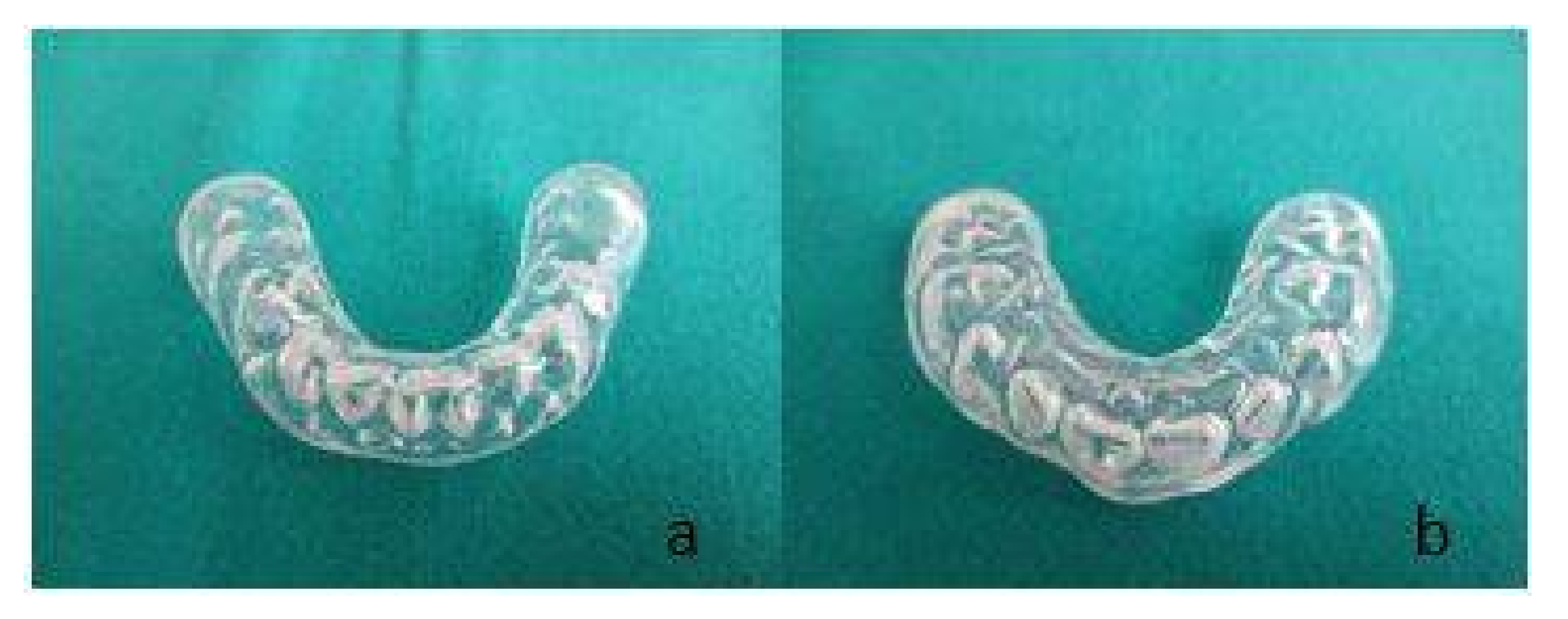
The parents subscribed the informed consent about the treatment and a removable dental appliance was fabricated (Figure 1).
A standard impression of the maxillary and mandibular arches with irreversible hydrocolloid were obtained. In this case, the patient showed a degree of collaboration to allow the intraoral alginate impression. The oral appliance was realized by employing thermoforming disc materials, using a positive pressure thermoformer. This appliance consisted of a mandibular and maxillary mouthguard aiming to prevent traumatic injuries of the tongue and lip. The bite plate of the appliance was smooth, with no indentations.
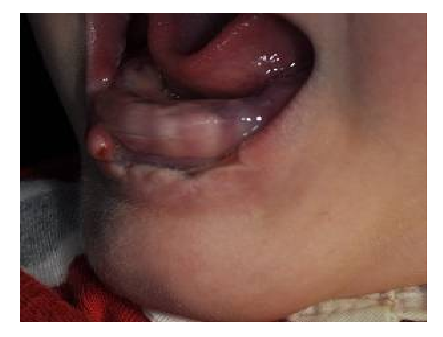
The parents were instructed and trained about insertion, removal, and cleaning of the appliance. Initially, the child could not tolerate the appliance inside this mouth, but once he accepted it, it reduced the self-inflicted damage of the lower lip and tongue. (Figure 2) The appliance was used as much as possible, all the day, in particular when SIB was severe.
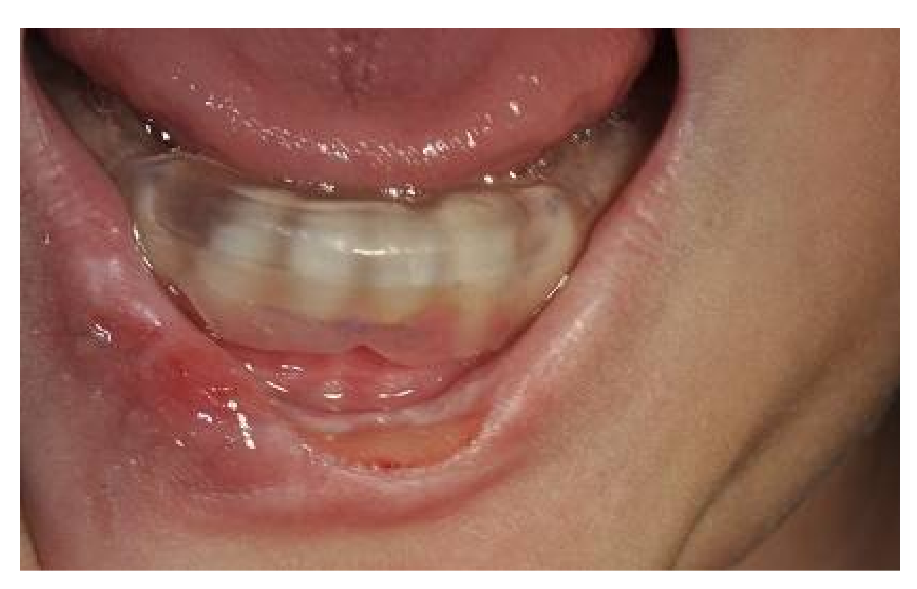
The child was re-examined after one week: biting of the lips and tongue was improved immediately after the insertion of the appliance. Initial healing of the lesion was observed. After two and four weeks, positive results were seen (Figure 3). The lesion had resolved completely. The parents were satisfied, SIB seemed to be under control, and the child could be treated initially with a conservative treatment before employing more invasive approaches.
3. Discussion
There are several studies on the pathogenesis of neurological disorders of LNS, such as self-mutilation, but the mechanisms of these disorders remain unclear.
Oral SIB is a difficult problem for the clinician to manage. There are no standard methods for the prevention of self-mutilation in LNS patients. Treatment modalities include drug therapy, extraction of teeth, oral appliances, and orthognathic surgery. The kind of treatment chosen strongly depends on the intensity of the self-injury behaviour. Both the general state of health of the patients with oral self-injury and the characteristics of the specific lesion are very variable, making it difficult to establish a common clinical protocol. A treatment approach is the extraction of teeth in primary or both dentitions. It may be an effective means to confront the severe medical problems arising from the persistent self-destruction, but it is an invasive approach and introduces a major oral disability.
According to the published literature, there is the possibility to treat oral self-injury with an intraoral appliance, that can restrict self-injurious biting and protect the tissues of individuals with SIB who use their teeth to inflict damage on oral and perioral structures. The appliance does not treat the source of the problem, but is an effective means of controlling self-mutilation. The intraoral appliance allows the protection of the oral soft tissue, including the buccal mucosa, tongue, and lips. The removable dental appliance which is described in this report is easy to fabricate and easy to be removed by the parents of affected children for everyday cleaning. The construction procedure must be as simple and short as possible, because of the limited cooperation ability of these kind of patients. Operators’ persuasive capacity, family, and operators’ efforts to change the negative behaviour of the patients are crucial.
Irreversible hydrocolloid impressions of the dental arches can be obtained in spite of a patient’s lack of compliance. Some patients with LNS show a sufficient degree of collaboration that allows the intraoral impressions, but other LNS patients do not cooperate, and sedation is sometime necessary to take the impressions.
4. Conclusions
Appropriate preventive methods have to be developed for each individual patient on the basis of the observation of each single case. Oral appliances represent a conservative solution for SIB and an alternative to more invasive approaches. They can be the initial solution for the management of oral self-injury in LNS patients.
Author Contributions
Giuliana Caserta treated the patient and wrote the manuscript; Patrizia Defabianis supervised the treatment, reviewed the manuscript, and approved the final manuscript as submitted.
Conflicts of Interest
The authors declare no conflict of interest.
References
- Seegmiller, J.E.; Rosenbloom, F.M.; Kelly, W.N. Enzyme defect associated with a sex-linked human neurological disorder and excessive purine synthesis. Science 1967, 155, 1682–1684. [Google Scholar] [CrossRef] [PubMed]
- Lesch, M.; Nyhan, W.L. A familiar disorder of uric acid metabolism and central nervous system function. Am. J. Med. 1964, 36, 561–570. [Google Scholar] [CrossRef]
- Shoptaw, J.T.; Reznik, J.I. Lesch–Nyhan syndrome: Report of three cases in one family. J. Dent. Child. 1978, 45, 403–407. [Google Scholar]
- Bundick, J. Lesch–Nyhan syndrome. J. Dent. Child. 1969, 36, 277–280. [Google Scholar]
- Nyhan, W.L. The Lesch–Nyhan syndrome. Annu. Rev. Med. 1973, 24, 41–60. [Google Scholar] [CrossRef] [PubMed]
- Fernald, C.D. The Lesch–Nyhan syndrome: Cerebral palsy, mental retardation, and self mutilation. J. Pediatr. Psychol. 1976, 1, 51–55. [Google Scholar] [CrossRef]
- Nyhan, W.L. Behavior in the Lesch Nyhan syndrome. J. Autism Child. Schizophr. 1976, 6, 235–252. [Google Scholar] [CrossRef] [PubMed]
- Scully, C. The orofacial manifestations of the Lesch–Nyhan syndrome. Int. J. Oral Surg. 1981, 10, 380–383. [Google Scholar] [CrossRef]
- Steadman, R.H.; McIntosh, G. Lesch-Nyhan syndrome. J. Oral Maxillofac. Surg. 1982, 40, 750–752. [Google Scholar] [CrossRef]
- Dicks, J.L. Lesch–Nyhan syndrome: A treatment-planning dilemma. Paediatr. Dent. 1982, 4, 127–130. [Google Scholar]
- Newcombe, D.S. Treatment of X-linked primary hyperuricemia with allopurinol. JAMA 1966, 198, 315–317. [Google Scholar] [CrossRef] [PubMed]
- Baumeister, A.A.; Frye, G.D. The biochemical basis of the behavioral disorder in the Lesch–Nyhan syndrome. Neurosci. Biobehav. Rev. 1985, 9, 169–178. [Google Scholar] [CrossRef]
- Van Moffaert, M. Management of self-mutilation. Psychother. Psychosom. 1989, 51, 180–186. [Google Scholar] [CrossRef] [PubMed]
- Olson, L.; Houlihan, D. A review of behavioral treatments used for Lesch–Nyhan syndrome. Behav. Modif. 2000, 24, 202–222. [Google Scholar] [CrossRef] [PubMed]
- Roach, P.S.; Delgado, M.; Anderson, L.; Iannaccone, S.T.; Burns, D.K. Carbamazepine trial for Lesch–Nyhan self-mutilation. J. Child Neurol. 1996, 11, 476–478. [Google Scholar] [CrossRef] [PubMed]
- Gutierrez, C.; Pellene, A.; Micheli, F. Botulinum toxin: Treatment of self-mutilation in patients with Lesch–Nyhansyndrome. Clin. Neuropharmacol. 2008, 31, 180–183. [Google Scholar] [CrossRef] [PubMed]
- Dabrowski, E.; Smathers, S.A.; Ralstrom, C.S.; Nigro, M.A.; Leleszi, J.P. Botulinum toxin as a novel treatment for self-mutilation in Lesch–Nyhan syndrome. Dev. Med. Child Neurol. 2005, 47, 636–639. [Google Scholar] [PubMed]
- Budnick, J. The Lesch–Nyhan syndrome. ASDC J. Dent. Child. 1967, 36, 277–280. [Google Scholar]
- Deon, L.L.; Kalichman, M.A.; Booth, C.L.; Slavin, K.V.; Gaebler-Spira, D.J. Pallidal deep-brain stimulation associated with complete remission of self-injurious behaviors in a patient with Lesch–Nyhan syndrome: A case report. J. Child. Neurol. 2012, 27, 117–120. [Google Scholar] [CrossRef] [PubMed]
- Piedimonte, F.; Andreani, J.C.; Piedimonte, L.; Micheli, F.; Graff, P.; Bacaro, V. Remarkable clinical improvement with bilateral globus pallidus internus deep brain stimulation in a case of Lesch–Nyhan disease: Five-year follow-up. Neuromodulation 2015, 18, 118–122. [Google Scholar] [CrossRef] [PubMed]
Figure 1.
(a) Total lower and (b) upper resin bites.

Figure 2.
Healing of the lesion one week after the beginning of the appliance therapy.

Figure 3.
The same lesion as in Figure 2 after four weeks: the recovery of the lip tissues is evident.
Figure 3.
The same lesion as in Figure 2 after four weeks: the recovery of the lip tissues is evident.
© 2018 by the authors. Licensee MDPI, Basel, Switzerland. This article is an open access article distributed under the terms and conditions of the Creative Commons Attribution (CC BY) license (http://creativecommons.org/licenses/by/4.0/).
Share and Cite
MDPI and ACS Style
Caserta, G.; Defabianis, P. Management and Prevention of Oral Self-Injuries in Lesch–Nyhan Syndrome. Reports 2018, 1, 8. https://doi.org/10.3390/reports1010008
AMA Style
Caserta G, Defabianis P. Management and Prevention of Oral Self-Injuries in Lesch–Nyhan Syndrome. Reports. 2018; 1(1):8. https://doi.org/10.3390/reports1010008
Chicago/Turabian StyleCaserta, Giuliana, and Patrizia Defabianis. 2018. "Management and Prevention of Oral Self-Injuries in Lesch–Nyhan Syndrome" Reports 1, no. 1: 8. https://doi.org/10.3390/reports1010008