Article

Incidence, Risk Factors, and Prevention of Deep Vein Thrombosis in Acute Ischemic Stroke Patients (IRIS-DVT Study): A Systematic Review and Meta-Analysis

1
Global Health Neurology Lab, Sydney, NSW 2150, Australia
2
UNSW Medicine and Health, South Western Sydney Clinical Campuses, University of New South Wales (UNSW), Sydney, NSW 2033, Australia
3
NSW Brain Clot Bank, NSW Health Pathology, Sydney, NSW 2170, Australia
4
Clinical Sciences Stream, Ingham Institute for Applied Medical Research, Sydney, NSW 2170, Australia
5
Department of Neurology & Neurophysiology, Liverpool Hospital & South Western Sydney Local Health District (SWSLHD), Sydney, NSW 2170, Australia
6
Division of Cerebrovascular Medicine & Neurology, Department of Neurology, National Cerebral and Cardiovascular Center (NCVC), 6-1 Kishibeshimmachi, Suita 564-8565, Osaka, Japan
*
Author to whom correspondence should be addressed.
Clin. Transl. Neurosci. 2025, 9(4), 49; https://doi.org/10.3390/ctn9040049
Submission received: 7 September 2025 / Revised: 5 October 2025 / Accepted: 6 October 2025 / Published: 9 October 2025
(This article belongs to the Topic Neurological Updates in Neurocritical Care)

Abstract

Background: Deep vein thrombosis (DVT) is a serious thromboinflammatory complication of acute ischemic stroke (AIS). The true incidence, mechanistic risk factors, and optimal prophylactic strategies remain uncertain, particularly in the era of reperfusion therapy. Methods: This systematic review and meta-analysis (IRIS-DVT) searched PubMed, Embase, Cochrane, Scopus, and Web of Science for studies reporting DVT incidence, risk factors, or prophylaxis in AIS (2004–2025). Random-effects models were used to generate pooled prevalence and effect estimates, and the certainty of evidence was graded using the GRADE framework. Results: Forty-two studies (n = 6,051,729 patients) were included. The pooled prevalence of DVT was 7% (95% CI, 6–9%), approximately seventy-fold higher than in the general population, with wide heterogeneity influenced by screening timing and diagnostic modality. Pathophysiological risk factors included higher stroke severity (NIHSS; SMD 0.41; 95% CI, 0.38–0.43), older age (SMD 0.32; 95% CI, 0.18–0.46), elevated D-dimer (SMD 0.55; 95% CI, 0.38–0.72), female sex (OR 1.33; 95% CI, 1.19–1.50), and malignancy (OR 2.69; 95% CI, 1.56–5.22), supported by moderate-certainty evidence. Respiratory infection and admission hyperglycemia showed weaker, low-certainty associations. Traditional vascular risk factors (hypertension, diabetes, atrial fibrillation, dyslipidemia) were not significantly related to DVT risk. Evidence for prophylaxis with low-molecular-weight heparin, direct oral anticoagulants, or intermittent pneumatic compression was limited and graded very low certainty. Conclusions: DVT complicates approximately one in fourteen AIS cases, reflecting a distinct thromboinflammatory process driven more by acute neurological severity, systemic hypercoagulability, and malignancy than by conventional vascular risk factors. Early systematic screening (≤72 h) and consistent use of mechanical prophylaxis are warranted. Dedicated AIS-specific mechanistic and interventional trials are urgently needed to refine prevention strategies and improve post-stroke outcomes.

1. Introduction

Deep vein thrombosis (DVT) is a serious and potentially life-threatening complication of acute ischemic stroke (AIS) [1]. It forms part of the broader spectrum of venous thromboembolism (VTE) [2], in which embolization to the pulmonary circulation may result in pulmonary embolism (PE)—a major contributor to post-stroke morbidity and mortality [3]. While PE is the most overtly fatal manifestation, its origins often lie in unrecognized distal or proximal DVT, highlighting the importance of early detection and prevention within stroke care pathways [4].
Over the past two decades, the advent of intravenous thrombolysis (IVT) and endovascular thrombectomy (EVT) has markedly improved neurological outcomes after AIS. Yet the risk of thromboembolic events has not declined proportionally [5,6,7]. Stroke-related immobility, systemic inflammation, endothelial dysfunction, and delayed initiation of prophylaxis collectively sustain a pro-thrombotic milieu even in modern, protocol-driven units [8]. Reported incidence rates of DVT in AIS vary widely across studies, ranging from below 1% to over 20% [8,9,10,11,12,13], reflecting methodological heterogeneity in screening protocols, imaging sensitivity, and regional prophylaxis practices rather than true biological variation [14,15].
Despite its clinical relevance, current knowledge of DVT after AIS remains fragmented. Most available studies are limited by retrospective or single-center designs, small sample sizes, or by extrapolating findings from non-stroke medical populations. As a result, the true burden of DVT in contemporary stroke care—and its complex interaction with reperfusion therapies, systemic inflammatory factors, and prophylactic strategies—remains uncertain. The comparative effectiveness of pharmacological prophylaxis (e.g., low-molecular-weight heparin, direct oral anticoagulants) [16,17,18,19] and mechanical approaches (e.g., intermittent pneumatic compression, inferior vena cava filters) [20,21,22,23,24] in AIS patients has not been comprehensively evaluated in a stroke-specific context. These limitations have hindered accurate risk stratification [4] and contributed to the ongoing inconsistency of international guideline recommendations, leaving clinicians without a unified, evidence-based prevention framework [25,26,27].
The Investigating the Incidence, Risk Factors, and Prophylactic Strategies for Deep Vein Thrombosis in Acute Ischemic Stroke Patients (IRIS-DVT) study was designed to address this critical evidence gap. It systematically synthesizes global data to define the incidence, determinants, and preventive strategies for DVT after AIS; examines how modern reperfusion therapies such as IVT and EVT alter thromboembolic risk through changes in mobility, procedural factors, and timing of prophylaxis; and evaluates the comparative efficacy and certainty of both pharmacological and mechanical interventions [5,28]. Beyond quantifying incidence and risk, the IRIS-DVT framework reconceptualizes DVT as a surrogate marker of systemic thromboinflammatory activation predisposing to PE and other VTE-related outcomes [1,29]. The IRIS-DVT study sought to generate evidence-based insights that can guide early risk assessment, inform precision-based prophylactic strategies, harmonize guideline recommendations, and improve long-term outcomes for patients with AIS.

2. Materials and Methods

2.1. Literature Search and Study Selection

We conducted a comprehensive systematic search of PubMed, Embase, Cochrane Library, Scopus, and Web of Science from January 2004 to May 2025. Search strategies combined terms related to “deep vein thrombosis” (DVT), “acute ischemic stroke” (AIS), “risk factors”, and “prophylactic interventions”, with filters for human studies in adults (≥18 years). A detailed search strategy is provided in the Supplementary Information. References of relevant reviews and meta-analyses were also screened to identify additional eligible studies.
Titles and abstracts were independently screened by two reviewers, with full-text reviews performed for potentially eligible studies. Discrepancies were resolved by consensus. A PRISMA (Preferred Reporting Items for Systematic Reviews and Meta-Analyses) flow diagram illustrates the selection process (Figure 1). This study was registered in Open Science Framework (OSF; registration ID: buxr8).

2.2. Eligibility Criteria

Studies were included if they: (1) enrolled adult patients with AIS; (2) reported the incidence or prevalence of DVT, risk factors for DVT after AIS, or the use and effectiveness of prophylactic interventions (pharmacological or mechanical); and (3) were designed as randomized controlled trials (RCTs), cohort studies, case–control studies, or other observational designs. Systematic reviews and meta-analyses were screened for relevant primary studies but were not pooled directly to avoid duplication. Case reports, small case series (<20 patients), pediatric populations, and non-English publications were excluded.

2.3. Data Extraction

All article titles and abstracts were initially reviewed in Endnote (Clarivate Analytics, London, UK) to exclude studies that did not meet the eligibility criteria. The remaining articles underwent full-text examination to confirm suitability for inclusion in the systematic review or meta-analysis. Data extraction was performed using a standardized sheet, capturing study-level demographics (author, country, publication year, registry or trial name, study design, number of centers), intervention characteristics (IVT, EVT, or both), though treatment-specific data were limited and inconsistently reported, and patient demographics (age, sex). Clinical and biological predictors collected included hypertension, diabetes mellitus, hyperlipidaemia, obesity, atrial fibrillation, tobacco and alcohol use, drug abuse, coronary artery disease, malignancy, respiratory infection, stroke severity measured by the National Institutes of Health Stroke Scale (NIHSS), admission glucose, D-dimer, low-density lipoprotein, and fibrinogen. Clinical outcomes were classified as patients who developed DVT versus those without DVT. When covariates or outcomes were incompletely reported, analyses were restricted to available cases; no imputation was undertaken for missing predictor variables, and such studies were excluded from specific pooled analyses.

2.4. Methodological Quality Assessment of Included Studies

The methodological quality assessment of included studies was conducted using the modified Jadad analysis (MJA) [30], completed independently by the primary researcher (Table S3). The risk of biases in results due to funding was also evaluated, based on the declaration of funding sources and conflicts of interest, extracted from each individual study (Table S4).

2.5. Certainty of Evidence Assessment (Grading)

The certainty of evidence across outcomes was assessed using the GRADE (Grading of Recommendations, Assessment, Development, and Evaluation) framework. The outcomes: incidence of DVT, risk factors, and prophylactic strategies were independently evaluated across five domains: risk of bias, inconsistency, indirectness, imprecision, and publication bias. Randomized controlled trials were initially rated as high certainty, whereas observational studies were rated as low certainty; evidence was subsequently downgraded for methodological limitations, heterogeneity, or imprecision, and upgraded in cases of large effect sizes, consistent associations, or dose–response relationships. Final ratings were categorized as high, moderate, low or very low.

2.6. Statistical Analyses

All statistical analyses were performed using STATA v13.0 (StataCorp, College Station, TX, USA). Baseline characteristics of included cohorts were extracted from each study. Where necessary, means and standard deviations (SDs) were estimated from medians and interquartile ranges (IQRs) using the method of Wan et al. [31], and combined with Bessel’s correction to ensure unbiased SD estimates. The pooled prevalence of DVT among patients with AIS was calculated using the metaprop command, applying a random-effects meta-analysis of proportions with exact 95% confidence intervals (CIs) obtained using the cimethod (exact) and ftt options. Associations between clinical or biological factors and DVT were synthesized using DerSimonian and Laird (DL) random-effects models, generating pooled odds ratios (ORs) for categorical variables and standardized mean differences (SMDs) for continuous variables.
Subgroup analyses were conducted according to reperfusion therapy type (IVT, EVT, or both), stroke territory (anterior, posterior, mixed), study design (retrospective, prospective, or mixed), diagnostic modality, timing of DVT screening, geographical region, and mid-point year of data collection. Pharmacological prophylaxis (low-molecular-weight heparin, unfractionated heparin, direct oral anticoagulants) and mechanical prophylaxis (intermittent pneumatic compression, compression stockings, inferior vena cava filters) were grouped according to reported use, and pooled effect sizes were estimated using random-effects models where sufficient data were available. Temporal trend heterogeneity was assessed by stratifying prevalence according to midpoint year of data collection. Apparent fluctuations were interpreted primarily as methodological variation (diagnostic sensitivity, coding practices, and prophylaxis availability) rather than true secular shifts.
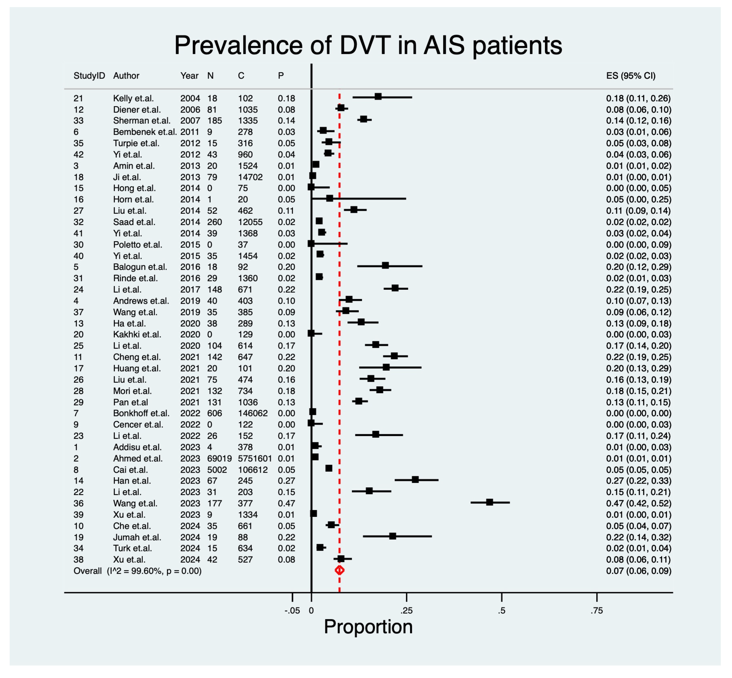
Forest plots were generated to display pooled effect sizes, weights, and heterogeneity estimates. Sensitivity analyses were performed using the metaninf command to assess the influence of individual studies on overall estimates. Between-study heterogeneity was quantified using the I2 statistic, with thresholds of <30% (low), 30–50% (moderate), 50–75% (substantial), and >75% (severe). Cochran’s Q test and Tau2 were additionally reported. Publication bias was assessed using funnel plots and Egger’s regression test (metabias and metafunnel commands). For selected predictors with consistent reporting, diagnostic accuracy was further assessed using summary receiver operating characteristic (SROC) curves and Fagan’s nomograms to illustrate discriminative performance and post-test probability. Funnel plot asymmetry was interpreted in conjunction with Egger’s p-values to evaluate bias risk. All statistical tests were two-tailed, and significance was set at p < 0.05. Detailed outputs of sensitivity analyses, funnel plots, SROC curves, and Fagan’s nomograms are provided in Figures S11–S45. Pooled summary estimates for prophylactic interventions and continuous biomarkers are presented in Tables S7 and S8. Heterogeneity metrics (I2, τ2) and study weights are displayed in Figure 2 and Figure 3.

3. Results

3.1. Description of Included Studies

A total of 3903 records were initially identified through database searches (PubMed, Embase, Cochrane, Scopus, and Web of Science) and an additional 183 records via citation searching and web searches. After removing 1307 duplicates automatically and 766 manually, 2013 records were screened. Of these, 1561 were excluded for irrelevance or being systematic reviews. A further 12 reports were not retrieved. Finally, 438 full-text articles were assessed for eligibility, and 393 were excluded (391 for focusing on other VTEs and 2 for insufficient data).
Ultimately, 42 studies, encompassing 6,051,729 AIS patients, were included in the meta-analysis (PRISMA flow diagram, Figure 1). These studies varied in design, including retrospective cohorts and randomized controlled trials, and spanned a wide geographical and temporal range. Further details regarding study characteristics, sample sizes, and outcome measures are summarized. Baseline characteristics of the included studies are summarized in Table 1, with detailed study-level data presented in Table S6. Discrete and continuous risk factors for deep vein thrombosis (DVT) following acute ischemic stroke are summarized in Table 2 and Table 3, respectively. Table 4 presents the key predictors of DVT after stroke, while Table 5 outlines the effectiveness of various prophylactic interventions in preventing DVT among patients with acute ischemic stroke. A comprehensive summary of the certainty and quality of evidence across all key outcomes is provided in Table 6.

3.2. Pooled Prevalence of DVT in AIS

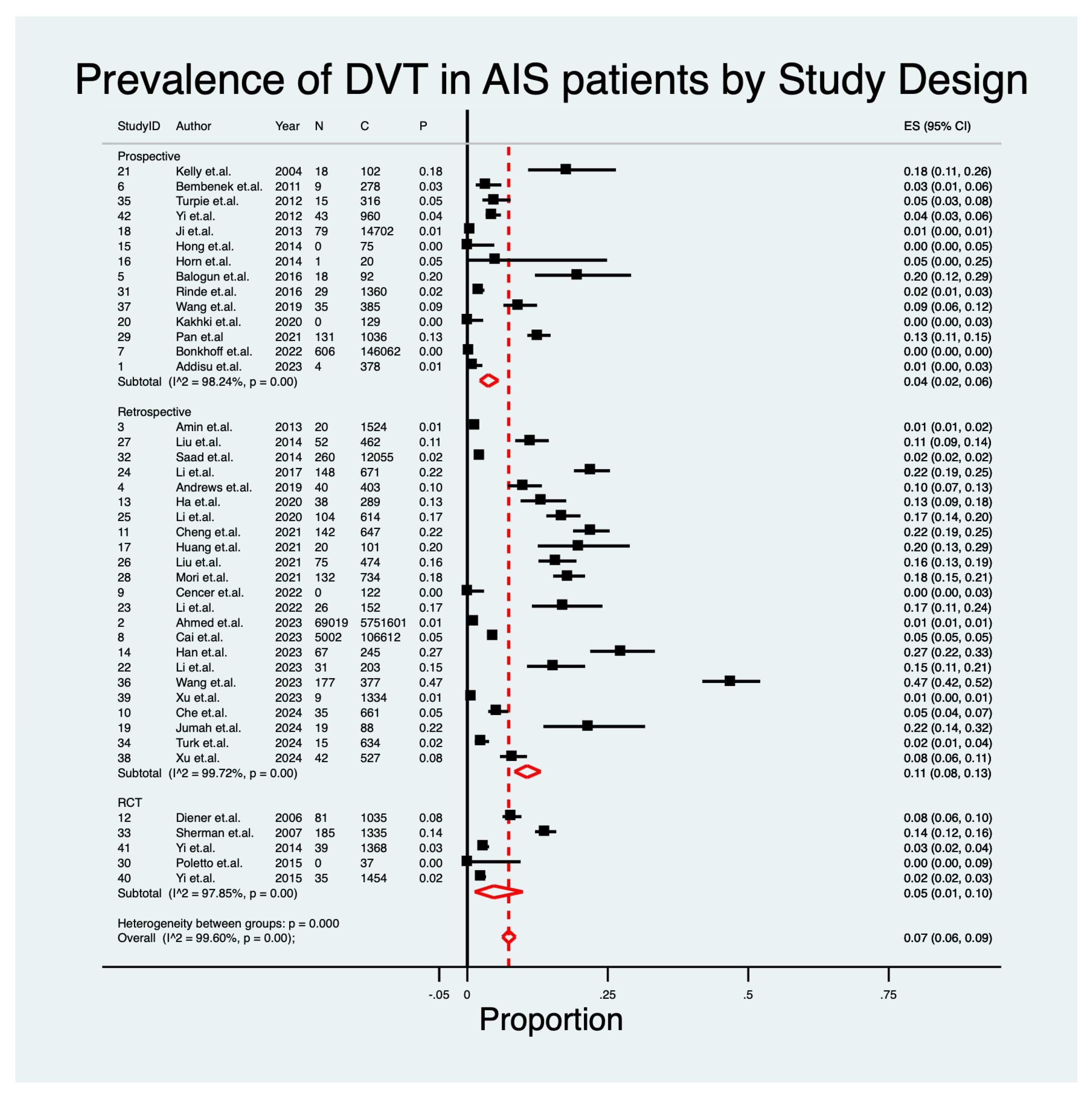
A total of 42 studies [5,10,32,33,34,35,36,37,38,39,40,41,42,43,44,45,46,47,48,49,50,51,52,53,54,55,56,57,58,59,60,61,62,63,64,65,66,67,68,69,70,71,72] reporting on the prevalence of DVT in patients with AIS, encompassing 6,051,729 patients, were included. The overall pooled prevalence of DVT was 7% (95% CI: 0.06–0.09; p < 0.001), with very high heterogeneity (I2 = 99.6%) (Table 3; Figure 2). Early systematic screening within 72 h identified DVT in up to 23% of patients, underscoring the importance of detection timing. Subgroup analyses revealed considerable variability in the reported prevalence of DVT among AIS patients. When stratified by study design, retrospective studies (N = 23) [5,33,34,35,39,40,41,42,44,47,49,52,53,54,55,56,57,58,62,64,66,68,69] showed the highest pooled prevalence of 11% (95% CI: 0.08–0.13; I2 = 99.7%), whereas randomized controlled trials (N = 5) [10,43,60,63,70,72] and prospective studies (N = 14) [32,36,37,38,45,46,48,50,51,59,61,65,67,71] reported lower estimates of 5% (95% CI: 0.01–0.10; I2 = 97.9%) and 4% (95% CI: 0.02–0.06; I2 = 97.8%), respectively (Table 3; Figure 3 and Figure S4). Temporal stratification demonstrated fluctuations across years of data collection, with more recent studies in 2023 [49,68] estimating a prevalence of 9% (95% CI: 0.07–0.12), compared to higher values in 2022 [41,52,64,66] (14%, 95% CI: 0.01–0.36) and much lower rates in earlier years, such as 2012 [45,46,60,72] (1%) and 2013 [70] (2%). Older studies from 2004 [51] and 2006 [63,65] reported markedly higher prevalence rates of 18% and 12%, respectively (Table 3; Figure S3). These temporal differences likely reflect heterogeneity in design, population size, diagnostic methods, and prophylaxis practices, rather than true secular trends (Figure S46). Prevalence estimates fluctuated markedly across time, reflecting methodological variability rather than true secular trends. The unexpectedly low prevalence observed in 2012–2013 (≈1–2%) likely reflects methodological artifacts, including reliance on administrative coding rather than systematic imaging, regional differences in reporting, and smaller cohort sizes in those years, rather than a true secular decline in thrombotic risk.
For clarity, ‘early systematic screening’ refers to active ultrasound or imaging surveillance performed within 72 h of stroke onset, irrespective of symptoms.
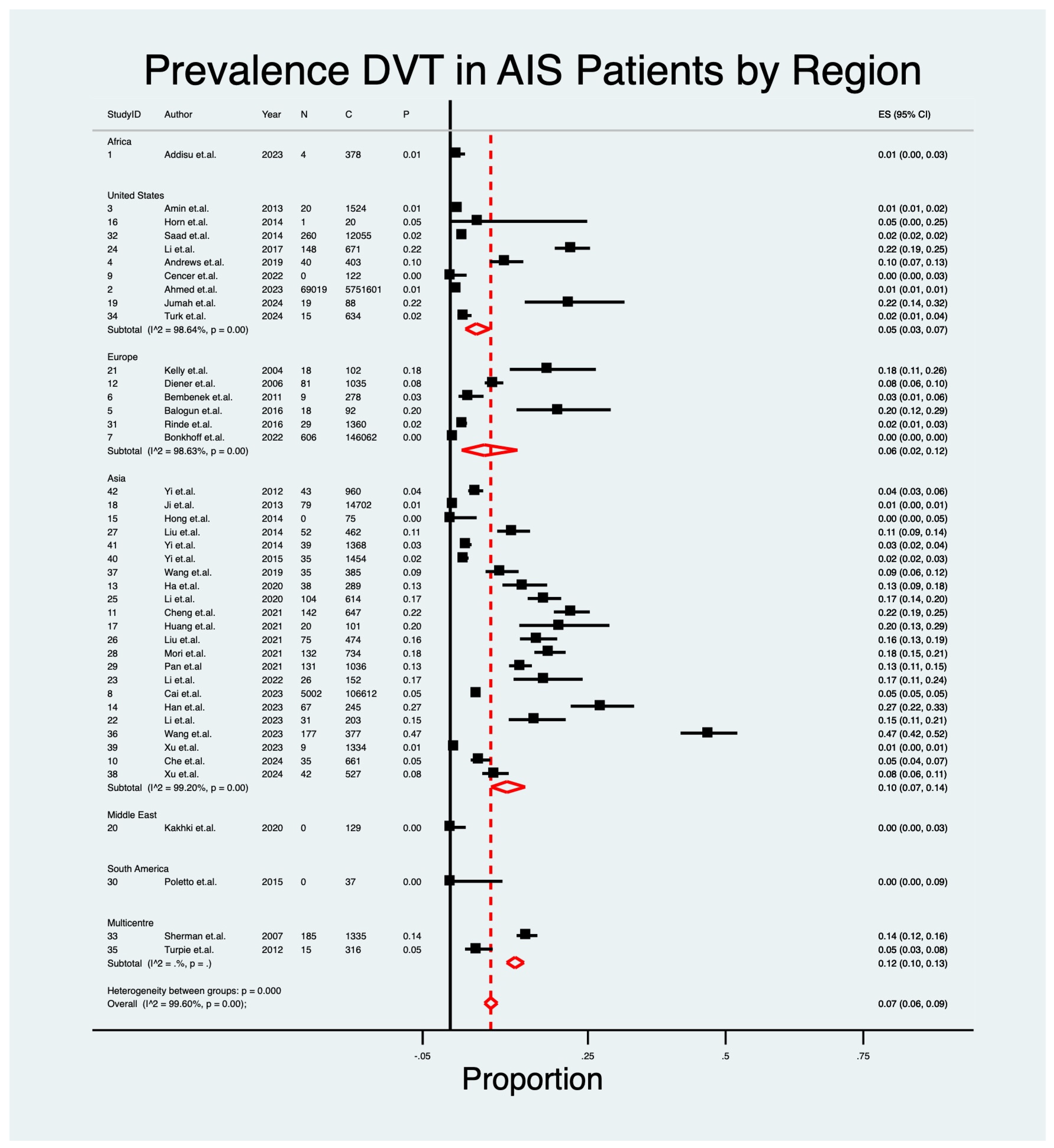
Regional analysis also highlighted variation, with studies from Asia [5,39,41,42,44,45,47,48,52,53,55,56,57,58,59,66,67,68,69,70,71,72] (N = 22) reporting a pooled prevalence of 10% (95% CI: 0.07–0.14), compared to 6% in Europe [36,37,38,51,61] (N = 6, 95% CI: 0.02–0.12) and 5% in North America [33,34,35,40,46,49,54,62,64] (N = 9, 95% CI: 0.03–0.07), while single studies from the Middle East [50], South America [60], and Africa [32] yielded very low prevalence estimates of 0–1% (Table 3; Figure 4 and Figure S2). The consistently higher prevalence reported in Asian cohorts may reflect systematic early ultrasound screening, inclusion of more severe stroke populations, and smaller single-center designs, whereas North American and European estimates often relied on record-based surveillance with less sensitive methods. Thus, geographic variation is more likely methodological than biological, though differences in thromboprophylaxis protocols and patient demographics may contribute. The timing of DVT screening further influenced estimates, with studies performing early screening within 72 h of stroke [49,54,57,58,66] reporting the highest pooled prevalence (23%, 95% CI: 0.12–0.35), compared with intermediate windows of 1–2 weeks [5,36,37,42,43,44,47,55,56,59,63,70,71,72,73] (12–13%) and routine in-hospital surveillance without systematic screening (2%, 95% CI: 0.02–0.03) (Table 3; Figure 5, Figures S5 and S47) [32,33,34,35,38,39,40,41,45,46,48,50,53,61,62,67,68,69]. Diagnostic modality also proved influential: magnetic resonance direct thrombus imaging (MRDTI) [51] detected the highest prevalence (18%, 95% CI: 0.11–0.26), followed by compression Doppler ultrasonography [5,10,36,42,43,44,52,56,58,59,61,63,66,67] (15%, 95% CI: 0.08–0.24) and color Doppler ultrasound [37,39,41,45,47,54,55,57,65,68] (9%, 95% CI: 0.05–0.13). In contrast, duplex ultrasound alone [64,70,71,72] identified a prevalence of 3% (95% CI: 0.02–0.04), and studies relying on clinical records [32,33,34,35,38,40,46,48,49,50,53,60,62,69] reported the lowest prevalence (2%, 95% CI: 0.01–0.02) (Table 3; Figure S6).

3.3. Predictive Indicators of DVT

Meta-analysis of discrete risk factors revealed that female sex [5,36,39,42,44,47,52,57,68] was significantly associated with higher odds of DVT (OR 1.33, 95% CI: 1.19–1.50; p < 0.001; I2 = 50.4%) (Figure S8). In contrast, traditional vascular risk factors such as hypertension [5,36,42,44,47,52,57,68,74] (OR 0.79, 95% CI: 0.52–1.21; p = 0.28; I2 = 77.2%), diabetes mellitus [5,36,39,42,44,47,52,57,68,74] (OR 1.06, 95% CI: 0.90–1.25; p = 0.49; I2 = 65.4%), and hyperlipidemia [36,39,44,52,57] (OR 0.99, 95% CI: 0.65–1.50; p = 0.96; I2 = 65.2%) showed no significant associations (Figures S8 and S9). Although atrial fibrillation [5,32,36,39,42,52,57,74] suggested a trend toward increased risk (OR 1.68, 95% CI: 0.93–3.05; p = 0.09), the results did not reach statistical significance and were highly heterogeneous (I2 = 97.7%) (Figure S9). Tobacco use [5,36,39,44,52,57,68] showed an apparent inverse association (OR 0.77, 95% CI: 0.62–0.95; p = 0.016; I2 = 24.2%), but this was likely artefactual, graded very low certainty, and should be interpreted cautiously (Figure S10). It is also possible that tobacco users represented a younger, less comorbid subset of AIS patients, or that competing risks such as early cardiovascular mortality limited detection of DVT in this group. Nonetheless, these alternative explanations underscore the likelihood of residual confounding, and the finding should not be interpreted as biologically protective. Other factors, including alcohol use [5,52,57,68] (OR 0.79, 95% CI: 0.52–1.20; p = 0.27; I2 = 33.1%) and coronary artery disease [5,36,47,52,57,74] (OR 1.16, 95% CI: 0.87–1.56; p = 0.30; I2 = 11.1%), were not significantly associated (Figure S10). By contrast, malignancy [5,42,44,52,74] (OR 2.69, 95% CI: 1.56–5.22; p = 0.022; I2 = 48.2%) and respiratory infection [5,42,47,52,57] (OR 2.30, 95% CI: 1.17–4.53; p = 0.016; I2 = 48.7%) emerged as strong predictors of DVT (Figure S9). The association of respiratory infection with DVT may be pathophysiologically plausible, as systemic inflammation, cytokine activation, and prolonged immobility during infection can amplify hypercoagulability in the acute stroke setting. These mechanisms could explain the nearly two-fold increase in risk observed, despite the low certainty of evidence. Discrete predictive factors are summarized in Table 2.
Table 1. Baseline clinical and methodological characteristics of studies included in the IRIS-DVT meta-analysis.
IDAuthorYearCohort SizeCrude Prevalence of DVT
n (n%)
CountryStudy DesignPrimary Stroke
Treatment
Immobilization Post Stroke?DVT Diagnosis ModalityDiagnosis Days Post Thrombectomy (Median)Chemical DVT ProphylaxisPhysical DVT Prophylaxis
1Addisu et al. [32]20233784 (1.1)EthiopiaRetrospectiveNo Acute ReperfusionUnspecifiedMedical NotesDuring HospitalizationUnspecifiedUnspecified
2Ahmed et al. [33]20235,751,60169,019 (1.2)United StatesRetrospectiveIV tPA or MTUnspecifiedMedical NotesDuring HospitalizationUnspecifiedUnspecified
3Amin et al. [34]2013152420 (1.3)United StatesRetrospectiveNAUnspecifiedMedical NotesDuring HospitalizationVariableVariable
4Andrews et al. [35]201940340 (9.9)United StatesRetrospectiveMTUnspecifiedMedical NotesDuring HospitalizationUnspecifiedUnspecified
5Balogun et al. [36]20169218 (19.6)United KingdomRetrospectiveNo Acute ReperfusionUnspecifiedCDUWithin 2 WeeksAntiplatelet Not Routine
6Bembenek et al. [37]20112699 (3.2)PolandProspectiveUnspecifiedUnspecifiedColor Doppler UltrasoundWithin 2 WeeksVariableUnspecified
7Bonkhoff et al. [38]2022146,062606 (0.4)GermanyRetrospectiveIV tPA when possibleUnspecifiedMedical NotesDuring HospitalizationUnspecifiedUnspecified
8Cai et al. [39]2023106,6125002 (4.7)ChinaRetrospectiveNo Acute ReperfusionUnspecifiedColor Doppler UltrasoundDuring HospitalizationVariableVariable
9Cencer et al. [40]20221220 (0.0)United StatesRetrospectiveIV tPAUnspecifiedMedical NotesDuring HospitalizationUnspecifiedUnspecified
10Che et al. [41]202466135 (5.3)ChinaRetrospectiveEVTUnspecifiedCDUDuring HospitalizationUnspecifiedUnspecified
11Cheng et al. [42]2021431142 (21.9)ChinaRetrospectiveVariableUnspecifiedCUSWithin 2 WeeksUnspecifiedUnspecified
12Diener et al. [43]2006103581 (7.8)MulticenterRCTVariableUnspecifiedCDUWithin 2 WeeksAntiplatelet or AnticoagulantUnspecified
13Ha et al. [44]202028938 (13.1)KoreaRetrospectiveIV tPA when possibleUnspecifiedCUSWithin 1 WeekNot RoutineNot Routine
14Han et al. [5]202324567 (27.3)ChinaRetrospectiveEVTYesCDUWithin 2 WeeksAntiplatelet IPC
15Hong et al. [45]2014750 (0.0)KoreaProspectiveVariableVariableCDUDuring HospitalizationUnspecifiedUnspecified
16Horn et al. [46]2014201 (5.0)United StatesProspectiveMTYesMedical NotesDuring HospitalizationUnspecifiedUnspecified
17Huang et al. [47]202110120 (19.8)ChinaRetrospectiveNAUnspecifiedCDUWithin 2 WeeksNot RoutineUnspecified
18Ji et al. [48]201314,70279 (0.5)ChinaRetrospectiveUnspecifiedUnspecifiedMedical NotesDuring HospitalizationUnspecifiedUnspecified
19Jumah et al. [49]20248819 (21.6)United StatesRetrospectiveVariableUnspecifiedMedical NotesWithin 72 hIV HeparinUnspecified
20Kakhki et al. [50]20201290 (0.0)IranRetrospectiveUnspecifiedUnspecifiedMedical NotesDuring HospitalizationUnspecifiedUnspecified
21Kelly et al. [75]200410218 (17.6)United KingdomProspectiveUnspecifiedUnspecifiedMRDTIWithin 1 MonthAspirinGCS
22Li et al. [52]202323431 (15.3)ChinaRetrospectiveUnspecifiedUnspecifiedCUSWithin 72 hUnspecifiedUnspecified
23Li et al. [53]202215226 (17.1)ChinaRetrospectiveIV tPAUnspecifiedMedical NotesDuring HospitalizationUnspecifiedUnspecified
24Li et al. [54]2017671148 (22.1)United StatesRetrospectiveUnspecifiedUnspecifiedCDUWithin 2 WeeksNone UsedNone Used
25Li et al. [55]2020614104 (16.9)ChinaRetrospectiveMTUnspecifiedCDUWithin 2 WeeksUnspecifiedUnspecified
26Liu et al. [57]202147475 (15.8)ChinaRetrospectiveIV tPAYesCDUWithin 72 hAntiplatelet Unspecified
27Liu et al. [56]201446252 (11.3)ChinaRetrospectiveVariableUnspecifiedCUSWithin 2 WeeksAntiplatelet or AnticoagulantUnspecified
28Mori et al. [58]2021734132 (18.0)JapanRetrospectiveUnspecifiedUnspecifiedCUSWithin 72 hUnspecifiedUnspecified
29Pan et al. [59]20211036131 (12.6)ChinaRetrospectiveVariableUnspecifiedCDUWithin 2 WeeksAntiplatelet or AnticoagulantUnspecified
30Poletto et al. [60]2015370 (0.0)BrazilRCTIV tPA when possibleUnspecifiedMedical NotesWithin 3 MonthsUnspecifiedUnspecified
31Rinde et al. [61]2016136029 (2.1)NorwayRetrospectiveVariableUnspecifiedCUSDuring HospitalizationUnspecifiedUnspecified
32Saad et al. [62]201412,055260 (2.2)United StatesRetrospectiveMTUnspecifiedMedical NotesDuring HospitalizationUnspecifiedUnspecified
33Sherman et al. [63]20071762185 (13.9)MulticenterRCTVariableYesCUSWithin 2 WeeksAntiplatelet or AnticoagulantUnspecified
34Turk et al. [64]202463415 (2.4)United StatesRetrospectiveUnspecifiedUnspecifiedDuplex UltrasonographyWithin 24 hVariableUnspecified
35Turpie et al. [65]201231615 (4.7)MulticenterProspectiveUnspecifiedUnspecifiedCDUWithin 1 MonthNot RoutineUnspecified
36Wang et al. [66]2023377177 (46.9)ChinaRetrospectiveIV tPAUnspecifiedCUSWithin 72 hUnspecifiedUnspecified
37Wang et al. [67]201938535 (9.1)NAProspectiveUnspecifiedUnspecifiedCUSDuring hospitalizationUnspecifiedIPC
38Xu et al. [68]202436942 (8.0)ChinaRetrospectiveUnspecifiedUnspecifiedCDUDuring hospitalizationUnspecifiedUnspecified
39Xu et al. [69]202313349 (0.7)ChinaRetrospectiveIV tPAUnspecifiedMedical NotesDuring hospitalizationUnspecifiedUnspecified
40Yi et al. [70]2015145435 (2.4)ChinaRCTUnspecifiedUnspecifiedDuplex UltrasonographyWithin 2 WeeksAntiplatelet Unspecified
41Yi et al. [72]2014136839 (2.9)ChinaRCTUnspecifiedUnspecifiedDuplex UltrasonographyWithin 2 WeeksAntiplatelet or AnticoagulantUnspecified
42Yi et al. [71]201296043 (4.5)ChinaProspectiveUnspecifiedUnspecifiedDuplex UltrasonographyWithin 2 WeeksUnspecifiedUnspecified
Abbreviations: NA: not available; DVT: deep vein thrombosis; MT: mechanical thrombectomy; IV tPA: intravenous tissue plasminogen activator; RCT: randomized controlled trial; IPC: intermittent pneumatic compression; CUS: compression ultrasound; CDU: color Doppler ultrasound; MRDTI: magnetic resonance direct thrombus imaging.
Table 2. Discrete risk factors for deep vein thrombosis after acute ischemic stroke: pooled effect estimates.
IDAuthorDVTFemaleHTNDMHLAFSmokingAlcoholCADMAL RI
n (n%)n (n%)n (n%)n (n%)n (n%)n (n%)n (n%)n (n%)n (n%)n (n%)n (n%)
YesNoYesNoYesNoYesNoYesNoYesNoYesNoYesNoYesNoYesNo
1Addisu et al. [32]4
(1.1)
--------102
(27.0)
276
(73.0)
----------
5Balogun et al. [36]18
(19.6)
18
(19.6)
74
(80.4)
36
(39.1)
56
(60.9)
18
(19.6)
74
(80.4)
18
(19.6)
74
(80.4)
18
(19.6)
74
(80.4)
18
(19.6)
74
(80.4)
--18
(19.6)
74
(80.4)
----
8Cai et al. [39]5002
(4.7)
5002
(4.7)
101,610
(95.3)
--5002
(4.7)
101,610
(95.3)
5002
(4.7)
101,610
(95.3)
5002
(4.7)
101,610
(95.3)
5002
(4.7)
101,610
(95.3)
--------
11Cheng et al. [42]96
(22.3)
96
(22.3)
335
(77.7)
96
(22.3)
335
(77.7)
96
(22.3)
335
(77.7)
--96
(22.3)
335
(77.7)
------96
(22.3)
335
(77.7)
96
(22.3)
335
(77.7)
14Ha et al. [44]38
(13.1)
114
(39.4)
175
(60.6)
208
(72.0)
81
(28.0)
87
(30.1)
202
(69.9)
195
(67.5)
94
(32.5)
--130
(45.0)
159
(55.0)
----5
(1.7)
284
(98.3)
--
15Han et al. [5]67
(27.3)
87
(35.5)
158
(64.5)
142
(58.0)
103
(42.0)
42
(17.1)
203
(82.9)
--102
(41.6)
143
(58.4)
77
(31.4)
168
(68.6)
52
(21.2)
193
(78.8)
10
(4.1)
235
(95.9)
15
(6.1)
230
(93.9)
152
(62.0)
93
(38.0)
18Huang et al. [47]20
(19.8)
29
(28.7)
72
(71.3)
83
(82.2)
18
(17.8)
15
(14.9)
86
(85.1)
--------23
(22.8)
78
(77.2)
0
(0.0)
101
(100.0)
66
(65.3)
35
(34.7)
23Li et al. [52]31
(15.3)
31
(13.2)
203
(86.8)
31
(13.2)
203
(86.8)
31
(13.2)
203
(86.8)
31
(13.2)
203
(86.8)
31
(13.2)
203
(86.8)
31
(13.2)
203
(86.8)
31
(13.2)
203
(86.8)
31
(13.2)
203
(86.8)
31
(13.2)
203
(86.8)
31
(13.2)
203
(86.8)
27Liu et al. [57]75
(15.8)
142
(30.0)
332
(70.0)
284
(59.9)
190
(40.1)
120
(25.3)
354
(74.7)
194
(40.9)
280
(59.1)
54
(11.4)
420
(88.6)
216
(45.6)
258
(54.4)
185
(39.0)
289
(61.0)
33
(7.0)
441
(93.0)
--45
(9.5)
429
(90.5)
40Xu et al. [68]29
(7.9)
217
(58.8)
152
(41.2)
269
(72.9)
100
(27.1)
164
(44.4)
205
(55.6)
----73
(19.8)
296
(80.2)
105
(28.5)
264
(71.5)
------
Abbreviations: n: number of patients; AF: atrial fibrillation; CAD: coronary artery disease; DM: diabetes mellitus; HTN: hypertension; HL: hyperlipidemia; MAL: malignancy/cancer diagnosis; RI: respiratory infection.
Table 3. Pooled prevalence of deep vein thrombosis in acute ischemic stroke: summary effects and heterogeneity across studies.
IDAuthorDVTAgeNIHSS ScoreD-DimerAdmission GlucoseLDLFibrinogen
n (n%)FR (Mean ± SD)FR (Mean ± SD)FR (Mean ± SD)FR (Mean ± SD)FR (Mean ± SD)FR (Mean ± SD)
YesNoYesNoYesNoYesNoYesNoYesNo
5Balogun et al. [36]18 (19.6)69.7 (13.4)69.1 (14.5)15.6 (7.3)12.8 (7.3)2.6 (1.9)1.4 (1.3)----3.9 (1.4)3.9 (1.4)
8Cai et al. [39]5002 (4.7)69.8 (11.7)67.2 (12.1)7.6 (7.1)5.3 (5.6)--6.8 (3.2)6.6 (2.9)2.8 (1.5)2.8 (1.3)--
11Cheng et al. [42]96 (22.3)73.4 (8.4)68.9 (12.0)--2.2 (1.9)1.6 (1.3)6.0 (2.4)6.0 (2.2)--3.6 (1.6)3.6 (1.5)
14Ha et al. [44]38 (13.1)71 (12.0)68.4 (11.2)7.4 (5.4)4.5 (3.8)12 (20.4)8.5 (13.4)------
15Han et al. [5]67 (27.3)72.1 (9.1)67.08 (11.7)16 (4.5)14.5 (5.7)2.8 (2.4)1.7
(1.7)
6.6 (1.8)6.5 (1.7)2.1 (0.7)2.2 (0.8)0.032 (0.0)0.032 (0.0)
18Huang et al. [47]20 (19.8)65 (16.4)66 (16.2)19.7 (10.6)16.9 (10.2)3.1 (5.3)1.6 (3.3)6.8 (3.2)6.6 (2.9)1.9 (1.2)2.0 (1.2)0.0025 (0.0)0.0029 (0.0)
23Li et al. [52]31 (15.3)64.7 (11.7)60.2 (12.0)--1.9 (1.7)0.8 (1.3)6.1 (1.9)6.3 (2.3)1.9 (0.6)1.7 (0.6)4.3 (1.2)4.1 (1.3)
27Liu et al. [57]75 (15.8)69.8 (9.8)62.7 (11.6)9.7 (5.3)8.0 (4.7)--8.0 (2.9)8.1 (3.1)2.92 (0.9)3.0 (0.8)3.15 (0.7)3.1 (0.7)
Abbreviations: n: number of patients; NIHSS: National Institute of Health Stroke Scale; LDL: low-density lipoprotein.
Continuous predictors also demonstrated important associations. Older age [5,36,39,42,44,47,52,57] was consistently linked with higher DVT risk (SMD 0.32, 95% CI: 0.18–0.46; p < 0.001; I2 = 60.8%) (Figure S13), while stroke severity, as measured by the NIHSS [5,36,39,44,47,57], showed a particularly robust relationship (SMD 0.41, 95% CI: 0.38–0.43; p < 0.001; I2 = 0%) (Figure S13). Laboratory markers further reinforced this pattern: elevated D-dimer levels [5,36,42,44,47,52] (SMD 0.55, 95% CI: 0.38–0.72; p < 0.001; I2 = 34.8%) and, to a lesser extent, higher admission glucose [5,39,42,52,57] (SMD 0.07, 95% CI: 0.04–0.09; p < 0.001; I2 = 0%) were significantly associated with DVT (Figures S13 and S14). By contrast, LDL cholesterol [5,39,47,52,57] (SMD −0.03, 95% CI: −0.12 to 0.09; p = 0.73; I2 = 27%) and fibrinogen [5,36,42,47,52,57] (SMD 0.01, 95% CI: −0.11 to 0.13; p = 0.87; I2 = 0%) showed no significant associations (Figure S14). Further details on pooled heterogeneity estimates for continuous predictors, including D-dimer, admission glucose, LDL, and fibrinogen, are summarized in Table S8.
Overall, the most consistent predictors of DVT after AIS were stroke severity (NIHSS score), malignancy, female sex, older age, and elevated D-dimer levels, all supported by moderate-certainty evidence. Respiratory infection and admission glucose also showed associations but with lower certainty. In contrast, traditional vascular risk factors such as hypertension, diabetes mellitus, hyperlipidemia, and atrial fibrillation were not significantly associated, and LDL cholesterol and fibrinogen showed no meaningful relationship. The observed inverse association with tobacco use was likely confounded and should not be interpreted as protective. These findings suggest that clinical focus should shift toward neurological severity, cancer status, and selected biomarkers rather than conventional vascular comorbidities when stratifying DVT risk in AIS patients (Table 4 and Table 5; Figures S8–S14).
Across 11 RCTs and cohort studies reporting prophylactic interventions, intermittent pneumatic compression (IPC) reduced DVT risk (pooled OR 0.62; 95% CI 0.41–0.93), while pharmacological agents such as LMWH showed a trend toward benefit (pooled OR 0.78; 95% CI 0.55–1.11), both graded low certainty (Tables S7 and S8).

3.4. Certainty of Evidence (GRADE)

The certainty of evidence across outcomes ranged from moderate to very low (Table 6). Moderate-certainty evidence supported the association of higher NIHSS scores, increasing age, elevated D-dimer levels, female sex, and malignancy with increased risk of DVT in AIS. Respiratory infection and admission glucose were supported by low-certainty evidence due to heterogeneity and modest effect sizes. The apparent inverse association with tobacco use was graded with very low certainty, reflecting likely residual confounding and inconsistency. Similarly, LDL and fibrinogen showed no significant associations and were rated very low. Evidence on the effectiveness of prophylactic interventions (e.g., LMWH, IPC) in AIS cohorts was insufficient and highly heterogeneous, warranting a very low certainty rating. Overall, while some predictors such as NIHSS and malignancy demonstrate robust and consistent associations, most other outcomes remain supported by low- to very low-certainty evidence.
Table 4. Predictors of deep vein thrombosis after acute ischemic stroke: meta-analysis of categorical and continuous variables.
SubgroupNPooled Prevalence Rate
(from Meta-Analysis)
95% CIz-Scorep-ValueI2 τ2
Overall427%0.06–0.0921.76.p < 0.0199.60%0.02
Study Design
Retrospective2311%0.08–0.1315.96p < 0.0199.72%-
Prospective144%0.02–0.067.54p < 0.0198.24%-
RCT55%0.01–0.103.70p < 0.0197.85%-
Region
Asia2210%0.07–0.143.24p < 0.0199.20-
Europe66%0.02–0.124.20p < 0.0198.63-
North America95%0.03–0.077.18p < 0.0198.64-
Middle East10%0.00–0.030.00---
Africa11%0.00–0.033.24---
South America10%0.00–0.090.00---
Multiple212%0.10–0.1326.77---
DVT Screening Post Stroke Screening
Within 24 h12%0.02–0.046.90---
Within 72 h523%0.12–0.356.66p < 0.0196.97%-
Within 1 Week113%0.09–0.1811.69---
Within 2 Weeks1412%0.08–0.179.40p < 0.0197.89%-
Within 1 Month27%0.05–109.63---
Within 3 Months10%0.00–0.090.00---
During hospitalization182%0.02–0.038.98p < 0.0199.75%-
Diagnosis Modality
MRDTI118%0.11–0.267.86---
CDU416%0.09–0.137.09p < 0.0195.25%-
CUS915%0.08–0.246.79p < 0.0198.55%-
Duplex Ultrasound43%0.02–0.0412.07p < 0.0165.85%-
Color Doppler Ultrasound109%0.05–0.137.09p < 0.0197.80%-
Medical Notes142%0.01–0.028.49p < 0.0199.14%-
Temporal Trends
202329%0.07–0.1213.68--
2022414%0.01–0.362.71p < 0.01-
202128%0.06–0.1012.69--
202038%0.00–0.242.34--
201979%0.05–0.138.36p < 0.01-
201819%0.06–0.1211.10--
201720%0.00–0.0038.41--
2016413%0.04–0.234.45p < 0.01-
201312%0.02–0.0310.96--
201241%0.00–0.031.71p = 0.14-
201122%0.02–0.0224.91--
201014%0.03–0.0612.29--
200913%0.01–0.065.19--
200821%0.00–0.0117.71--
200723%0.02–0.0413.40--
2006212%0.10–0.1326.77--
2004118%0.11–0.267.86--
200318%0.06–0.1017.3--
Abbreviations N: number of studies; CI: confidence interval; RCT: randomized controlled trial; MRDTI: magnetic resonance direct thrombus imaging; CDU: compression Doppler ultrasound; CUS: compression ultrasound.
Table 5. Effectiveness of prophylactic interventions for preventing deep vein thrombosis in acute ischemic stroke patients.
Summary Effects Heterogeneity ¶Heterogeneity
Variance Estimates
REDL
OutcomeN
(Studies)
n
(Cohort)
Effect MeasureEffect (OR/SMD)[95% CI]Tests of Overall Effectz ScoreCochrane’s QHI2 ≤ * τ2Φ
Female91,439,000OR1.332[1.185; 1.498] p < 0.001 4.797 16.14 1.4250.40%0.0077
Respiratory Infection41054OR2.301[1.169; 4.529] p = 0.016 2.411 5.851.39648.70%0.1019
Malignancy51,335,226OR2.69[1.557; 5.215] p = 0.022 2.298 7.721.38948.20%0.3959
Atrial Fibrillation81,442,445OR1.684[0.930; 3.049] p = 0.085 1.721 310.936.66597.70%0.5145
Coronary Artery Disease71,335,554OR1.164[0.871; 1.556] p = 0.304 1.028 6.751.06111.10%0.0286
Peripheral Vascular Disease41,437,577OR1.477[0.665; 3.283] p = 0.339 0.957 186.797.89198.40%0.5098
Diabetes Mellitus101,442,824OR1.06[0.898; 1.250] p = 0.493 0.685 26.021.765.40%0.0188
Hyperlipidemia5107,701OR0.989[0.653; 1.497] p = 0.957 −0.05411.491.69565.20%0.125
Hypertension91,336,167OR0.791[0.515; 1.214] p = 0.283 −1.07435.112.09577.20%0.2682
Alcohol Use41322OR0.789[0.517; 1.203] p = 0.271 −1.1024.491.22333.10%0.0618
Tobacco Use7108,315OR0.767[0.618; 0.952 p = 0.016 −2.4027.911.14824.20%0.0216
NIHSS Score6113,033SMD0.405[0.377; 0.433] p < 0.001 28.446 4.590.9580%0
D-Dimer61662SMD0.551[0.378; 0.723] p < 0.001 6.24 7.661.23834.80%0.0157
LDL5112,861SMD−0.34[−0.126; 0.088] p = 0.734 5.48 5.481.1727%0.0047
Admission Glucose5113,267SMD0.066[0.039; 0.094] p < 0.001 4.687 1.30.5690%0
Age8113,825SMD0.32[0.181; 0.460] p < 0.001 4.494 17.841.59660.80%0.0197
Fibrinogen61884SMD0.01[−0.112; 0.133] p = 0.869 0.165 2.430.6970%0
Abbreviations: N, number of studies; n, number of patients; OR, odds ratio; CI, confidence interval; REDL, DerSimonian and Laird random-effects method; Q, heterogeneity measure calculated with 95% CIs based on the noncentral χ2 (common-effect) distribution for Cochran’s Q test; H, relative excess in Cochran’s Q over its degrees of freedom; NIHSS, National Institutes of Health Stroke Scale/Score; LDL, low-density lipoprotein; I2, proportion of total variation in effect estimates attributable to between-study heterogeneity (based on Cochran’s Q test); τ2, between-study variance for subgroup heterogeneity comparisons; *, values of I2 are expressed as percentages; ¶, heterogeneity values calculated with 95% CIs based on the gamma (random-effects) distribution for Q; Φ, heterogeneity variance estimates (τ2) derived from the DerSimonian and Laird method.
Table 6. GRADE summary of evidence on incidence, risk factors, and prophylaxis of deep vein thrombosis in acute ischemic stroke (IRIS-DVT study).
A. Incidence/Prevalence
OutcomeNo. of Studies (N)Patient Number (n)Effect Estimate (95% CI)Risk of BiasInconsistencyIndirectnessImprecisionPublication BiasCertainty of Evidence (GRADE)Reasons for Downgrade/Upgrade
Prevalence of DVT in AIS42 6,051,729Pooled prevalence: 7% (95% CI 5–9%)LowModerate (regional and temporal heterogeneity)LowMinimalPossible⬤⬤⬤◯ ModerateDowngraded: heterogeneity; Upgraded: large sample size, precise estimates
B. Risk Factors
PredictorNo. of Studies (N)Patient Number (n)Effect Estimate (OR/SMD, 95% CI)Risk of BiasInconsistencyIndirectnessImprecisionPublication BiasCertainty of Evidence (GRADE)Reasons for Downgrade/Upgrade
Stroke severity (NIHSS)6 107,795SMD 0.41 (0.38–0.43)LowVery low (I2 = 0%)LowMinimalUnlikely⬤⬤⬤◯ ModerateDowngraded: observational designs; Upgraded: strong, consistent effect
Age8108,676SMD 0.32 (0.18–0.46)LowModerate (I2 ≈ 61%)LowMinimalUnlikely⬤⬤⬤◯ ModerateDowngraded: inconsistency; Upgraded: large sample size
Female sex9108,847OR 1.33 (1.19–1.50)LowModerate (I2 ≈ 50%)LowAdequatePossible⬤⬤⬤◯ ModerateDowngraded: inconsistency; Upgraded: robust effect
D-dimer elevation61590SMD 0.55 (0.38–0.72)LowLow–moderate (I2 ≈ 35%)LowMinimalPossible⬤⬤⬤◯ ModerateDowngraded: possible bias; Upgraded: strong effect
Malignancy51199OR 2.69 (1.56–5.22)LowModerate (I2 ≈ 48%)LowSomewhat wide CIPossible⬤⬤⬤◯ ModerateDowngraded: inconsistency; Upgraded: large effect
Respiratory infection51485OR 2.30 (1.17–4.53)ModerateModerate (I2 ≈ 49%)LowWide CILikely⬤⬤◯◯ LowDowngraded: inconsistency, imprecision, bias
Admission hyperglycemia5108,212SMD 0.07 (0.04–0.09)LowLow (I2 = 0%)LowSmall effectPossible⬤⬤◯◯ LowDowngraded: trivial effect size, possible bias
Tobacco use (inverse)7108,315OR 0.77 (0.62–0.95)HighLow (I2 ≈ 24%)HighCI near nullLikely⬤◯◯◯ Very LowDowngraded: confounding, indirectness, bias
LDL cholesterol5107,666SMD −0.03 (−0.12–0.09)ModerateLow (I2 ≈ 27%)ModerateNull effect, small nLikely⬤◯◯◯ Very LowDowngraded: imprecision, indirectness
Fibrinogen61775SMD 0.01 (−0.11–0.13)ModerateLow (I2 = 0%)ModerateWide CI incl. nullPossible⬤◯◯◯ Very LowDowngraded: imprecision, indirectness
C. Prophylaxis
InterventionNo. of Studies (N)Patient Number (n)Effect Estimate (OR/SMD, 95% CI)Risk of BiasInconsistencyIndirectnessImprecisionPublication BiasCertainty of Evidence (GRADE)Reasons for Downgrade/Upgrade
Pharmacological prophylaxis (Anticoagulants)41066Heterogeneous, no stable pooled estimateHigh (small, observational)Very low (I2 = 0%)LowWide CILikely⬤◯◯◯ Very LowDowngraded: high risk of bias, small observational
Pharmacological prophylaxis (Antiplatelets)51531Heterogeneous, no stable pooled estimateHigh (small, observational)Very low (I2 = 0%)LowWide CILikely⬤◯◯◯ Very LowDowngraded: high risk of bias, small observational
IPC (intermittent pneumatic compression)3732Trend toward reduced DVT; effect inconsistentModerateModerate–highLowModeratePossible⬤◯◯◯ Very LowDowngraded: inconsistency, imprecision, small observational
This table presents pooled estimates, certainty of evidence, and rationale for grading according to the GRADE framework, with outcomes stratified into incidence/prevalence, risk factors, and prophylactic interventions. Certainty of evidence was assessed across five domains: risk of bias, inconsistency, indirectness, imprecision, and publication bias. Based on 42 studies (n > 6 million), the pooled prevalence of DVT in AIS was 7% (95% CI 5–9%), approximately seventy times higher than in the general population. Among risk factors, moderate-certainty evidence supports stroke severity (NIHSS), older age, female sex, elevated D-dimer, and malignancy as consistent predictors, while low-certainty evidence was found for respiratory infection and admission hyperglycemia; tobacco use showed an inverse association but with very low certainty, likely due to confounding, and LDL cholesterol and fibrinogen were not significantly associated. For prophylaxis, evidence for pharmacological interventions (LMWH, UFH, DOACs) was highly heterogeneous and graded very low certainty, while IPC showed a directional trend toward reduced DVT incidence but remains very low certainty due to limited and inconsistent RCT data. Overall, the strongest and most reliable predictors of DVT in AIS were NIHSS, age, D-dimer, female sex, and malignancy (moderate certainty), whereas preventive strategies remain under-investigated with substantial uncertainty. Abbreviations: AIS, Acute Ischemic Stroke; CI, Confidence Interval; DVT, Deep Vein Thrombosis; DOACs, Direct Oral Anticoagulants; GRADE, Grading of Recommendations, Assessment, Development and Evaluation; IPC, Intermittent Pneumatic Compression; I2, I-squared statistic (heterogeneity measure); LMWH, Low Molecular Weight Heparin; NIHSS, National Institutes of Health Stroke Scale; OR, Odds Ratio; RCT, Randomized Controlled Trial; SMD, Standardized Mean Difference; UFH, Unfractionated Heparin.

3.5. Prophylactic Interventions

Evidence regarding prophylactic strategies for DVT prevention in AIS was heterogeneous and limited. Pharmacological measures such as low-molecular-weight heparin (LMWH) and direct oral anticoagulants, along with mechanical approaches including intermittent pneumatic compression (IPC), were variably reported across studies. Pooled estimates did not allow firm conclusions regarding their effectiveness, given wide heterogeneity, limited sample sizes, and inconsistent reporting of outcomes (Tables S7 and S8; Figures S12, S19, S31, S39 and S45). As such, the certainty of evidence for prophylaxis effectiveness in AIS remains very low. Pooled summary effects for pharmacological prophylaxis (LMWH, UFH, DOACs) and mechanical interventions (IPC, stockings, IVC filters) are presented in Table S7. While pooled analyses (Table S7, Figures S12, S19, S31, S39 and S45) did not allow firm conclusions, a directional trend toward reduced DVT incidence with LMWH and IPC was observed, albeit with high heterogeneity. Pharmacological prophylaxis estimates were particularly inconsistent, while IPC showed more reproducible effects in smaller RCTs. These analyses demonstrated high heterogeneity and limited certainty, consistent with our main findings.

3.6. Sensitivity and Bias Analyses

Sensitivity analyses, performed by sequentially excluding individual studies, did not materially alter pooled prevalence or risk factor estimates, confirming the robustness of the main findings (Figures S15–S21). Assessment of publication bias using funnel plots and Egger’s regression revealed potential small-study effects for some predictors; however, patterns were inconsistent and did not systematically affect the overall conclusions (Figures S22–S33). Sensitivity analyses confirmed the robustness of the pooled estimates (see Figures S15–S21). Funnel plot and Egger’s regression results are illustrated in Figures S22–S33. Diagnostic performance measures, including SROC and Fagan analyses, are provided in Figures S34–S45. Detailed heterogeneity outputs for prophylactic strategies and continuous predictors are reported in Tables S7 and S8.

4. Discussion

The IRIS-DVT study provides the most comprehensive synthesis to date on thrombotic complications in AIS, addressing a longstanding evidence gap. By focusing exclusively on AIS, rather than grouping with hemorrhagic or mixed stroke subtypes as earlier reviews did, this meta-analysis delivers a more precise and clinically applicable understanding of DVT in stroke care. Our pooled prevalence estimates of 7%, seventy times higher than the general population, confirms AIS as a distinct high-risk condition that warrants systematic preventive strategies [76].
A key finding is the impact of screening timing on prevalence. Systematic imaging within 72 h identified DVT in nearly one in four patients, compared with substantially lower rates when screening was delayed or unsystematic. This suggests that a large proportion of thrombi remain clinically silent unless actively sought. Current AHA/ASA (2021) and ESO (2016) guidelines recommend prophylaxis in immobilized AIS patients but provide no clear direction on optimal detection windows [77,78,79]. Our results argue for early systematic screening within the first 72 h, and no later than two weeks, to minimize underdiagnosis (Figure S47). This is supported by smaller cohort studies and carries direct implications for updating practice guidelines [49,54,57,58,66].
Regional differences may partly reflect genetic predispositions such as Factor V Leiden or prothrombin G20210A variants, which are rare in East Asian but common in European cohorts, influencing baseline thrombotic risk [80]. Moreover, variability in post-thrombectomy immobilization and delayed prophylaxis initiation may accentuate risk after reperfusion therapies [81]. Dedicated studies stratifying by ancestry, thrombophilia, and treatment modality are warranted to clarify these interactions. Temporal patterns further contextualize our findings. The decline in prevalence around 2006 coincided with the introduction of mandatory hospital-wide VTE assessments and prophylaxis protocols in the US, UK, and France, while the rise after 2015 paralleled the global adoption of EVT following pivotal trials [82,83] (Figure S46). Although most included studies did not report treatment modality, these temporal shifts strongly suggest evolving practice patterns influence DVT risk. EVT may contribute through longer procedural times, femoral access, and immobility, whereas IVT-treated patients face deferred prophylaxis in the first 24 h [5,84,85,86,87]. These observations [88] underscore an urgent need for dedicated studies examining reperfusion-specific thrombotic risk, which current guidelines do not yet address.
Among predictors, stroke severity (NIHSS) emerged as the most consistent and powerful risk factor, reinforcing the clinical intuition that severely affected, immobilized patients require early prophylaxis. Elevated D-dimer, admission hyperglycaemia, and older age also showed significant associations, reflecting systemic hypercoagulability and metabolic stress [89,90]. Interestingly, LDL and fibrinogen, which are established pro-thrombotic markers in other contexts, did not demonstrate significant associations in AIS populations. This may reflect underreporting, heterogeneity in laboratory measurement, or confounding from acute-phase responses, rather than true absence of pathophysiological relevance. Malignancy, respiratory infection, and female sex were robust categorical predictors, while traditional vascular risk factors, including hypertension, diabetes, hyperlipidaemia, and atrial fibrillation, were not significantly associated. This divergence highlights that AIS-related thrombosis is shaped by acute systemic and neurological stressors rather than chronic comorbidities. The apparent inverse association with tobacco use is likely artefactual, reflecting residual confounding or selection bias, and should not influence practice.
Despite broad use of low-molecular-weight heparin, unfractionated heparin, direct oral anticoagulants, and IPC, evidence on prophylaxis effectiveness in AIS remains limited and heterogeneous. Our findings align with the CLOTS 3 trial [91], which demonstrated IPC reduces proximal DVT, supporting its consistent endorsement in guidelines. By contrast, pharmacological prophylaxis remains debated, with bleeding risk often outweighing uncertain benefit. Both AHA/ASA [78] and ESO guidelines [79] recommend IPC as first-line and acknowledge uncertainty around anticoagulants; our GRADE assessment concurs, rating evidence for prophylaxis as very low certainty (Table 6). Our interpretation of prophylactic effectiveness and continuous biomarker predictors is supported by the extended analyses in Tables S7 and S8, which confirm the limited certainty and heterogeneity underlying these associations.
The certainty of evidence across outcomes ranged from moderate to very low. Moderate-certainty evidence supported NIHSS, age, D-dimer, malignancy, and female sex as reliable predictors, while respiratory infection and glucose were supported by low certainty. Tobacco use, LDL, fibrinogen, and all prophylactic interventions were very low certainty. These gradings suggest that while certain predictors can confidently inform risk stratification, most associations remain tentative and highlight critical evidence gaps.
The implications for clinical practice are immediate. Screening should be standardized, with early ultrasound in high-risk patients and no later than two weeks for all immobilized AIS patients. Risk stratification should focus on stroke severity, systemic illness (malignancy, infection), and biomarkers such as D-dimer and glucose. IPC should be applied universally to immobilized patients, while pharmacological prophylaxis should be individualized according to bleeding risk. These findings call for refinement of existing guidelines, particularly in the EVT era, by incorporating timing of screening and stratified prophylaxis into routine stroke care.
This study has notable strengths, including its unprecedented sample size, inclusion of both categorical and continuous predictors, robust sensitivity analyses, and adherence to PRISMA/MOOSE methodology (Table S2). The use of the GRADE framework adds transparency and enhances clinical relevance. Limitations include high heterogeneity, reflecting variability in study design, populations, diagnostic methods, and prophylaxis practices. Treatment modality was poorly reported, limiting conclusions regarding IVT- and EVT-specific risks. Evidence on prophylaxis was sparse and inconsistent, and most studies were observational, leaving potential for residual confounding. Our conclusions regarding prophylaxis and continuous predictors should be interpreted in light of the additional analyses provided in Tables S7 and S8 and Figures S11–S45.

5. Conclusions

In conclusion, the IRIS-DVT study establishes AIS as a distinct high-risk population for DVT, with an estimated prevalence seven times higher than that of the general population. Our GRADE assessment shows that the most reliable predictors of DVT, stroke severity (NIHSS), malignancy, female sex, older age, and elevated D-dimer, are supported by moderate-certainty evidence, while other associations such as respiratory infection and admission glucose are of low certainty, and most prophylactic interventions remain backed only by very low-certainty data. These gradings underscore that while some risk factors can be confidently used for stratification, others demand cautious interpretation and further study. For clinical practice, the implications are immediate. AIS patients should undergo systematic DVT screening within 72 h of stroke onset, and no later than two weeks, with particular priority given to those with high NIHSS scores, malignancy, infection, or elevated D-dimer. Intermittent pneumatic compression should be applied consistently in immobilized patients, while pharmacological prophylaxis should be individualized according to bleeding risk. By integrating these findings into evidence-based care pathways, clinicians can reduce the burden of DVT and pulmonary embolism in AIS. At the same time, major evidence gaps persist, particularly around optimal prophylaxis in reperfusion-treated patients and the effectiveness of anticoagulants in this setting. Addressing these uncertainties through adequately powered, AIS-specific trials will be crucial for refining guidelines and improving outcomes. The IRIS-DVT study provides comprehensive evidence base and a roadmap for translating these findings into actionable strategies in stroke medicine.

Supplementary Materials

The following supporting information can be downloaded at https://www.mdpi.com/article/10.3390/ctn9040049/s1: Table S1: PRISMA-2020 Checklist; Table S2: MOOSE Checklist; Table S3: Modified Jadad Analysis for Methodological Quality; Table S4: Funding Bias Scores for Studies; Table S5: Outputs from Egger’s Test for Publication Bias for Predictive Indicators; Table S6: Summary Effects and Heterogeneity from Meta-analysis of Discrete Risk Factors Associated with Deep Vein Thrombosis in Acute Ischemic Stroke Patients; Table S7: Summary Effects and Heterogeneity from Meta-analysis of Medications Associated with Deep Vein Thrombosis in Acute Ischemic Stroke Patients; Table S8: Summary Effects and Heterogeneity from Meta-analysis of Continuous Predictive Markers Associated with Deep Vein Thrombosis in Acute Ischemic Stroke Patients; Figure S1: Forest Plots of Prevalence of Deep Vein Thrombosis (DVT) in Acute Ischemic Stroke (AIS); Figure S2: Forest Plots of Prevalence of Deep Vein Thrombosis (DVT) in Acute Ischemic Stroke (AIS) Stratified by Country; Figure S3: Forest Plots of Prevalence of Deep Vein Thrombosis (DVT) in Acute Ischemic Stroke (AIS) Stratified by Data End Date; Figure S4: Forest Plots of Prevalence of Deep Vein Thrombosis (DVT) in Acute Ischemic Stroke (AIS) Stratified by Study Design; Figure S5: Forest Plots of Prevalence of Deep Vein Thrombosis (DVT) in Acute Ischemic Stroke (AIS) Stratified by DVT Screening Window; Figure S6: Forest Plots of Prevalence of Deep Vein Thrombosis (DVT) in Acute Ischemic Stroke (AIS) Stratified by DVT Diagnosis Modality; Figure S7: Forest Plots of Prevalence of Deep Vein Thrombosis (DVT) in Acute Ischemic Stroke (AIS) Stratified by Primary Stroke Treatment; Figure S8: Forest Plots of Discrete Predictive Indicators of Deep Vein Thrombosis (DVT) in Acute Ischemic Stroke (AIS) Patients. (1); Figure S9: Forest Plots of Discrete Predictive Indicators of Deep Vein Thrombosis (DVT) in Acute Ischemic Stroke (AIS) Patients. (2); Figure S10: Forest Plots of Discrete Predictive Indicators of Deep Vein Thrombosis (DVT) in Acute Ischemic Stroke (AIS) Patients. (3); Figure S11: Forest Plots of Discrete Predictive Indicators of Deep Vein Thrombosis (DVT) in Acute Ischemic Stroke (AIS) Patients. (4); Figure S12: Forest Plots of Medication Use Related Predictive Indicators of Deep Vein Thrombosis (DVT) in Acute Ischemic Stroke (AIS) Patients; Figure S13: Forest Plots of Continuous Predictive Indicators of Deep Vein Thrombosis (DVT) in Acute Ischemic Stroke (AIS) Patients. (1); Figure S14: Forest Plots of Continuous Predictive Indicators of Deep Vein Thrombosis (DVT) in Acute Ischemic Stroke (AIS) Patients. (2); Figure S15: Sensitivity Analysis of Discrete Predictive Indicators of Deep Vein Thrombosis (DVT) in Acute Ischemic Stroke (AIS) Patients. (1); Figure S16: Sensitivity Analysis of Discrete Predictive Indicators of Deep Vein Thrombosis (DVT) in Acute Ischemic Stroke (AIS) Patients. (2); Figure S17: Sensitivity Analysis of Discrete Predictive Indicators of Deep Vein Thrombosis (DVT) in Acute Ischemic Stroke (AIS) Patients. (3); Figure S18: Sensitivity Analysis of Discrete Predictive Indicators of Deep Vein Thrombosis (DVT) in Acute Ischemic Stroke (AIS) Patients. (4); Figure S19: Sensitivity Analysis of Medication Use Related Predictive Indicators of Deep Vein Thrombosis (DVT) in Acute Ischemic Stroke (AIS) Patients; Figure S20: Sensitivity Analysis of Continuous Predictive Indicators of Deep Vein Thrombosis (DVT) in Acute Ischemic Stroke (AIS) Patients. (1); Figure S21: Sensitivity Analysis of Continuous Predictive Indicators of Deep Vein Thrombosis (DVT) in Acute Ischemic Stroke (AIS) Patients. (2); Figure S22: Graphs of Egger’s Regression Test for Meta-analysis on the Association between Deep Vein Thrombosis (DVT) and Risk Factors in Acute Ischemic Stroke (AIS) Patients. (1); Figure S23: Graphs of Egger’s Regression Test for Meta-analysis on the Association between Deep Vein Thrombosis (DVT) and Risk Factors in Acute Ischemic Stroke (AIS) Patients. (2); Figure S24: Graphs of Egger’s Regression Test for Meta-analysis on the Association between Deep Vein Thrombosis (DVT) and Risk Factors in Acute Ischemic Stroke (AIS) Patients. (3); Figure S25: Graphs of Egger’s Regression Test for Meta-analysis on the Association between Deep Vein Thrombosis (DVT) and Risk Factors in Acute Ischemic Stroke (AIS) Patients. (4); Figure S26: Graphs of Egger’s Regression Test for Meta-analysis on the Association between Deep Vein Thrombosis (DVT) and Medication Use in Acute Ischemic Stroke (AIS) Patients; Figure S27: Funnel Plots of Discrete Predictive Indicators of Deep Vein Thrombosis (DVT) in Acute Ischemic Stroke (AIS) Patients. (1); Figure S28: Funnel Plots of Discrete Predictive Indicators of Deep Vein Thrombosis (DVT) in Acute Ischemic Stroke (AIS) Patients. (2); Figure S29: Funnel Plots of Discrete Predictive Indicators of Deep Vein Thrombosis (DVT) in Acute Ischemic Stroke (AIS) Patients. (3); Figure S30: Funnel Plots of Discrete Predictive Indicators of Deep Vein Thrombosis (DVT) in Acute Ischemic Stroke (AIS) Patients. (4); Figure S31: Funnel Plots of Medication Use Related Predictive Indicators of Deep Vein Thrombosis (DVT) in Acute Ischemic Stroke (AIS) Patients; Figure S32: Funnel Plots of Continuous Predictive Indicators of Deep Vein Thrombosis (DVT) in Acute Ischemic Stroke (AIS) Patients. (1); Figure S33: Funnel Plots of Continuous Predictive Indicators of Deep Vein Thrombosis (DVT) in Acute Ischemic Stroke (AIS) Patients. (2); Figure S34: SROC for Meta-analysis on the Association between Deep Vein Thrombosis (DVT) and Risk Factors in Acute Ischemic Stroke (AIS) Patients. (1); Figure S35: SROC for Meta-analysis on the Association between Deep Vein Thrombosis (DVT) and Risk Factors in Acute Ischemic Stroke (AIS) Patients. (2); Figure S36: SROC for Meta-analysis on the Association between Deep Vein Thrombosis (DVT) and Risk Factors in Acute Ischemic Stroke (AIS) Patients. (3); Figure S37: SROC for Meta-analysis on the Association between Deep Vein Thrombosis (DVT) and Risk Factors in Acute Ischemic Stroke (AIS) Patients. (4); Figure S38: SROC for Meta-analysis on the Association between Deep Vein Thrombosis (DVT) and Risk Factors in Acute Ischemic Stroke (AIS) Patients. (5); Figure S39: SROC on the Association between Deep Vein Thrombosis (DVT) and Medication Use in Acute Ischemic Stroke (AIS) Patients; Figure S40: Fagan Analysis for Meta-analysis on the Association between Deep Vein Thrombosis (DVT) and Risk Factors in Acute Ischemic Stroke (AIS) Patients. (1); Figure S41: Fagan Analysis for Meta-analysis on the Association between Deep Vein Thrombosis (DVT) and Risk Factors in Acute Ischemic Stroke (AIS) Patients. (2); Figure S42: Fagan Analysis for Meta-analysis on the Association between Deep Vein Thrombosis (DVT) and Risk Factors in Acute Ischemic Stroke (AIS) Patients. (3); Figure S43: Fagan Analysis for Meta-analysis on the Association between Deep Vein Thrombosis (DVT) and Risk Factors in Acute Ischemic Stroke (AIS) Patients. (4); Figure S44: Fagan Analysis for Meta-analysis on the Association between Deep Vein Thrombosis (DVT) and Risk Factors in Acute Ischemic Stroke (AIS) Patients. (5); Figure S45: Fagan Analysis on the Association between Deep Vein Thrombosis (DVT) and Medication Use in Acute Ischemic Stroke (AIS) Patients; Figure S46: Scatter plot of prevalence of deep vein thrombosis across years of study publication; Figure S47: Line graph of prevalence of deep vein thrombosis by post-stroke screening window.

Author Contributions

S.M.M.B. is the Principal Investigator of the IRIS-DVT Study, conceptualized it, developed the overarching framework and supervised the Global Health Neurology Lab team. He provided intellectual leadership, validated key concepts, and oversaw all aspects of study design and manuscript development. S.M.M.B. encouraged Y. Y. to explore this topic and guided the synthesis and interpretation of findings. Y.Y. and S.M.M.B. jointly conducted the literature review, data collection, drafting of the manuscript, and critical revisions. D.C. contributed to the data collection, validation, and discussion during the drafting and revision process. All authors have read and agreed to the published version of the manuscript.

Funding

This study received no direct funding. S.M.M.B. received separate financial support through the Grant-in-Aid for Scientific Research (KAKENHI) funded by the Japan Society for the Promotion of Science (JSPS), Japanese Ministry of Education, Culture, Sports, Science and Technology (MEXT), Japan (Grant ID: 23KF0126). S.M.M.B. was also awarded the JSPS International Fellowship supported by MEXT and the Australian Academy of Science for the period 2023–2025 (Grant ID: P23712).

Institutional Review Board Statement

Not applicable.

Informed Consent Statement

Not applicable.

Data Availability Statement

The original contributions presented in this study are included in the article. Further inquiries can be directed to the corresponding author.

Acknowledgments

We gratefully acknowledge the support of the JSPS International Fellowship (Grant ID: P23712) and the Grant-in-Aid for Scientific Research (KAKENHI) (Grant ID: 23KF0126).

Conflicts of Interest

S.M.M.B. reports leadership or fiduciary roles with the following organizations: National Cerebral and Cardiovascular Center (Osaka, Japan) as Visiting Director (2023–2025); Rotary District 9675 (Sydney, Australia) as District Chair for Diversity, Equity, and Inclusion; the Global Health and Migration Hub Community, Global Health Hub Germany (Berlin, Germany) as Chair and Founding Member; and editorial board memberships at PLOS One, BMC Neurology, Frontiers in Neurology, Frontiers in Stroke, Frontiers in Public Health, Journal of Aging Research, Neurology International, Diagnostics, and BMC Medical Research Methodology. He also serves as a Member of the College of Reviewers for the Canadian Institutes of Health Research (CIHR), Government of Canada; Director of Research for the World Headache Society (Bengaluru, India); Scientific Review Committee Member at Cardiff University Biobank (UK); Chair of the Rotary Reconciliation Action Plan (RAP), Rotary District 9675 (NSW, Australia); Healthcare and Medical Adviser for Japan Connect (Osaka, Japan); and Expert Adviser/Reviewer for the Cariplo Foundation (Milan, Italy). These roles are unrelated to the submitted work. Other authors (D.C., Y.Y.) declare no conflicts of interest. The funding bodies had no role in the design, data collection, interpretation, or preparation of this manuscript. The content is solely the responsibility of the authors and does not represent the official views of any affiliated or funding organizations.

References

  1. Khan, M.T.; Ikram, A.; Saeed, O.; Afridi, T.; Sila, C.A.; Smith, M.S.; Irshad, K.; Shuaib, A. Deep Vein Thrombosis in Acute Stroke—A Systemic Review of the Literature. Cureus 2017, 9, e1982. [Google Scholar] [CrossRef]
  2. Chen, D.; Bhaskar, S.M.M. Pulmonary Embolism in Acute Ischaemic Stroke: Evolving Evidence, Diagnostic Challenges, and a Novel Thromboinflammatory Axis Hypothesis. Int. J. Mol. Sci. 2025, 26, 6733. [Google Scholar] [CrossRef]
  3. Wenger, N.; Sebastian, T.; Engelberger, R.P.; Kucher, N.; Spirk, D. Pulmonary embolism and deep vein thrombosis: Similar but different. Thromb. Res. 2021, 206, 88–98. [Google Scholar] [CrossRef]
  4. Hayssen, H.; Cires-Drouet, R.; Englum, B.; Nguyen, P.; Sahoo, S.; Mayorga-Carlin, M.; Siddiqui, T.; Turner, D.; Yesha, Y.; Sorkin, J.D.; et al. Systematic review of venous thromboembolism risk categories derived from Caprini score. J. Vasc. Surg. Venous Lymphat. Disord. 2022, 10, 1401–1409.e1407. [Google Scholar] [CrossRef]
  5. Han, L.; Yang, J.M.; Qian, W.Y.; Xu, X.P.; Tung, T.H.; Liu, Y.; Wang, F. Risk factors for lower extremity deep vein thrombosis in acute stroke patients following endovascular thrombectomy: A retrospective cohort study. Front. Neurol. 2023, 14, 1249365. [Google Scholar] [CrossRef] [PubMed]
  6. Bhaskar, S.; Stanwell, P.; Cordato, D.; Attia, J.; Levi, C. Reperfusion therapy in acute ischemic stroke: Dawn of a new era? BMC Neurol. 2018, 18, 8. [Google Scholar] [CrossRef] [PubMed]
  7. Mosconi, M.G.; Paciaroni, M. Treatments in Ischemic Stroke: Current and Future. Eur. Neurol. 2022, 85, 349–366. [Google Scholar] [CrossRef]
  8. Kushner, A.; West, W.P.; Khan Suheb, M.Z.; Pillarisetty, L.S. Virchow Triad. In StatPearls; StatPearls Publishing LLC.: Treasure Island, FL, USA, 2025. [Google Scholar]
  9. Tondel, B.G.; Morelli, V.M.; Hansen, J.B.; Braekkan, S.K. Risk factors and predictors for venous thromboembolism in people with ischemic stroke: A systematic review. J. Thromb. Haemost. 2022, 20, 2173–2186. [Google Scholar] [CrossRef]
  10. Abramowitz, H.B.; Gertz, S.D. Venous stasis, deep venous thrombosis and airline flight: Can the seat be fixed? Ann. Vasc. Surg. 2007, 21, 267–271. [Google Scholar] [CrossRef] [PubMed]
  11. Nesheim, M. Thrombin and fibrinolysis. Chest 2003, 124, 33S–39S. [Google Scholar] [CrossRef]
  12. Samama, M.M.; Cohen, A.T.; Darmon, J.Y.; Desjardins, L.; Eldor, A.; Janbon, C.; Leizorovicz, A.; Nguyen, H.; Olsson, C.G.; Turpie, A.G.; et al. A comparison of enoxaparin with placebo for the prevention of venous thromboembolism in acutely ill medical patients. Prophylaxis in Medical Patients with Enoxaparin Study Group. N. Engl. J. Med. 1999, 341, 793–800. [Google Scholar] [CrossRef]
  13. Lieberman, J.S.; Borrero, J.; Urdaneta, E.; Wright, I.S. Thrombophlebitis and cancer. JAMA 1961, 177, 542–545. [Google Scholar] [CrossRef]
  14. Stevens, S.M.; Woller, S.C.; Baumann Kreuziger, L.; Doerschug, K.; Geersing, G.J.; Klok, F.A.; King, C.S.; Murin, S.; Vintch, J.R.E.; Wells, P.S.; et al. Antithrombotic Therapy for VTE Disease: Compendium and Review of CHEST Guidelines 2012–2021. Chest 2024, 166, 388–404. [Google Scholar] [CrossRef]
  15. Linnemann, B.; Beyer-Westendorf, J.; Espinola-Klein, C.; Mühlberg, K.S.; Müller, O.J.; Klamroth, R. Management of Deep Vein Thrombosis: An Update Based on the Revised AWMF S2k Guideline. Hamostaseologie 2024, 44, 97–110. [Google Scholar] [CrossRef]
  16. Onwuzo, C.; Olukorode, J.; Sange, W.; Tanna, S.J.; Osaghae, O.W.; Hassan, A.; Kristilere, H.; Orimoloye, D.A.; Omokore, O.; Ganiyu, B.; et al. A Review of the Preventive Strategies for Venous Thromboembolism in Hospitalized Patients. Cureus 2023, 15, e48421. [Google Scholar] [CrossRef]
  17. Wang, M.; Zeraatkar, D.; Obeda, M.; Lee, M.; Garcia, C.; Nguyen, L.; Agarwal, A.; Al-Shalabi, F.; Benipal, H.; Ahmad, A.; et al. Drug-drug interactions with warfarin: A systematic review and meta-analysis. Br. J. Clin. Pharmacol. 2021, 87, 4051–4100. [Google Scholar] [CrossRef]
  18. Horlocker, T.T. Low molecular weight heparin and neuraxial anesthesia. Thromb. Res. 2001, 101, V141–V154. [Google Scholar] [CrossRef] [PubMed]
  19. Pernod, G.; Joly, M.; Sonnet, B. Direct oral anticoagulant (DOAC) versus low-molecular-weight heparin (LMWH) for the treatment of cancer-associated thrombosis (which agent for which patient). J. Med. Vasc. 2020, 45, 6S17–6S23. [Google Scholar] [CrossRef]
  20. Sachdeva, A.; Dalton, M.; Lees, T. Graduated compression stockings for prevention of deep vein thrombosis. Cochrane Database Syst. Rev. 2018, 11, CD001484. [Google Scholar] [CrossRef] [PubMed]
  21. Makedonov, I.; Kahn, S.R.; Galanaud, J.P. Prevention and Management of the Post-Thrombotic Syndrome. J. Clin. Med. 2020, 9, 923. [Google Scholar] [CrossRef] [PubMed]
  22. Mol, G.C.; Dronkers, C.E.A.; van de Ree, M.A.; van der Pas, S.L.; Tegelberg-Stassen, M.; Sanders, F.B.M.; Koppen, S.; de Weerdt, O.; Koster, T.; Hovens, M.M.C.; et al. Elastic compression stockings one year after DVT diagnosis: Who might discontinue? Thromb. Res. 2019, 173, 35–41. [Google Scholar] [CrossRef] [PubMed]
  23. Asmar, S.; Michael, G.; Gallo, V.; Weinberg, M.D. The Role of IVC Filters in the Management of Acute Pulmonary Embolism. J. Clin. Med. 2024, 13, 1494. [Google Scholar] [CrossRef]
  24. Li, X.; Haddadin, I.; McLennan, G.; Farivar, B.; Staub, D.; Beck, A.; Thompson, D.; Partovi, S. Inferior vena cava filter––Comprehensive overview of current indications, techniques, complications and retrieval rates. Vasa 2020, 49, 449–462. [Google Scholar] [CrossRef]
  25. Speth, J. Guidelines in Practice: Prevention of Venous Thromboembolism. Aorn J. 2023, 118, 321–328. [Google Scholar] [CrossRef] [PubMed]
  26. Kakkos, S.K.; Caprini, J.A.; Geroulakos, G.; Nicolaides, A.N.; Stansby, G.; Reddy, D.J.; Ntouvas, I. Combined intermittent pneumatic leg compression and pharmacological prophylaxis for prevention of venous thromboembolism. Cochrane Database Syst. Rev. 2016, 9, Cd005258. [Google Scholar] [CrossRef] [PubMed]
  27. Badireddy, M.; Mudipalli, V.R. Deep Venous Thrombosis Prophylaxis. In StatPearls; StatPearls Publishing LLC.: Treasure Island, FL, USA, 2025. [Google Scholar]
  28. Chen, Z.J.; Li, X.F.; Liang, C.Y.; Cui, L.; Yang, L.Q.; Xia, Y.M.; Cao, W.; Gao, B.L. Comparison of Prior Bridging Intravenous Thrombolysis With Direct Endovascular Thrombectomy for Anterior Circulation Large Vessel Occlusion: Systematic Review and Meta-Analysis. Front. Neurol. 2021, 12, 602370. [Google Scholar] [CrossRef]
  29. Dennis, M.; Sandercock, P.; Graham, C.; Forbes, J.; Collaboration, C.T.; Smith, J. The Clots in Legs Or sTockings after Stroke (CLOTS) 3 trial: A randomised controlled trial to determine whether or not intermittent pneumatic compression reduces the risk of post-stroke deep vein thrombosis and to estimate its cost-effectiveness. Health Technol. Assess. 2015, 19, 1–90. [Google Scholar] [CrossRef]
  30. Jadad, A.R.; Moore, R.A.; Carroll, D.; Jenkinson, C.; Reynolds, D.J.; Gavaghan, D.J.; McQuay, H.J. Assessing the quality of reports of randomized clinical trials: Is blinding necessary? Control Clin. Trials 1996, 17, 1–12. [Google Scholar] [CrossRef] [PubMed]
  31. Wan, X.; Wang, W.; Liu, J.; Tong, T. Estimating the sample mean and standard deviation from the sample size, median, range and/or interquartile range. BMC Med. Res. Methodol. 2014, 14, 135. [Google Scholar] [CrossRef]
  32. Addisu, Z.D.; Mega, T.A. Clinical Characteristics and Treatment Outcomes of Acute Ischemic Stroke with Atrial Fibrillation Among Patients Admitted to Tertiary Care Hospitals in Amhara Regional State: Retrospective-Cohort Study. Vasc. Health Risk Manag. 2023, 19, 837–853. [Google Scholar] [CrossRef]
  33. Ahmed, R.; Mhina, C.; Philip, K.; Patel, S.D.; Aneni, E.; Osondu, C.; Lamikanra, O.; Akano, E.O.; Anikpezie, N.; Albright, K.C.; et al. Age- and Sex-Specific Trends in Medical Complications After Acute Ischemic Stroke in the United States. Neurology 2023, 100, e1282–e1295. [Google Scholar] [CrossRef]
  34. Amin, A.N.; Lin, J.; Thompson, S.; Wiederkehr, D. Rate of deep-vein thrombosis and pulmonary embolism during the care continuum in patients with acute ischemic stroke in the United States. BMC Neurol. 2013, 13, 17. [Google Scholar] [CrossRef]
  35. Andrews, C.E.; Mouchtouris, N.; Fitchett, E.M.; Al Saiegh, F.; Lang, M.J.; Romo, V.M.; Herial, N.; Jabbour, P.; Tjoumakaris, S.I.; Rosenwasser, R.H.; et al. Revascularization and functional outcomes after mechanical thrombectomy for acute ischemic stroke in elderly patients. J. Neurosurg. 2020, 132, 1182–1187. [Google Scholar] [CrossRef] [PubMed]
  36. Balogun, I.O.; Roberts, L.N.; Patel, R.; Pathansali, R.; Kalra, L.; Arya, R. Clinical and laboratory predictors of deep vein thrombosis after acute stroke. Thromb. Res. 2016, 142, 33–39. [Google Scholar] [CrossRef]
  37. Bembenek, J.; Karlinski, M.; Kobayashi, A.; Czlonkowska, A. Early stroke-related deep venous thrombosis: Risk factors and influence on outcome. J. Thromb. Thrombolysis 2011, 32, 96–102. [Google Scholar] [CrossRef]
  38. Bonkhoff, A.K.; Rubsamen, N.; Grefkes, C.; Rost, N.S.; Berger, K.; Karch, A. Development and Validation of Prediction Models for Severe Complications After Acute Ischemic Stroke: A Study Based on the Stroke Registry of Northwestern Germany. J. Am. Heart Assoc. 2022, 11, e023175. [Google Scholar] [CrossRef]
  39. Cai, W.; Zhang, R.; Wang, Y.; Li, Z.; Liu, L.; Gu, H.; Yang, K.; Yang, X.; Wang, C.; Wang, A.; et al. Predictors and outcomes of deep venous thrombosis in patients with acute ischemic stroke: Results from the Chinese Stroke Center Alliance. Int. Angiol. 2023, 42, 503–511. [Google Scholar] [CrossRef]
  40. Cencer, S.; Tubergen, T.; Packard, L.; Gritters, D.; LaCroix, H.; Frye, A.; Wills, N.; Zachariah, J.; Wees, N.; Khan, N.; et al. Shorter Intensive Care Unit Stay (12 Hours) Post Thrombolysis Is Safe and Reduces Length of Stay for Minor Stroke Patients. Neurohospitalist 2022, 12, 504–507. [Google Scholar] [CrossRef]
  41. Che, F.; Wang, A.; Ju, Y.; Liu, L.; Ma, N.; Cheng, Z.; Duan, H.; Zhao, X.; Geng, X. Prevalence and Impact of Medical Complications on Clinical Outcomes in Acute Ischemic Stroke Patients After Endovascular Therapy––Data From a Comprehensive Stroke Unit in China. World Neurosurg. 2024, 182, e386–e399. [Google Scholar] [CrossRef] [PubMed]
  42. Cheng, H.R.; Huang, G.Q.; Wu, Z.Q.; Wu, Y.M.; Lin, G.Q.; Song, J.Y.; Liu, Y.T.; Luan, X.Q.; Yuan, Z.Z.; Zhu, W.Z.; et al. Individualized predictions of early isolated distal deep vein thrombosis in patients with acute ischemic stroke: A retrospective study. BMC Geriatr. 2021, 21, 140. [Google Scholar] [CrossRef] [PubMed]
  43. Diener, H.C.; Ringelstein, E.B.; von Kummer, R.; Landgraf, H.; Koppenhagen, K.; Harenberg, J.; Rektor, I.; Csanyi, A.; Schneider, D.; Klingelhofer, J.; et al. Prophylaxis of thrombotic and embolic events in acute ischemic stroke with the low-molecular-weight heparin certoparin: Results of the PROTECT Trial. Stroke 2006, 37, 139–144. [Google Scholar] [CrossRef]
  44. Ha, S.H.; Kim, Y.J.; Heo, S.H.; Chang, D.I.; Kim, B.J. Prediction of deep vein thrombosis by ultrasonography and D-dimer in Asian patients with ischemic stroke. BMC Neurol. 2020, 20, 257. [Google Scholar] [CrossRef]
  45. Hong, J.M.; Lee, J.S.; Song, H.J.; Jeong, H.S.; Choi, H.A.; Lee, K. Therapeutic hypothermia after recanalization in patients with acute ischemic stroke. Stroke 2014, 45, 134–140. [Google Scholar] [CrossRef] [PubMed]
  46. Horn, C.M.; Sun, C.H.; Nogueira, R.G.; Patel, V.N.; Krishnan, A.; Glenn, B.A.; Belagaje, S.R.; Thomas, T.T.; Anderson, A.M.; Frankel, M.R.; et al. Endovascular Reperfusion and Cooling in Cerebral Acute Ischemia (ReCCLAIM I). J. Neurointerv. Surg. 2014, 6, 91–95. [Google Scholar] [CrossRef] [PubMed]
  47. Huang, Y.; Guo, C.; Song, K.; Li, C.; Ding, N. Association of clinical and laboratory variables with in-hospital incidence of deep vein thrombosis in patients after acute ischemic stroke: A retrospective study. Medicine 2021, 100, e24601. [Google Scholar] [CrossRef] [PubMed]
  48. Ji, R.; Wang, D.; Shen, H.; Pan, Y.; Liu, G.; Wang, P.; Wang, Y.; Li, H.; Wang, Y.; China National Stroke Registry (CNSR) Investigators. Interrelationship among common medical complications after acute stroke: Pneumonia plays an important role. Stroke 2013, 44, 3436–3444. [Google Scholar] [CrossRef]
  49. Jumah, A.; Fu, S.; Albanna, A.J.; Agarwal, U.; Fana, M.; Choudhury, O.; Idris, A.; Elfaham, A.; Iqbal, Z.; Schultz, L.; et al. Early vs late anticoagulation in acute ischemic stroke with indications outside atrial fibrillation. J. Stroke Cerebrovasc. Dis. 2024, 33, 107757. [Google Scholar] [CrossRef]
  50. Kakhki, R.D.; Dehghanei, M.; ArefNezhad, R.; Motedayyen, H. The Predicting Role of Neutrophil- Lymphocyte Ratio in Patients with Acute Ischemic and Hemorrhagic Stroke. J. Stroke Cerebrovasc. Dis. 2020, 29, 105233. [Google Scholar] [CrossRef]
  51. Kelly, J.; Rudd, A.; Lewis, R.R.; Coshall, C.; Moody, A.; Hunt, B.J. Venous thromboembolism after acute ischemic stroke: A prospective study using magnetic resonance direct thrombus imaging. Stroke 2004, 35, 2320–2325. [Google Scholar] [CrossRef]
  52. Li, F.; Wei, C.; Huo, S.; Liu, X.; Du, J. Predictors of deep-vein thrombosis for acute stroke at admission to a rehabilitation unit: A retrospective study. Front. Neurol. 2023, 14, 1137485. [Google Scholar] [CrossRef]
  53. Li, G.; Wang, C.; Wang, S.; Hao, Y.; Xiong, Y.; Zhao, X. Clinical Significance and Dynamic Change of Coagulation Parameters in Ischemic Stroke Patients Treated with Intravenous Thrombolysis. Clin. Appl. Thromb. Hemost. 2022, 28, 10760296221121287. [Google Scholar] [CrossRef]
  54. Li, S.Y.; Feng, L.; Xiao, M.J.; Chen, S.Y.; He, J.C.; Wang, Z. Derivation and Validation of a Clinical Prediction Scale for Isolated Distal Deep Venous Thrombosis in Patients after Acute Ischemic Stroke. J. Stroke Cerebrovasc. Dis. 2017, 26, 2087–2092. [Google Scholar] [CrossRef] [PubMed]
  55. Li, W.; Ding, J.; Sui, X.; Qi, Z.; Wu, L.; Sun, C.; Ji, K.; Ma, Q.; Ji, X.; Liu, K.J. Prognosis and risk factors for reocclusion after mechanical thrombectomy. Ann. Clin. Transl. Neurol. 2020, 7, 420–428. [Google Scholar] [CrossRef] [PubMed]
  56. Liu, L.P.; Zheng, H.G.; Wang, D.Z.; Wang, Y.L.; Hussain, M.; Sun, H.X.; Wang, A.X.; Zhao, X.Q.; Dong, K.H.; Wang, C.X.; et al. Risk assessment of deep-vein thrombosis after acute stroke: A prospective study using clinical factors. CNS Neurosci. Ther. 2014, 20, 403–410. [Google Scholar] [CrossRef] [PubMed]
  57. Liu, Z.; Liu, D.; Guo, Z.N.; Jin, H.; Sun, T.; Ni, C.; Yan, X. Incidence and Risk Factors of Lower-Extremity Deep Vein Thrombosis After Thrombolysis Among Patients with Acute Ischemic Stroke. Pharmgenomics Pers. Med. 2021, 14, 1107–1114. [Google Scholar] [CrossRef]
  58. Mori, T.; Yoshioka, K.; Tanno, Y. Frequency of deep vein thrombosis at admission for acute stroke and associated factors: A cross-sectional study. Thromb. J. 2021, 19, 62. [Google Scholar] [CrossRef]
  59. Pan, X.; Wang, Z.; Chen, Q.; Xu, L.; Fang, Q. Development and Validation of a Nomogram for Lower Extremity Deep Venous Thrombosis in Patients after Acute Stroke. J. Stroke Cerebrovasc. Dis. 2021, 30, 105683. [Google Scholar] [CrossRef]
  60. Poletto, S.R.; Rebello, L.C.; Valenca, M.J.; Rossato, D.; Almeida, A.G.; Brondani, R.; Chaves, M.L.; Nasi, L.A.; Martins, S.C. Early mobilization in ischemic stroke: A pilot randomized trial of safety and feasibility in a public hospital in Brazil. Cerebrovasc. Dis. Extra 2015, 5, 31–40. [Google Scholar] [CrossRef]
  61. Rinde, L.B.; Smabrekke, B.; Mathiesen, E.B.; Lochen, M.L.; Njolstad, I.; Hald, E.M.; Wilsgaard, T.; Braekkan, S.K.; Hansen, J.B. Ischemic Stroke and Risk of Venous Thromboembolism in the General Population: The Tromso Study. J. Am. Heart Assoc. 2016, 5, e004311. [Google Scholar] [CrossRef]
  62. Saad, A.; Adil, M.M.; Patel, V.; Owada, K.; Winningham, M.J.; Nahab, F. Clinical outcomes after thrombectomy for acute ischemic stroke on weekends versus weekdays. J. Stroke Cerebrovasc. Dis. 2014, 23, 2708–2713. [Google Scholar] [CrossRef]
  63. Sherman, D.G.; Albers, G.W.; Bladin, C.; Fieschi, C.; Gabbai, A.A.; Kase, C.S.; O’Riordan, W.; Pineo, G.F.; Investigators, P. The efficacy and safety of enoxaparin versus unfractionated heparin for the prevention of venous thromboembolism after acute ischaemic stroke (PREVAIL Study): An open-label randomised comparison. Lancet 2007, 369, 1347–1355. [Google Scholar] [CrossRef]
  64. Al Turk, M.; Abraham, M. Incidence of Symptomatic Venous Thromboembolisms in Stroke Patients. J. Intensive Care Med. 2024, 39, 895–899. [Google Scholar] [CrossRef] [PubMed]
  65. Turpie, A.G.; Hull, R.D.; Schellong, S.M.; Tapson, V.F.; Monreal, M.; Samama, M.M.; Chen, M.; Yusen, R.D.; Investigators, E. Venous thromboembolism risk in ischemic stroke patients receiving extended-duration enoxaparin prophylaxis: Results from the EXCLAIM study. Stroke 2013, 44, 249–251. [Google Scholar] [CrossRef]
  66. Wang, Y.; Cao, M.; Liu, X.; Sun, Y.; Wang, Y.; Jin, R.; Zhang, W.; Shao, B. Nomogram Prediction for Lower Extremity Deep Vein Thrombosis in Acute Ischemic Stroke Patients Receiving Thrombolytic Therapy. Clin. Appl. Thromb. Hemost. 2023, 29, 10760296231171603. [Google Scholar] [CrossRef] [PubMed]
  67. Wang, Y.; Shi, Y.; Dong, Y.; Dong, Q.; Ye, T.; Fang, K. Clinical Risk Factors of Asymptomatic Deep Venous Thrombosis in Patients With Acute Stroke. Clin. Appl. Thromb. Hemost. 2019, 25, 1076029619868534. [Google Scholar] [CrossRef]
  68. Xu, H.; Yin, Q. Construction and validation of a prediction model for acute ischemic stroke patients with concomitant deep vein thrombosis. Medicine 2024, 103, e40754. [Google Scholar] [CrossRef]
  69. Xu, J.; Chen, X.; Xie, Y.; Wang, Y.; Chen, S.; Dong, Q.; Dong, Y.; Fang, K. Low-dose vs. standard-dose alteplase for Chinese patients with acute ischemic stroke: A propensity score analysis. Front. Neurol. 2023, 14, 1120547. [Google Scholar] [CrossRef]
  70. Yi, X.; Chi, W.; Wang, C.; Zhang, B.; Lin, J. Low-molecular-weight heparin or dual antiplatelet therapy is more effective than aspirin alone in preventing early neurological deterioration and improving the 6-month outcome in ischemic stroke patients. J. Clin. Neurol. 2015, 11, 57–65. [Google Scholar] [CrossRef]
  71. Yi, X.; Lin, J.; Han, Z.; Zhou, X.; Wang, X.; Lin, J. The incidence of venous thromboembolism following stroke and its risk factors in eastern China. J. Thromb. Thrombolysis 2012, 34, 269–275. [Google Scholar] [CrossRef] [PubMed]
  72. Yi, X.; Lin, J.; Wang, C.; Zhang, B.; Chi, W. Low-molecular-weight heparin is more effective than aspirin in preventing early neurologic deterioration and improving six-month outcome. J. Stroke Cerebrovasc. Dis. 2014, 23, 1537–1544. [Google Scholar] [CrossRef]
  73. Li, Z.; Ni, J. Role of microRNA-26a in the diagnosis of lower extremity deep vein thrombosis in patients with bone trauma. Exp. Ther. Med. 2017, 14, 5069–5074. [Google Scholar] [CrossRef]
  74. Zhu, X.; Zhang, T.; Zhou, L.; Yin, X.; Dong, Q. Stratification of venous thromboembolism risk in stroke patients by Caprini score. Ann. Palliat. Med. 2020, 9, 631–636. [Google Scholar] [CrossRef] [PubMed]
  75. Kinoda, A.; Macznik, A.; Kimura, T.; Muramoto, Y.; Katsumata, Y.; Sato, K. 1-Year Prevalence and Factors Related to Injuries and Illnesses in Japanese Judo Collegiate Athletes. J. Funct. Morphol. Kinesiol. 2024, 9, 148. [Google Scholar] [CrossRef] [PubMed]
  76. Wolf, S.; Barco, S.; Di Nisio, M.; Mahan, C.E.; Christodoulou, K.C.; Ter Haar, S.; Konstantinides, S.; Kucher, N.; Klok, F.A.; Cannegieter, S.C.; et al. Epidemiology of deep vein thrombosis. Vasa 2024, 53, 298–307. [Google Scholar] [CrossRef]
  77. Hirsh, J.; Hoak, J. Management of Deep Vein Thrombosis and Pulmonary Embolism. Circulation 1996, 93, 2212–2245. [Google Scholar] [CrossRef]
  78. Kleindorfer, D.O.; Towfighi, A.; Chaturvedi, S.; Cockroft, K.M.; Gutierrez, J.; Lombardi-Hill, D.; Kamel, H.; Kernan, W.N.; Kittner, S.J.; Leira, E.C.; et al. 2021 Guideline for the Prevention of Stroke in Patients With Stroke and Transient Ischemic Attack: A Guideline From the American Heart Association/American Stroke Association. Stroke 2021, 52, e364–e467. [Google Scholar] [CrossRef]
  79. Dennis, M.; Caso, V.; Kappelle, L.J.; Pavlovic, A.; Sandercock, P.; For the European Stroke Organisation. European Stroke Organisation (ESO) guidelines for prophylaxis for venous thromboembolism in immobile patients with acute ischaemic stroke. Eur. Stroke J. 2016, 1, 6–19. [Google Scholar] [CrossRef]
  80. De Stefano, V.; Chiusolo, P.; Paciaroni, K.; Leone, G. Epidemiology of factor V Leiden: Clinical implications. Semin. Thromb. Hemost. 1998, 24, 367–379. [Google Scholar] [CrossRef]
  81. Krishnan, R.; Mays, W.; Elijovich, L. Complications of Mechanical Thrombectomy in Acute Ischemic Stroke. Neurology 2021, 97, S115–S125. [Google Scholar] [CrossRef] [PubMed]
  82. Gladstone, D.J.; Black, S.E. Update on intravenous tissue plasminogen activator for acute stroke: From clinical trials to clinical practice. Cmaj 2001, 165, 311–317. [Google Scholar]
  83. Jadhav, A.P.; Jovin, T.G. Endovascular therapy for acute ischemic stroke: The standard of care. Brain Circ. 2016, 2, 178–182. [Google Scholar] [CrossRef] [PubMed]
  84. Douds, G.L.; Hellkamp, A.S.; Olson, D.M.; Fonarow, G.C.; Smith, E.E.; Schwamm, L.H.; Cockroft, K.M. Venous thromboembolism in the Get With The Guidelines-Stroke acute ischemic stroke population: Incidence and patterns of prophylaxis. J. Stroke Cerebrovasc. Dis. 2014, 23, 123–129. [Google Scholar] [CrossRef] [PubMed]
  85. Sartori, M.; Favaretto, E.; Cosmi, B. Relevance of immobility as a risk factor for symptomatic proximal and isolated distal deep vein thrombosis in acutely ill medical inpatients. Vasc. Med. 2021, 26, 542–548. [Google Scholar] [CrossRef]
  86. Navarrete, S.; Solar, C.; Tapia, R.; Pereira, J.; Fuentes, E.; Palomo, I. Pathophysiology of deep vein thrombosis. Clin. Exp. Med. 2023, 23, 645–654. [Google Scholar] [CrossRef]
  87. Anderson, F.A., Jr.; Spencer, F.A. Risk factors for venous thromboembolism. Circulation 2003, 107, I9–I16. [Google Scholar] [CrossRef]
  88. Huang, S.; Xu, J.; Kang, H.; Guo, W.; Ren, C.; Wehbe, A.; Song, H.; Ma, Q.; Zhao, W.; Ding, Y.; et al. A Comprehensive Prediction Model for Futile Recanalization in AIS Patients Post-Endovascular Therapy: Integrating Clinical, Imaging, and No-Reflow Biomarkers. Aging Dis. 2024, 15, 2852–2862. [Google Scholar] [CrossRef] [PubMed]
  89. Matsuo, T.; Kobayashi, H.; Kario, K.; Suzuki, S. Fibrin D-dimer in thrombogenic disorders. Semin. Thromb. Hemost. 2000, 26, 101–107. [Google Scholar] [CrossRef]
  90. Wang, J.; Feng, A.; Xu, J.; Liu, Y.; Li, F.; Sun, Y.; Sun, H.; Yang, F.; Zhao, J.; Tang, Y. D-dimer and its Combination with Blood Lipid on Prognosis of Patients with Acute Ischemic Stroke. J. Stroke Cerebrovasc. Dis. 2020, 29, 105394. [Google Scholar] [CrossRef]
  91. Collaboration, C.T.; Dennis, M.; Sandercock, P.; Reid, J.; Graham, C.; Forbes, J.; Murray, G. Effectiveness of intermittent pneumatic compression in reduction of risk of deep vein thrombosis in patients who have had a stroke (CLOTS 3): A multicentre randomised controlled trial. Lancet 2013, 382, 516–524. [Google Scholar] [CrossRef]
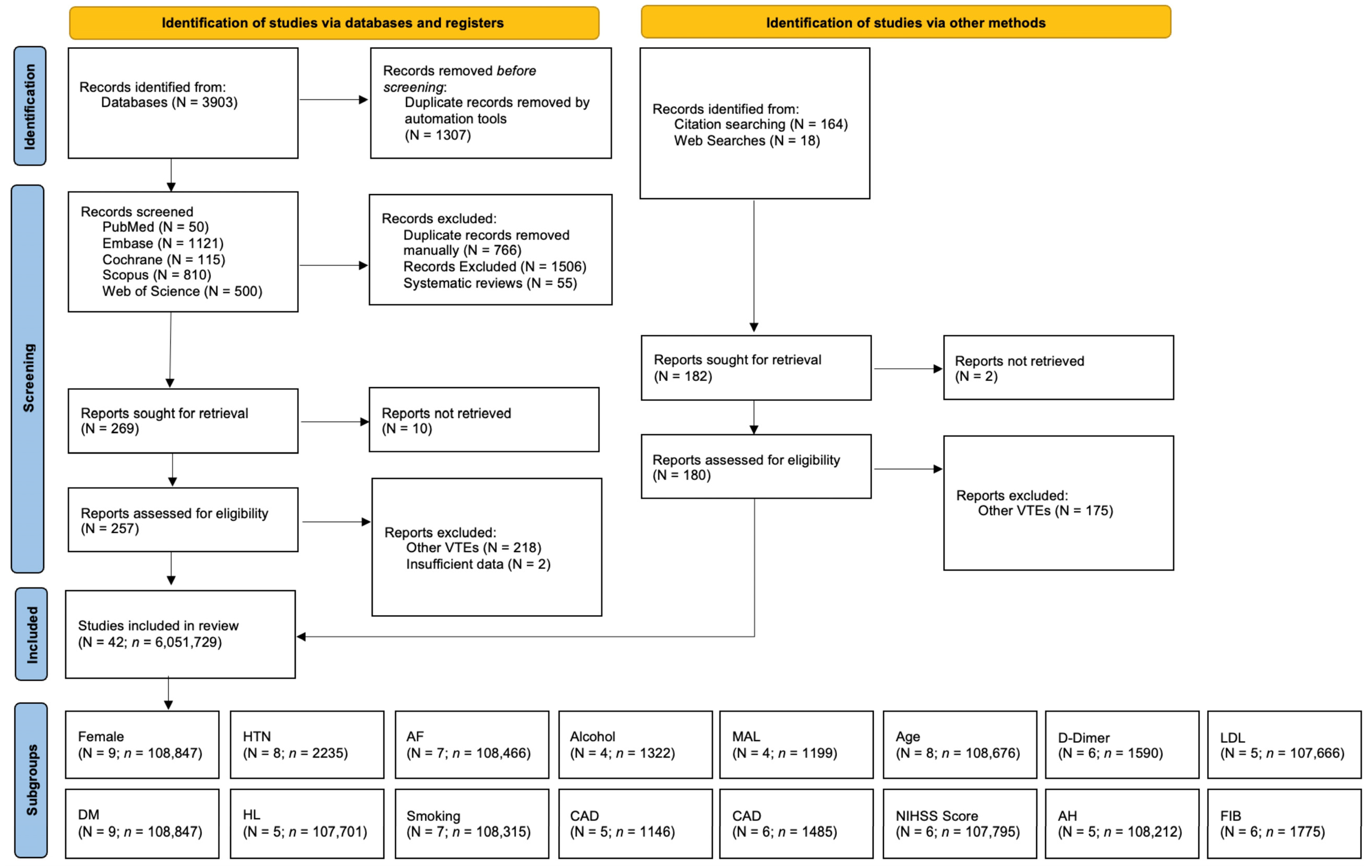
Figure 1. PRISMA flow diagram of study selection for the IRIS-DVT meta-analysis. Abbreviations: DVT: deep vein thrombosis; VTE: venous thromboembolism; HTN: hypertension; DM: diabetes mellitus; HL: hyperlipidemia; AF: atrial fibrillation; CAD: coronary artery disease; MAL: malignancy; NIHSS: National Institute of Health Stroke Scale; LDL: low density lipoprotein; AH: admission hyperglycemia; FIB: fibrinogen.
Ctn 09 00049 g001
Figure 2. Forest plot of pooled prevalence of deep vein thrombosis in acute ischemic stroke patients.
Ctn 09 00049 g002
Figure 3. Forest plot of pooled prevalence of deep vein thrombosis stratified by study design. Abbreviation: DVT: Deep venous thrombosis; AIS: Acute ischemic stroke.
Ctn 09 00049 g003
Figure 4. Forest plot of pooled prevalence of deep vein thrombosis stratified by geographical region. Abbreviation: DVT: Deep venous thrombosis; AIS: Acute ischemic stroke.
Ctn 09 00049 g004
Figure 5. Forest plot of pooled prevalence of deep vein thrombosis stratified by timing of post-stroke screening. Abbreviation: DVT: Deep venous thrombosis; AIS: Acute ischemic stroke.
Ctn 09 00049 g005
Disclaimer/Publisher’s Note: The statements, opinions and data contained in all publications are solely those of the individual author(s) and contributor(s) and not of MDPI and/or the editor(s). MDPI and/or the editor(s) disclaim responsibility for any injury to people or property resulting from any ideas, methods, instructions or products referred to in the content.