Review

Evolution of Pharmacologic Induction of Burst Suppression in Adult TBI: Barbiturate Coma Versus Modern Sedatives

by
Đula Đilvesi
1,2,
Teodora Tubić
1,3,
Sanja Maričić Prijić
1,3 and
Jagoš Golubović
1,2,*
1
Faculty of Medicine, University of Novi Sad, Hajduk Veljkova 3, 21000 Novi Sad, Serbia
2
Department of Neurosurgery, University Clinical Centre of Vojvodina, Hajduk Veljkova 1, 21000 Novi Sad, Serbia
3
Department for Anesthesia, Intensive Care and Pain Management, University Clinical Centre of Vojvodina, Hajduk Veljkova 1, 21000 Novi Sad, Serbia
*
Author to whom correspondence should be addressed.
Clin. Transl. Neurosci. 2025, 9(4), 53; https://doi.org/10.3390/ctn9040053
Submission received: 30 October 2025 / Revised: 15 November 2025 / Accepted: 17 November 2025 / Published: 19 November 2025
(This article belongs to the Topic Neurological Updates in Neurocritical Care)

Abstract

Background: Severe traumatic brain injury (TBI) often leads to elevated intracranial pressure (ICP) that requires aggressive management. Inducing burst suppression with deep sedation is an established therapy for refractory intracranial hypertension. Traditionally, barbiturate coma has been used to achieve burst-suppression EEG in TBI patients, but alternative sedative agents (propofol, midazolam, ketamine, dexmedetomidine) are increasingly utilized in modern neurocritical care. This review compares barbiturates with these alternatives for inducing burst suppression in adult TBI, focusing on protocols, mechanisms, efficacy in controlling ICP, safety profiles, and impacts on neurological outcomes. Methods: A search of the literature was performed, including clinical trials, observational studies, and guidelines on deep sedation for ICP control in adult TBI. Studies comparing high-dose barbiturates to other sedatives (propofol, midazolam, ketamine, dexmedetomidine) in the context of burst suppression or severe TBI management were included. Data on sedative protocols (dosing and EEG targets), mechanisms of action, ICP-lowering efficacy, complications, and patient outcomes were extracted and analyzed qualitatively. Results: High-dose barbiturates (e.g., pentobarbital or thiopental) and propofol are both effective at inducing burst-suppression EEG and reducing ICP via cerebral metabolic suppression. Barbiturate coma remains a third-tier intervention reserved for ICP refractory to other treatments. Propofol infusion has become first-line for routine ICP control due to rapid titratability and shorter half-life, though it can also achieve burst suppression at high doses. Midazolam infusions provide sedation and seizure prophylaxis but yield less metabolic suppression and ICP reduction compared to barbiturates or propofol, and are associated with longer ventilation duration and delirium. Ketamine, once avoided for fear of raising ICP, has shown neutral or lowering effects on ICP when used in ventilated TBI patients, thanks to its analgesic properties and maintenance of blood pressure; however, ketamine alone does not reliably produce burst-suppression patterns. Dexmedetomidine offers sedative and anti-delirium benefits with minimal respiratory depression, but it is generally insufficient for deep burst-suppressive sedation and has only a modest effect on ICP. In comparative clinical evidence, propofol and barbiturates both effectively lower ICP, but neither has demonstrated clear improvement in long-term neurological outcome when used prophylactically. Early routine use of barbiturate coma may increase complications (hypotension, immunosuppression), and thus, current practice restricts it to refractory cases. Modern sedation protocols emphasize using the minimal necessary sedation to maintain ICP < 22 mmHg, with continuous EEG monitoring to titrate therapy to a burst-suppression target (commonly 2–5 bursts per minute) when deep coma is employed. Conclusions: In adult TBI patients with intracranial hypertension, propofol-based sedation is favored for first-line ICP control and can achieve burst suppression if needed, whereas high-dose barbiturates are reserved for ICP crises unresponsive to standard measures. Compared to barbiturates, alternative agents (propofol, midazolam, ketamine, dexmedetomidine) offer differing advantages: propofol provides potent, fast-acting metabolic suppression; midazolam adds anticonvulsant sedation for prolonged use at the cost of slower wake-up; ketamine supports hemodynamics and analgesia; dexmedetomidine aids lighter sedation and delirium control. The choice of agent is guided by the clinical scenario, balancing ICP reduction needs against side effect profiles. While all sedatives can transiently reduce ICP, careful monitoring and a tiered therapy approach are essential, as no sedative has conclusively improved long-term neurological outcomes in TBI. EEG monitoring for burst suppression and meticulous titration is required when employing barbiturate or propofol coma. Ongoing research into optimal combinations and protocols may further refine sedation strategies to improve safety and outcomes in severe TBI.

1. Introduction

Traumatic brain injury (TBI) is a leading cause of mortality and disability in young adults worldwide. One hallmark of severe TBI is elevated intracranial pressure (ICP) resulting from cerebral edema, contusions, or other mass lesions. Elevated ICP can reduce cerebral perfusion pressure and precipitate secondary brain injury. Controlling ICP is, therefore, a central goal in TBI management to prevent herniation and additional ischemic damage. Along with surgical interventions and other medical therapies, sedation is a fundamental component of neurocritical care for TBI. Appropriate sedation can decrease cerebral metabolic activity and cerebral blood flow (CBF), thereby lowering ICP. Sedation also prevents agitation, pain, and ventilator asynchrony, which can otherwise spike ICP. In the acute phase of severe TBI, patients are typically intubated and mechanically ventilated, necessitating sedative agents to ensure patient comfort and compliance with therapy [1,2,3].
In cases of refractory intracranial hypertension, an aggressive level of sedation aiming to induce an electroencephalographic burst suppression pattern is sometimes utilized. Burst suppression refers to a characteristic EEG pattern of high-voltage bursts alternating with isoelectric suppressed intervals, indicating profound depression of cortical activity. Achieving burst suppression through high-dose anesthetic drugs maximally reduces cerebral metabolic rate of oxygen (CMRO2)-a strategy intended to “rest” the injured brain and control ICP when standard measures fail. Historically, barbiturate coma (using agents such as pentobarbital or thiopental) has been the prototypical method to induce burst suppression in TBI patients with intractable ICP elevation. Early studies in the 1980s demonstrated that high-dose barbiturates could lower ICP by ~30–50% through potent cerebro-metabolic suppression and vasoconstriction, and this became an entrenched rescue therapy for refractory cases [4,5,6].
However, barbiturate coma is also associated with significant risks, notably systemic hypotension leading to reduced CPP, immunosuppression with increased infection risk, metabolic disturbances, and prolonged recovery times due to the drug’s long half-life. Moreover, clinical trials have not shown a clear improvement in long-term outcomes with routine barbiturate use. As a result, modern TBI management guidelines (such as the Brain Trauma Foundation 4th Edition) do not recommend prophylactic barbiturate infusion and advise reserving barbiturate coma for ICP elevations refractory to all other standard medical and surgical interventions. Over the past two decades, critical care practice has increasingly shifted toward alternative sedatives that might achieve similar ICP control with more favorable pharmacological profiles [4,6,7,8].
Among these, propofol (an intravenous anesthetic) has become a mainstay sedative in neurocritical care. Propofol has a rapid onset and short duration of action, allowing fine titration. Like barbiturates, propofol is a GABA_A receptor agonist that can produce deep anesthesia and burst suppression at high doses, while also lowering CMRO2 and CBF. Many centers use high-dose propofol infusions as an alternative to barbiturates for burst suppression therapy, given propofol’s easier titratability and quicker recovery. Other sedative agents commonly employed in the ICU include midazolam (a benzodiazepine), ketamine (a dissociative NMDA-receptor antagonist), and dexmedetomidine (a selective alpha-2 adrenergic agonist). Each of these agents has distinct mechanisms and physiological effects that could influence intracranial dynamics. Midazolam provides anxiolysis and anticonvulsant effects but tends to accumulate with prolonged use, potentially prolonging ventilation. Ketamine has sympathomimetic properties that support blood pressure and was historically avoided in TBI due to concerns of raising ICP, but recent evidence suggests that ketamine does not increase ICP in adequately ventilated patients and may even be beneficial. Dexmedetomidine produces a unique arousable sedation with minimal respiratory depression and may help in attenuating agitation and delirium; it usually cannot induce a coma-level sedation by itself, but it can be adjunctive in sedation strategies [9,10,11,12].
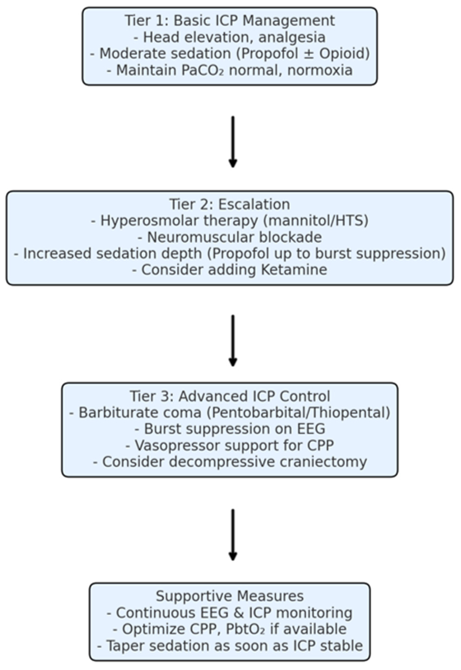
Modern sedation protocols in neurocritical care emphasize a tailored approach: using the lowest effective dose of sedation to achieve ICP control and patient comfort, while avoiding over-sedation that can complicate neurological assessment and lead to complications (like pneumonia and prolonged coma). Neuromonitoring, such as continuous EEG (or processed indices like BIS), is often employed when deep sedation is used to guide titration and ensure the desired depth (e.g., burst suppression) is reached without excessive drug dosing. Sedation is also integrated into a tiered ICP management strategy: basic measures (head elevation, analgesia, moderate sedation) form first-tier therapy, escalated interventions (hyperosmolar therapy, moderate hyperventilation, paralysis) are second-tier, and drastic measures like barbiturate coma or decompressive craniectomy are third-tier options if ICP remains uncontrollable [1,6,13].
Given the pivotal but potentially double-edged role of sedation in severe TBI, it is important to understand how barbiturates compare to alternative sedatives in achieving burst suppression and controlling ICP, and how these choices influence patient outcomes. This article provides a structured review of the evidence on barbiturates versus other agents (propofol, midazolam, ketamine, dexmedetomidine) for inducing burst suppression in adult TBI patients. We compare their mechanisms of action, protocols for administration and monitoring, efficacy in reducing ICP, safety and side-effect profiles, and impact on neurologic outcomes. We also discuss how these agents fit into current neurocritical care protocols and practice, aiming to guide clinicians in optimizing sedation strategies for severe TBI [2,3,14,15,16].

2. Materials and Methods

2.1. Search Strategy and Study Selection

We conducted a comprehensive literature search to identify clinical studies and reviews comparing barbiturates with alternative sedative agents for ICP control or burst suppression in adult TBI. The search was performed in PubMed/MEDLINE and Scopus (date range: 1980 through August 2025), using combinations of keywords and MeSH terms such as “traumatic brain injury,” “intracranial pressure,” “sedation,” “barbiturate coma,” “propofol,” “benzodiazepines,” “midazolam,” “ketamine,” “dexmedetomidine,” “burst suppression,” and “EEG monitoring”. We also manually reviewed reference lists of relevant articles and international TBI management guidelines for additional citations.
Studies were eligible for inclusion if they met the following criteria: (1) Population: adults (age ≥ 16) with severe TBI (typically defined by Glasgow Coma Scale ≤ 8 or requiring ICU admission) and elevated ICP; (2) Intervention: use of high-dose barbiturates to induce deep sedation or burst suppression; (3) Comparison: an alternative sedative regimen (e.g., propofol-based sedation, benzodiazepine sedation, ketamine or dexmedetomidine use), or studies evaluating the effects of these agents on ICP/clinical outcomes; (4) Outcomes: reporting on ICP control, physiological effects (CBF, metabolism), complications (hypotension, organ dysfunction, infections), and/or patient outcomes (mortality, Glasgow Outcome Scale or similar neurological outcome at follow-up). We included randomized controlled trials, prospective and retrospective cohort studies, case series if larger studies were lacking, and relevant systematic or narrative reviews. Although all included cohorts were classified as “adult,” the primary studies often combined young adults and older adults without stratifying their responses to sedative therapy. Where available, we extracted age ranges and confirmed that elderly patients (>65 years) were included in several datasets; however, none of the included studies provided separate dosing, pharmacokinetic adjustments, or ICP-response analyses by age subgroup. Because of this, elderly patients were not analyzed separately in our review. We acknowledge that geriatric patients may metabolize barbiturates, benzodiazepines, and propofol differently, and future studies should more clearly distinguish age-related physiological differences in sedative response. We excluded animal studies and clinical studies focused on pediatric TBI, as well as papers not available in English. For completeness, when high-level evidence was sparse, we also incorporated consensus guideline recommendations and pathophysiological data from key pharmacological studies.

2.2. Data Extraction and Synthesis

From each included study or source, we extracted pertinent data regarding sedation protocols (drug dosing, titration method, use of EEG or BIS monitoring, target burst suppression criteria), mechanism of action of the sedative agents, changes observed in intracranial dynamics (ICP values, cerebral perfusion pressure, cerebral blood flow, metabolic rate), any measured clinical endpoints (such as control of refractory ICP episodes, duration of ICP crisis, mortality, functional outcome scores), and reported adverse events (hemodynamic instability, propofol infusion syndrome, infectious complications, etc.). Given the heterogeneity of study designs and outcomes, a quantitative meta-analysis was not feasible. Instead, we performed a qualitative synthesis, grouping findings under thematic categories (e.g., ICP reduction efficacy, safety profile, neurological outcomes) to facilitate comparison across agents.
Two reviewers (authors of this paper) independently screened the titles and abstracts for relevance, and consensus was used to resolve any inclusion disagreements. The strength of evidence was considered when formulating conclusions and recommendations: for example, randomized trial or meta-analysis data were given more weight than observational studies or expert opinion.

3. Results

3.1. Sedative Mechanisms of Action and Burst Suppression Induction

Barbiturates: Barbiturates such as pentobarbital and thiopental are potent central nervous system depressants that primarily enhance GABA_A receptor activity, leading to increased inhibitory neurotransmission. At high doses, barbiturates induce general anesthesia and an EEG burst-suppression pattern, reflecting near-complete cortical silence. They also inhibit excitatory neurotransmission (glutamate AMPA/kainate receptors) and reduce cerebral metabolic demand. By lowering CMRO2, barbiturates cause coupled reductions in CBF (via cerebral autoregulation) and cerebral blood volume, which results in decreased ICP. The burst suppression itself is a marker of maximal metabolic suppression: for instance, achieving ~4 bursts per minute on EEG indicates roughly a 50% reduction in CMRO2 from baseline. Barbiturates also have anticonvulsant properties, raising the seizure threshold and actively terminating epileptiform activity, which can be beneficial in TBI patients at risk of seizures or status epilepticus [17,18,19].
Propofol: Propofol acts as a GABA_A receptor agonist (at a distinct binding site from barbiturates) and also has NMDA receptor antagonism and other neuromodulatory effects. Its mechanism yields profound sedation and hypnosis. Propofol’s pharmacokinetics (high lipid solubility, rapid redistribution and metabolism) confer a fast onset and relatively short duration of action. Like barbiturates, propofol produces dose-dependent EEG slowing; at moderate doses, one sees delta-wave dominance, and at high doses, propofol can generate burst suppression patterns very similar to those from barbiturate coma. Propofol reliably decreases CMRO2 and CBF in tandem (up to ~30–40% reduction at deep anesthesia levels), thereby lowering ICP if cerebral autoregulation is intact. Propofol is also a strong antiseizure agent and is often used to halt refractory status epilepticus via burst-suppression coma. A unique aspect of propofol is its antioxidant and anti-inflammatory properties (it scavenges free radicals and may reduce lipid peroxidation in neural cells), which have led to speculation that propofol sedation could confer some neuroprotective benefit beyond ICP control. In addition to its established antioxidant and anti-inflammatory effects during later phases of brain injury, several experimental and early clinical studies suggest that propofol may exert beneficial cellular effects even in the acute phase of TBI. Proposed mechanisms include rapid suppression of excitotoxic calcium influx, attenuation of early oxidative stress, and modulation of pro-inflammatory cytokine release within hours after injury. Although these effects do not yet translate into proven improvements in long-term outcome, they may partially contribute to the early stabilization seen during propofol-based ICP control [20,21,22].
Midazolam: Midazolam is a benzodiazepine that potentiates GABA_A receptor activity by increasing the frequency of channel opening in the presence of GABA. It provides sedative, anxiolytic, and anticonvulsant effects. However, midazolam alone has a ceiling to how deep a coma it can induce; typically, it does not produce burst suppression as readily as propofol or barbiturates unless given in very high (often impractical) doses. Midazolam does reduce cerebral metabolic rate and CBF, but to a lesser degree than equipotent doses of propofol or barbiturates. Thus, the ICP-lowering effect of midazolam is generally milder. On EEG, midazolam tends to induce diffuse slowing but not an isoelectric pattern in usual clinical dosing. Midazolam’s pharmacokinetic profile in critical illness is variable-it is highly lipophilic and accumulates in peripheral tissues, leading to a prolonged sedative effect with continuous infusions (especially beyond 2–3 days). It is metabolized hepatically and has active metabolites. These factors mean midazolam can result in delayed emergence from sedation when used long-term. Nonetheless, midazolam’s strong anticonvulsant property is valuable; it is often employed as a first-line continuous infusion for status epilepticus and can be part of sedation regimens in TBI when long-term sedation is needed or when hypotension precludes higher propofol doses [23,24,25,26].
Ketamine: Ketamine is an NMDA receptor antagonist that induces a dissociative anesthetic state. Unlike the GABA-agonist sedatives, ketamine increases neuronal inhibition by blocking excitatory glutamate transmission. It produces analgesia and a trance-like cataleptic state rather than traditional CNS depression. Importantly, ketamine stimulates sympathetic outflow, typically causing an increase in heart rate, blood pressure, and cardiac output. This hemodynamic effect often helps maintain or raise CPP, which can be advantageous in TBI patients, as hypotension is deleterious. Historically, ketamine raised concerns because older studies suggested it could raise ICP (likely due to increases in CBF from vasodilation and blood pressure elevation). However, newer investigations in ventilated TBI patients have shown that ketamine does not significantly elevate ICP, and in many cases, ICP stays the same or decreases slightly when ketamine is given (for example, during painful procedures or as part of a sedation regimen). The likely explanation is that any ketamine-induced rise in CBF is offset by concomitant reductions in sympathetic surges from pain and by ensuring adequate ventilation (preventing hypercapnia). Ketamine at typical sedative/analgesic doses (e.g., low-dose infusion 0.5–3 mg/kg/h or intermittent boluses) does not produce burst suppression on EEG; instead, EEG under ketamine shows a high-frequency activity pattern. At extremely high doses, ketamine can induce anesthesia and unconsciousness, but even then, a true burst-suppressed EEG is not characteristic. Thus, ketamine is not used as a sole agent to induce burst suppression. Instead, ketamine’s role is often as an adjunct: added to propofol or midazolam to provide analgesia and maintain blood pressure, thereby allowing lower doses of GABAergic sedatives. Notably, ketamine also has neuroprotective theoretical benefits by reducing excitotoxicity (through NMDA blockade) and has been used as a third-line agent in refractory status epilepticus as well [27,28,29,30].
Dexmedetomidine: Dexmedetomidine is a selective α2-adrenergic receptor agonist. It produces a unique form of sedation that mimics non-REM sleep, with patients often appearing calm and drowsy but arousable (termed “cooperative sedation”). The mechanism involves inhibition of noradrenergic neurons in the locus coeruleus, reducing sympathetic tone and inducing sedation and analgesia. Dexmedetomidine by itself typically can only achieve mild to moderate sedation (e.g., a target Richmond Agitation–Sedation Scale of ~0 to −2 in many patients). It cannot reliably induce deep coma or burst suppression even at high infusion rates, due to a plateau in its sedative effect (higher doses mainly cause more bradycardia and hypotension rather than deeper EEG suppression). As for intracranial effects, dexmedetomidine can modestly reduce ICP indirectly by blunting agitation and sympathetic surges; it may also lower CBF slightly through blood pressure reduction, but it does not significantly change CMRO2. Thus, dexmedetomidine is not indicated for treating refractory ICP or achieving burst suppression. Instead, its utility in TBI is as an adjunct for sedation in patients who are intermediate in the course: for example, a patient who is recovering and no longer needs heavy sedation might be transitioned to dexmedetomidine to reduce delirium and allow neurological assessments. Dexmedetomidine tends to preserve respiratory drive, so it can facilitate earlier weaning from mechanical ventilation or even be used in intubated patients with minimal respiratory reserve. Additionally, by decreasing the need for other sedatives, dexmedetomidine may reduce the incidence of ICU delirium. Its sympatholytic effect can cause bradycardia and hypotension, which require close monitoring but can be managed with dose adjustment or vasopressors if needed [12,31,32,33].
In summary, barbiturates and propofol share a similar mechanism (powerful GABA-mediated suppression), leading to burst suppression and maximal ICP reduction, whereas midazolam is a less potent metabolic suppressant, ketamine works via NMDA blockade with supportive hemodynamic effects, and dexmedetomidine provides sedation via alpha-2 agonism without the depth required for burst suppression. These mechanistic differences underlie variations in how each agent is used in practice for TBI sedation (Table 1).

3.2. Sedation Protocols and EEG Monitoring in TBI

When deploying deep sedation to control ICP, standardized protocols and careful monitoring are crucial to balance efficacy with safety. Key elements of such protocols include drug dosing strategies (loading and maintenance), goals for depth of sedation (often defined by EEG or clinical scales), and monitoring for side effects (Table 2). Continuous EEG monitoring is considered the gold standard for confirming burst suppression and adjusting sedative dosing accordingly [34,35,36].
Barbiturate Coma Protocol: In adults with refractory intracranial hypertension, a typical pentobarbital coma protocol begins with a loading dose to rapidly achieve therapeutic brain levels. One commonly used regimen is: pentobarbital 10 mg/kg IV loading over 30 min, followed by additional booster doses of 5 mg/kg IV every hour for 3 doses (total cumulative load ~25 mg/kg). This aggressive loading saturates the peripheral compartments and helps ensure the drug crosses the blood–brain barrier to reach an effective concentration. After or even during the loading phase, continuous infusion is started for maintenance, usually at 1 mg/kg/h (some protocols use 3–5 mg/kg/h initially and then taper down). The infusion rate is then titrated based on the ICP response and EEG findings. The EEG target is to induce a burst suppression pattern-commonly aiming for approximately 2–5 bursts per minute on the raw EEG. In practice, this might correspond to a burst-suppression ratio of ~70–90% (meaning the EEG is suppressed 70–90% of the time) if using processed EEG indices. Some protocols specifically direct that once burst suppression is achieved, the infusion rate can be adjusted to maintain a given burst frequency range. For example, if bursts become too infrequent (indicating deep beyond target, e.g., isoelectric EEG or less than 1 burst in 10 s), the infusion can be slightly reduced; conversely, if EEG shows continuous activity or more frequent bursts, the infusion is increased [17,37,38].
Because continuous full-montage EEG is not always available, ICUs often use simplified monitoring like the Bispectral Index (BIS) or other processed EEG monitors to guide titration. A BIS value in the range of ~10–20 typically corresponds to a deep hypnotic state with burst suppression. In published adult series of pentobarbital coma, achieving around BIS ~15 was associated with the desired burst suppression (roughly 3 bursts/min). It is important to note that during barbiturate coma, standard clinical signs (like a sedation scale or pupillary response) are not reliable to gauge depth-hence the reliance on EEG. Neurological examinations cannot be performed while a patient is in a barbiturate coma, aside from assessing brainstem reflexes if the sedation is not too profound. Therefore, ancillary monitoring such as ICP monitors, automated pupillometry, or brain tissue oxygen monitors (PbtO2) is often used to track the patient’s neurological status indirectly [4,39,40].
Infusion Maintenance and Taper: Once ICP is under control with barbiturate infusion, the protocol usually calls for continuing the coma for at least 24–48 h of sustained ICP control. If ICP spikes recur, dosing may escalate to deepen the suppression (limited by hemodynamics and toxicity). Barbiturate levels can be measured (therapeutic pentobarbital coma level often in the range of 30–40 µg/mL, though practice varies) to avoid severe toxicity. After a period of stable ICP, the barbiturate infusion is gradually tapered rather than stopped abruptly, to prevent rebound intracranial hypertension and facilitate elimination. A common taper schedule is to reduce the infusion by ~0.5 mg/kg/h every 6–12 h, closely watching ICP. Another strategy used is a step-down daily taper: for example, if a patient was on 3 mg/kg/h for maintenance, reduce to 2 mg/kg/h for 24 h, then 1.5 for 24 h, then 1 mg/kg/h, etc. Tapering can take several days. If ICP rises during the wean, the infusion may be raised again to re-establish control [1,41,42,43].
Propofol Infusion Protocol: Propofol is typically started earlier in severe TBI management as a first-line sedative. For routine sedation in intubated TBI patients, propofol might be started at a moderate dose (e.g., 20–50 mcg/kg/min, which is 1.2–3 mg/kg/h) and titrated to achieve a sedation goal (commonly a RASS of −4 to −5, or a state where the patient is unresponsive to anything except noxious stimuli). If ICP remains elevated despite this level of sedation, the propofol infusion can be escalated to high-dose ranges (50–100+ mcg/kg/min, i.e., up to 6–10 mg/kg/h) to deepen the sedation toward burst suppression. Propofol can induce burst suppression, usually at the upper end of dosing. Clinical signs like loss of motor response, plus an EEG showing bursts with intervening flat periods or a BIS value < 20, indicate the target depth is reached. One major difference from barbiturates is the speed of titration: propofol reaches steady state quickly, so infusion adjustments will affect the EEG within minutes rather than hours. This allows a quicker response to ICP changes-for instance, if a patient has an acute ICP spike, the propofol infusion rate can be doubled for a short period to deepen sedation rapidly. Conversely, if hypotension occurs, propofol can be temporarily reduced and another agent (like vasopressor or adjunct sedative) used to maintain ICP control [44,45,46].
The limitation with propofol is the risk of Propofol-Related Infusion Syndrome (PRIS) at high doses and prolonged durations. PRIS is a potentially fatal complication characterized by severe lactic acidosis, rhabdomyolysis, cardiac failure, and renal failure. TBI patients appear to be at particular risk, possibly because they often require high-dose propofol for ICP control and have catecholamine surges that propofol dampens (leading to a mismatch in metabolic demand). It is generally recommended to avoid exceeding 4–5 mg/kg/h for more than 48 h if possible. If very deep propofol sedation is needed beyond that time frame, many ICUs will lighten the propofol or switch to an alternative (e.g., transition to midazolam or add ketamine/dexmedetomidine to allow propofol dose reduction). During propofol infusion, monitoring for early signs of PRIS is critical: this includes checking for unexplained metabolic acidosis, hyperkalemia, rising creatine kinase, arrhythmias, or the classic greenish hue of the urine (due to phenolic metabolites). If PRIS is suspected, propofol must be weaned off immediately [47,48,49].
Midazolam Use: Midazolam infusions in severe TBI are often used in two scenarios: (1) adjunct to propofol-to achieve synergy at lower individual drug doses, or (2) replacement for propofol after 2–3 days, to avoid PRIS when ongoing sedation is needed. A typical midazolam infusion dose for deep sedation might range from 0.1 to 0.4 mg/kg/h (with boluses of 2–5 mg IV as needed to control breakthrough agitation or spikes in ICP). Midazolam’s effect is slower, so dose adjustments are made every 30–60 min rather than minutes. It is not common practice to aim for burst suppression with midazolam alone, as extremely high doses would be needed. Instead, if midazolam is the primary sedative, the goal might be deep sedation (RASS −4/−5) rather than an EEG endpoint, accepting that its ICP-lowering effect may not be as profound. Continuous EEG could still be used to ensure no subclinical seizures occur and to monitor depth (e.g., one might see a continuous delta slowing pattern at adequate sedation). If ICP remains refractory on midazolam, one would escalate to barbiturates or propofol; midazolam by itself is rarely considered a definitive therapy for refractory ICP but is part of the sedation toolkit [24,50].
Ketamine-Augmented Protocols: Ketamine is frequently introduced when hypotension limits the use of propofol or barbiturates. For example, a patient on high-dose propofol with borderline blood pressure could benefit from adding ketamine infusion (e.g., 0.5–1 mg/kg/h)-this often allows a reduction in propofol dose while maintaining or even improving ICP control, because ketamine prevents the pain or stimuli-induced ICP spikes and keeps blood pressure higher (thus improving CPP). Some protocols in refractory ICP include low-dose ketamine (like 1 mg/kg IV bolus followed by infusion) as an adjunct, even during barbiturate coma, to provide analgesia and potentially some neuroprotective effect. It is important to avoid giving ketamine as a rapid bolus in TBI without adequate ventilation and sedation, as transient increases in blood pressure and CBF could spike ICP; instead, it is given slowly or as an infusion. Another specific indication in neurocritical care is during stimulating procedures (suctioning, turning, or transport)-a bolus of ketamine (0.5 mg/kg) prior to these activities can blunt ICP elevations that such stimuli often provoke. Modern evidence has dispelled the old dogma against ketamine in head injury, and it is now considered a safe and effective adjunct in the armamentarium for managing severe TBI, albeit not as a sole agent for burst suppression [51,52,53].
Dexmedetomidine in Protocols: Dexmedetomidine is usually started at 0.2–0.7 mcg/kg/h (after a careful or skipped loading dose, since a full loading can cause bradycardia/hypotension). In TBI care, dexmedetomidine might be used in the weaning phase: for instance, after days of heavy sedation, as ICP stabilizes, clinicians may lighten propofol or turn off barbiturates and start dexmedetomidine to maintain a calmer state while neurological function returns. It can also be added at lower doses concurrently with propofol or midazolam to reduce their required dose and potentially mitigate delirium. If a patient is emerging and remains agitated but no longer needs deep coma, dexmedetomidine infusion can keep them lightly sedated without respiratory compromise, aiding in extubation and neurologic exams. There is no specific EEG target with dexmedetomidine-it does not cause burst suppression. Typically, one monitors using clinical scales (RASS ~ −2) or simply observation of patient comfort. One should watch for hypotension (especially in volume-depleted patients) and bradycardia; if mean arterial pressure drops too much under dexmedetomidine, a vasopressor or reducing the dose is necessary to maintain adequate CPP [54,55].
EEG Monitoring: Continuous EEG or quantitative EEG monitoring is strongly recommended whenever burst suppression therapy is being employed, whether via barbiturates or propofol. Achieving the right depth is critical: insufficient depth may fail to control ICP or seizures, while excessive depth exposes the patient to more drug toxicity without additional benefit. EEG allows real-time assessment of suppression. Typically, the goal is set as mentioned (around a few bursts per minute). There is some variability in practice: some experts target a “moderate” burst-suppression (e.g., 8–10 bursts/min) versus others target “deep” (2–5 bursts/min), depending on how refractory the situation is. In many protocols, bursts are defined as any EEG activity > 0.5–1 s long separated by at least 1 s of flat suppression. A suppression ratio (percentage of time suppressed) of ~70–90% is often the objective [38,56,57].
When using processed monitors like BIS, clinicians correlate the BIS or patient state index (PSI) with known EEG behavior. For instance, a BIS value below 20 usually indicates significant burst suppression or a near-isoelectric EEG. However, neuromuscular artifacts and other factors (like electrode noise) can affect BIS, so raw EEG confirmation is ideal. In fact, ICU cases have been reported where patients were found to be in burst suppression inadvertently because sedation was deeper than intended-highlighting the importance of EEG monitoring to avoid over-sedation. Besides EEG, other monitoring priorities during burst suppression therapy include invasive arterial blood pressure (for beat-to-beat blood pressure, as hypotension is common and must be managed immediately), core temperature (since both barbiturates and propofol impair thermoregulation and shivering, plus barbiturates may induce a mild hypothermia), and frequent metabolic panels (to catch acidosis, electrolyte shifts, or hepatic/renal impairment) [58,59,60].

3.3. Impact on Intracranial Pressure and Cerebral Physiology

One of the primary outcomes of interest is how effectively each sedative strategy lowers ICP and maintains ICP control in severe TBI. The evidence shows that both barbiturate and propofol coma can produce substantial reductions in ICP in patients who respond to the therapy, while midazolam and dexmedetomidine have more limited effects. Ketamine has a neutral to beneficial effect on ICP when used appropriately.
Barbiturates on ICP: High-dose barbiturates have been proven to lower ICP in a majority of patients with refractory intracranial hypertension. In clinical series, about 50–75% of severe TBI patients with uncontrollable ICP will exhibit a response (ICP decrease) after barbiturate coma is initiated. The magnitude of ICP reduction can be significant: studies have documented decreases of 10–15 mmHg in mean ICP, or cases of bringing ICP from dangerous levels (>30 mmHg) down to safer ranges (<20 mmHg) after reaching burst suppression. This ICP-lowering effect stems from the profound drop in cerebral metabolic rate-as metabolism falls, CBF also drops (assuming autoregulation is intact), leading to less intracranial blood volume. Furthermore, burst suppression might interrupt pathological cortical depolarizations and seizures that can worsen edema. However, a critical caveat is that barbiturate-induced systemic hypotension occurs in up to 30% or more of patients; if mean arterial pressure falls significantly, the net CPP may not improve despite a lower ICP. In other words, while ICP goes down, cerebral perfusion could be compromised unless vasoactive support is provided. Therefore, in practice, when barbiturate coma is started, patients often require concurrent vasopressor infusions (e.g., norepinephrine) to maintain an adequate CPP (typically ≥60–70 mmHg). The need for vasopressors and hemodynamic monitoring is an expected part of barbiturate therapy. Some studies have noted that responders to barbiturate (those whose ICP falls by > ICP 5 mmHg) had better survival than non-responders, which suggests that if the brain is still recruitable to metabolic suppression, outcomes might be somewhat better [4,53,61,62].
Propofol on ICP: Propofol infusion, even at moderate doses used for sedation, usually leads to a modest reduction in ICP in severe TBI patients. For example, sedation with propofol can lower ICP by a few mmHg compared to no sedation or lighter sedation, due to reduced patient activity and some metabolic suppression. At higher doses, propofol’s effect approximates that of barbiturates: profound propofol anesthesia with burst suppression can similarly drop ICP substantially and control ICP spikes. Propofol has the advantage that it reduces intracranial hypertension rapidly-after a bolus or increase in infusion, ICP can fall within minutes if the patient is responsive to sedation. Additionally, propofol tends to preserve cerebral autoregulation better than some other sedatives; studies have shown that reactivity to CO2 and flow-metabolism coupling remain intact, meaning propofol does not cause luxury perfusion or steal phenomena in the injured brain. This is beneficial because it ensures that regions of the brain still regulate their blood flow based on metabolic needs even under deep propofol sedation. Propofol, however, can cause significant hypotension as well by myocardial depression and vasodilation, especially when used in high doses. The drop in mean arterial pressure can be 10–20% or more. Clinicians mitigate this by fluid loading or vasopressors if needed, similar to barbiturates. One notable point: propofol’s effect on ICP might be partially offset if hypotension is not corrected, as cerebral perfusion pressure would drop, potentially triggering autoregulatory vasodilation and opposing the ICP-lowering effect. So, maintaining adequate perfusion is essential to fully realize the ICP benefit [20,46,63].
Comparative studies suggest no large difference in ICP control between propofol and barbiturates when each is titrated to burst suppression. Both are potent enough to maximize metabolic suppression. Some older hemodynamic studies (from the 1990s) compared thiopental and propofol in head-injured patients: they found that both drugs lowered CBF and CMRO2 similarly, although propofol caused less of a blood pressure drop at equivalent brain metabolic effect. Moreover, propofol’s shorter half-life allows intermittent neurological assessments if needed-for instance, stopping propofol for 15–30 min can allow a quick clinical exam before ICP rises, something not feasible with long-acting barbiturates [20,64,65].
Midazolam on ICP: Midazolam sedation has a less dramatic influence on ICP. It certainly helps control ICP indirectly by preventing agitation, coughing, and reducing metabolic demand modestly. Studies that compared midazolam to propofol for general ICU sedation in brain-injured patients found that ICP values were similar or only slightly higher with midazolam, suggesting midazolam can be adequate for baseline ICP management in many cases. However, in truly refractory ICP scenarios, midazolam likely cannot achieve enough metabolic suppression to bring down very high ICP. For example, in patients with ICP > 30 mmHg refractory to analgesia and moderate propofol, switching to heavy midazolam alone (without barbiturate) often will not suffice. One specific issue is midazolam’s propensity to accumulate-after some days, the patient can become excessively sedated without additional clinical benefit and waking them for a neuro exam can be delayed by days. Additionally, benzodiazepine use is strongly linked with delirium in ICU survivors; while delirium is a later issue, it is pertinent to outcomes and will be discussed later. If midazolam is used, it is often in combination with other agents (fentanyl for analgesia, maybe low-dose propofol, etc.), and it is usually considered a second-line sedative for ICP control if propofol is contraindicated (e.g., severe hemodynamic instability or prolonged sedation needed) [66,67,68].
Ketamine on ICP and CPP: Contemporary evidence indicates that ketamine does not increase ICP in the context of sedated, ventilated TBI patients. Several prospective studies have even shown instances of ICP decreasing after ketamine administration. For example, ketamine boluses given to patients with refractory intracranial hypertension often resulted in either no change or a slight reduction in ICP, while CPP improved due to increased mean arterial pressure. This is likely because ketamine’s analgesic effect prevents pain-induced ICP spikes and its blood pressure support improves cerebral perfusion (which can trigger reflex cerebral vasoconstriction, lowering ICP). Ketamine also does not lower CMRO2 in the way GABAergics do; it may slightly increase CMRO2 in some brain regions due to excitatory activity, but overall, the net effect in the injured brain appears not harmful under controlled ventilation. In short, ketamine is considered ICP-neutral to ICP-lowering in modern TBI care, overturning the historical view. It is thus a useful adjunct, especially in patients where maintaining blood pressure is challenging-it helps avoid the ICP rises that accompany hypotension or high-dose vasopressors (which can cause vasoconstriction peripherally but also some cerebral vessel constriction). It is worth noting that ketamine has bronchodilatory properties and preserves respiratory drive (at sub-anesthetic doses), which can be advantageous when weaning from ventilation or managing coexisting pulmonary issues [69,70].
Dexmedetomidine on ICP: Dexmedetomidine generally has minimal direct effect on ICP. It can indirectly assist by calming the patient, reducing coughing on the tube, and promoting sleep cycles, which can all prevent ICP spikes. If dexmedetomidine lowers blood pressure significantly, ICP could paradoxically rise due to lowered CPP, but usually any hypotension from dex is mild and manageable. One scenario where dexmedetomidine is useful for ICP is during emergence from anesthesia: for example, after barbiturate or propofol coma is tapered, adding dexmedetomidine can smooth the transition, so the patient does not become agitated (which would raise ICP). In summary, dexmedetomidine is ICP-neutral for practical purposes; it is included in protocols not to directly reduce ICP but to facilitate overall management (reducing doses of other sedatives, managing agitation) [3,71,72].
Across all agents, the most important factor is that sedation should be used as part of a multimodal ICP management plan. Sedation alone, no matter how deep, cannot resolve ICP issues stemming from large mass lesions or if the mean arterial pressure is very low. Thus, it is always in conjunction with other measures: CSF drainage if an EVD is present, osmotic therapy, and possibly neuromuscular blockade to remove muscle tone as a factor in ICP (e.g., some protocols give a paralytic infusion along with heavy sedation to eliminate even cough or posturing, further stabilizing ICP). The tiered approach typically ensures analgesia and moderate sedation (often propofol under 4 mg/kg/h plus fentanyl)-this addresses many cases of moderate ICP elevation. If ICP remains >22 mmHg persistently, escalate to second tier (hyperosmolar therapy, paralysis, mild hyperventilation). If still refractory, then third tier: which includes barbiturate or propofol coma, and/or decompressive craniectomy depending on patient factors. Barbiturates and decompression are sometimes used sequentially or together in extreme cases, but surgery is often considered if not already performed before committing to long-term barbiturate coma [1,6,62].

3.4. Clinical Efficacy and Neurological Outcomes

ICP Crisis Control: In terms of immediate efficacy, the success of any sedative strategy can be measured by its ability to control refractory ICP and prevent acute herniation or neurological deterioration. High-dose barbiturates have shown efficacy in aborting ICP crises about 65–75% of the time in uncontrolled series. Propofol, when used to a similar depth, likely has comparable success rates in ICP control (though fewer studies quantify this, since propofol is ubiquitous as a sedative). What is clear is that if a patient’s ICP cannot be controlled by even barbiturate coma, the prognosis is extremely poor without surgical intervention. So, these sedatives are indeed life-saving measures in certain scenarios, buying time and stability for the injured brain. Many case series from modern neuroICUs report that a subset of severe TBI patients are salvaged from near-terminal ICP elevation by barbiturate or propofol coma, allowing them to survive to later undergo rehabilitation [6,8,18].
Functional Outcomes and Mortality: The more controversial aspect is whether inducing burst-suppression sedation improves long-term neurological outcomes (such as functional recovery at 6 months) or mortality, beyond the short-term ICP control. The evidence suggests that routine use of barbiturate coma does not improve outcomes and may even worsen them if used indiscriminately. A Cochrane review and other systematic analyses of trials in severe head injury found no significant difference in mortality or favorable outcome rates between patients treated with barbiturates and those who were not, except that barbiturates were associated with a higher incidence of hypotensive events. In one often-cited randomized trial from the 1980s, barbiturate therapy lowered ICP in refractory cases but did not confer a survival advantage; patients in the barbiturate group had more hemodynamic instability. More recently, a propensity-score adjusted observational study indicated that early use of barbiturates (within the first 24 h of TBI ICU care) was associated with higher mortality compared to those managed without early barbiturate coma. This finding likely reflects both the severity of patients needing barbiturates and the detrimental side effects (like cardiovascular depression) tipping the risk-benefit scale when used very early. As a result, current guidelines strongly discourage prophylactic high-dose barbiturate infusion-meaning one should not start barbiturate coma before exhausting other measures or without clear indication (e.g., one should not initiate burst-suppression sedation in a severe TBI patient whose ICP is still manageable by lighter sedation and medical therapy, just hoping it will improve outcome-evidence says it will not, and it might harm) [18,73,74].
For propofol, no large trials have specifically evaluated propofol coma vs. not in TBI outcomes, because propofol is standard for sedation. However, indirectly, studies show that maintaining propofol sedation to control ICP does not hurt outcomes and is part of standard care. A small, randomized study comparing propofol vs. midazolam sedation in severe TBI found no significant difference in 6-month neurological outcomes, although propofol showed a trend towards better short-term neurological scores and lower markers of inflammation. Propofol’s rapid awakening allows earlier neuro exams and potentially earlier transition to rehab if the patient improves, which could positively influence outcomes by minimizing ICU complications like infections. That said, in very prolonged sedation cases, propofol cannot be continued indefinitely at high doses due to PRIS, so prolonged deep sedation almost inevitably means some exposure to barbiturates or benzodiazepines too [68,75,76].
Mid-term cognitive outcomes can be affected by the sedative choice as well. Deep sedation itself has been linked to long-term cognitive impairment in ICU survivors (post-ICU syndrome). Specifically, benzodiazepine-based sedation is associated with a higher incidence of delirium, which in turn correlates with worse cognitive outcomes months later. Dexmedetomidine, by contrast, is linked to less delirium. In TBI patients, outcome is mostly dictated by the primary brain injury severity, but sedation practices may modulate secondary injury and the recovery trajectory. Over-sedation can delay neurological assessments and lead to unnecessary prolongation of mechanical ventilation (with attendant risks like pneumonia, muscle deconditioning, etc.). For example, a patient kept in a barbiturate coma for many days might miss the window for certain neuro-prognostic evaluations or interventions. Thus, one principle is to use the shortest duration of deep sedation necessary. The front-line clinicians typically attempt to lighten sedation once ICP has been stable for at least 24 h, in order to assess the neurological exam. This was highlighted in consensus recommendations: if ICP is controlled, try to reduce sedation after a day and see if the patient can tolerate a wake-up for exam-the information gained can be crucial (the patient might be following commands, changing management towards early rehabilitation) [77,78,79].
Comparative Outcomes for Agents: There have been no studies showing how using one sedative agent over another (e.g., propofol vs. midazolam produces a dramatic difference in ultimate Glasgow Outcome Scale or functional independence. They are largely supportive therapies. One retrospective analysis found that patients sedated predominantly with midazolam had longer ICU stays and ventilation duration compared to those with propofol, but neurological outcomes were similar; midazolam use also correlated with more frequent need for additional ICP-lowering interventions (possibly because it was not as potent on ICP). Ketamine’s impact on outcome in TBI is still being studied; some hypothesize its maintenance of CPP could translate to better outcomes, but concrete data is limited. At the very least, ketamine does not worsen outcomes and is useful in avoiding hypotensive insults, which are known to worsen TBI prognosis. Dexmedetomidine in general ICU populations has shown reductions in delirium and maybe a slight mortality benefit in some studies, but in the neuro ICU, the data is less clear. Its use in TBI patients is rising, particularly to facilitate smoother wake-ups and earlier extubation, which could indirectly improve recovery and shorten ICU length of stay [14,53,80].
Seizure Control: Another facet of efficacy is prevention and control of seizures, which are common after TBI (both early post-traumatic seizures and later). Barbiturates and propofol are both effective in stopping refractory seizures, and they may be chosen specifically if a patient has developed status epilepticus on top of TBI. Midazolam is also a mainstay for seizure control, often as a continuous infusion for status epilepticus. Notably, TBI guidelines often recommend a week of prophylactic anti-epileptic medication (like phenytoin or levetiracetam) to prevent early seizures, but this is separate from sedation. Still, having a sedative that raises the seizure threshold can be beneficial. In this regard, phenobarbital (a barbiturate) and midazolam both reduce the incidence of late post-traumatic seizures when used for sedation, as some studies have observed. Dexmedetomidine does not have known anti-epileptic effects, and ketamine at sub-anesthetic doses can actually induce epileptiform EEG activity in some cases (though clinically it is rarely a problem, and at high doses of ketamine can even treat status epilepticus via NMDA blockade). Thus, in patients with combined refractory ICP and refractory status epilepticus, an approach is often to use multiple agents: e.g., high-dose pentobarbital for burst suppression addressing both ICP and seizures, plus perhaps midazolam or propofol as needed. These scenarios are complex but highlight that sedation choice may be influenced by the presence of seizures (favoring GABAergic drugs) [24,25,30].

3.5. Safety Profiles and Complications

Each sedative agent comes with a distinct profile of side effects and risks, which must be carefully weighed, especially in the already vulnerable TBI population that may have systemic injuries and labile physiology (Figure 1).
Barbiturates-Risks: The main acute risk of barbiturate coma is hemodynamic instability. By causing systemic vasodilation and myocardial depression, barbiturates often lead to hypotension. In a severely injured brain, hypotension can be devastating, as it lowers CPP and can exacerbate ischemic injury. Therefore, managing a patient in barbiturate coma typically necessitates fluid optimization and vasopressor support. Patients often need central venous pressure monitoring and arterial lines; some may require high-dose vasopressors, which come with their own risks (limb ischemia, arrhythmias). Barbiturates also cause myocardial suppression and can precipitate arrhythmias at high doses or if serum levels become very high. Another issue is immunosuppression: barbiturates inhibit neutrophil function and stress responses, so prolonged barbiturate coma is associated with higher rates of infections, particularly ventilator-associated pneumonia (VAP). In fact, some observational studies have noted that nearly half of patients on barbiturate coma developed pneumonia within days. There is also impaired gastrointestinal motility (ileus) and stress ulcer risk, so prophylactic measures are needed. Additionally, high-dose barbiturates can accumulate in adipose tissue-meaning even after stopping, the drug might leach out slowly, prolonging coma beyond what is needed. Recovery from a prolonged pentobarbital coma can take days to a week or more for full clearance, especially if the patient’s metabolism or temperature was lowered [45,73,81].
From a logistical standpoint, frequent blood level measurements and EEG monitoring add complexity. Barbiturates also require intubation and mechanical ventilation (they absolutely cannot be used without securing the airway), but in severe TBI, the patient is intubated already. There is a risk of drug interactions as well; for example, barbiturates induce hepatic enzymes and can alter the metabolism of other drugs (not a big acute issue, but relevant in polypharmacy). If the patient has other injuries (like hemorrhagic shock or unstable spine), the cardiovascular depression from barbiturates can complicate their management significantly [73].
Propofol-Risks: Propofol’s most feared complication is PRIS, as described earlier. The incidence of PRIS in neurocritical patients is not negligible, especially with doses above 4–5 mg/kg/h for longer than 48–72 h. Mortality of full-blown PRIS is high. To mitigate this, clinicians limit the dose and duration and monitor labs. Another common side effect of propofol is hypotension and bradycardia. Propofol often causes a dose-dependent drop in blood pressure, which may necessitate vasopressors. Unlike barbiturates, propofol is easier to quickly adjust or pause to address hypotension. Propofol infusion can also cause hypertriglyceridemia (since it is formulated in a lipid emulsion); with prolonged use, triglycerides should be monitored weekly or so, as extreme hypertriglyceridemia can cause pancreatitis. If triglycerides climb above ~500 mg/dL, switching sedatives might be indicated. Pain at the IV site and risk of infection in the propofol tubing (it’s a lipid medium) mean lines should be changed per protocol to avoid bacterial growth [68,82,83].
One subtle risk of deep propofol sedation is masking neurological changes-since propofol wears off quickly, some centers do daily wake-up trials even in brain injury patients (provided ICP is stable) to check neuro status. But if burst suppression is intentionally maintained, these wake-ups might not be carried out, so changes like a new hemiparesis or subtle seizures could go unnoticed without thorough monitoring [84].
Midazolam-Risks: Benzodiazepine sedation carries a high risk of delirium after withdrawal; patients often go through a period of agitation and confusion when midazolam is tapered off, especially if used for a long time. Midazolam also causes respiratory depression and hypotension (though usually less blood pressure drop than an equivalent deep dose of propofol). Accumulation can lead to a scenario of prolonged coma (“ICU sedation syndrome”) where the patient remains unarousable days after sedatives are stopped. This makes neurological prognostication challenging. In terms of interactions, midazolam is metabolized via CYP450 in the liver, so it can be affected by other drugs or organ dysfunction [85,86].
Another safety point: if kidney or liver function is impaired, midazolam’s active metabolite (alpha-hydroxymidazolam) can accumulate and prolong sedation. Flumazenil is an antagonist that can reverse benzodiazepine effects in theory, but its use in chronically sedated ICU patients is risky (it could precipitate seizures or severe agitation by sudden reversal). Thus, one usually has to just wait for midazolam to clear gradually [85].
Ketamine-Risks: The chief concerns with ketamine are different. Ketamine can cause psychiatric emergence phenomena (hallucinations, delirium) when used for anesthesia, but in the ICU setting, patients are usually on multiple sedatives, so this is less of an issue. It can increase salivation and bronchial secretions, so an anticholinergic (like glycopyrrolate) is sometimes given to counter that. Ketamine also raises blood pressure and heart rate; while beneficial in hypotensive patients, it could stress the heart in those with cardiac disease or trigger arrhythmia if they have underlying coronary issues. In TBI, many patients are young and tolerate this well, but older patients or those with comorbidities should be monitored for tachyarrhythmias or myocardial ischemia due to increased oxygen demand from tachycardia. Another risk is that ketamine, being a dissociative, might not fully prevent limb movement or muscle tone if used alone (patients can have nystagmus or spontaneous limb movements even when appearing sedated). For a paralyzed and ventilated patient, this is not relevant, but if not paralyzed, the dissociative state could lead to some thrashing that might concern providers (though the patient is not aware of it). Ketamine is mostly metabolized hepatically (into norketamine), so it is relatively safe in renal impairment, but caution is advised in severe liver dysfunction, as metabolism could slow [69,87].
Dexmedetomidine-Risks: The two main side effects of dexmedetomidine are bradycardia and hypotension. Dexmedetomidine often causes the heart rate to drop; in young healthy individuals, this can be significant sinus bradycardia, and cases of asystole have been reported in predisposed patients (especially if also on other vagotonic drugs or with high vagal tone). If a patient’s heart rate is already low (e.g., due to beta blockers or intrinsic) or they have high spinal cord injury causing bradycardia, dex can worsen it. Hypotension from dexmedetomidine arises from its central sympatholysis and peripheral vasodilation. Usually, reducing the infusion or a small dose of pressor will correct it. On the flip side, occasionally an initial hypertensive response can occur due to stimulation of peripheral alpha2 receptors before central effects kick in (this is why loading doses are often skipped in fragile patients). Dexmedetomidine should be used with caution in patients with severe cardiac conduction blocks or compromised ventricular function due to its effects on heart rate and output. Another consideration is withdrawal: if someone has been on dexmedetomidine for over a week at high doses, abrupt cessation can lead to rebound hypertension and agitation (due to a sudden increase in sympathetic tone). Tapering is recommended for prolonged use. Compared to other sedatives, dexmedetomidine has no direct respiratory suppression effect, which is a safety advantage-patients can breathe spontaneously and are easier to extubate. It also does not cause deep coma, so neurological examination can often be performed (a patient may rouse in response to a voice or gentle stimulation even on moderate-dose dex) [12,88].
Interference with Neurologic Exam and ICU Course: All sedatives, when used deeply, will complicate the neurologic exam. This is particularly salient in TBI, where we rely on examinations for prognostication and detecting events like seizures or worsening edema. Barbiturate coma completely obliterates the clinical exam (except brainstem reflexes if not fully suppressed), so one risk is that the clinical team could miss signs of improvement or deterioration. For instance, new intracranial events (like a new hemorrhage or stroke) might go unnoticed until a scheduled imaging, because the patient shows no exam change. Thus, frequent imaging and use of adjunct monitors (ICP monitor trends, CT scans every few days if needed, etc.) is a safety measure. Sedation also tends to prolong mechanical ventilation days, increasing the risk of complications such as pneumonia, blood clots, and ICU-acquired weakness. In this light, protocols encouraging daily sedation interruption (common in general ICU) are modified in the neuro ICU: sedation interruptions are only carried out when it is judged safe for the brain (stable ICP) and even then, they are performed carefully with readiness to re-sedate if ICP rises [46,89,90].
Comparing agents: propofol’s quick on/off means one can do daily wake-up trials (except if using it explicitly for ICP control, then you might not interrupt until safe). Midazolam and barbiturates preclude that because of their long effects. Dexmedetomidine, by design, allows patient interaction sooner. Some centers have reported that using dexmedetomidine to lightly sedate TBI patients led to shorter ventilation times and earlier mobilization, without worse ICP-but those are usually cases without extreme ICP issues [91].
Multi-Organ Effects: High-dose sedation can affect other organ systems too. For example, propofol and barbiturates both reduce GI motility; barbiturates can decrease hepatic blood flow and potentially affect liver function. All sedatives can impair renal perfusion indirectly by lowering blood pressure. Immune function depression, as noted with barbiturates, may also occur with benzodiazepines and propofol to some extent (sedation, in general, may attenuate stress responses beneficial for fighting infection, though it also attenuates harmful stress). There is a delicate balance between preventing the stress of ICP spikes (which cause catecholamine surges, hyperglycemia, etc., potentially harmful) and causing excess physiological suppression (leading to things like insulin resistance, reduced renal filtration, etc.). Beyond midazolam and ketamine, several other agents reviewed also undergo significant hepatic metabolism. Propofol is primarily hepatically metabolized and may accumulate in patients with severe hepatic dysfunction, although its extra-hepatic clearance through pulmonary and renal pathways provides partial compensation. Barbiturates depend heavily on hepatic biotransformation, and reduced hepatic perfusion-common in shock or multiorgan failure-may prolong drug clearance substantially. Dexmedetomidine is cleared through both hepatic metabolism and renal excretion of metabolites; impaired hepatic function can markedly reduce its clearance, increasing susceptibility to bradycardia and hypotension. These considerations reinforce the need to individualize sedative regimens based on organ function in critically ill TBI patients [50,92].
In sum, safety management involves: vigilant hemodynamic monitoring and support, infection surveillance, nutrition (sedated TBI patients need feeding via enteral route early to improve outcomes; sedation can impede GI feeding tolerance, but protocols encourage starting tube feeds as soon as feasible with pro-kinetics if needed), prophylaxis for DVT (since sedated patients cannot move-usually sequential compression plus low-dose heparin when safe), and stress ulcer prophylaxis. All these supportive measures must accompany heavy sedation.

4. Discussion

4.1. Integration of Sedation into Modern TBI Management

The comparative analysis of barbiturates and alternative sedatives highlights that sedation is a double-edged sword in TBI care. On one hand, adequate sedation is indispensable for severe TBI patients-it reduces metabolic demand, controls agitation, facilitates mechanical ventilation, and is one of the first-line measures to blunt elevated ICP. On the other hand, overly deep or prolonged sedation can lead to systemic complications and may not improve, or may even worsen, long-term neurological recovery if not judiciously used. Modern neurocritical care strives to strike a balance: using sedation strategically as part of a tiered ICP management algorithm and tailoring the agent and depth to the patient’s moment-to-moment needs [90,93].
Current expert consensus and guidelines support the following general approach: For all severe TBI patients in the ICU, begin with sedation and analgesia sufficient for comfort and basic ICP control. Typically, this means an infusion of propofol (for its rapid effect and easy titration) combined with an opioid analgesic (such as fentanyl or morphine) to manage pain from injuries, vent tube, etc. The target initially might be a RASS of around −3 to −4 (deep enough to prevent agitation but not a flat EEG), while maintaining the ability to elicit brainstem reflexes and perhaps a minimal motor response to pain. This level of sedation often suffices to maintain ICP below the treatment threshold (e.g., <22 mmHg) in many patients, especially when combined with head elevation, drainage of CSF if an EVD is placed, and osmotherapy as needed [1,94].
If ICP starts trending upward despite moderate sedation, the protocol moves to deepen sedation (tier one escalation). Propofol dose might be increased towards the upper recommended limit, and short-acting analgesics optimized. Here, the difference in sedation agents becomes relevant: propofol can be quickly escalated, whereas midazolam (if it were being used as primary) might require switching to propofol or adding propofol because of its slower titration. At this stage, one might also consider adding a second agent like hyperosmolar therapy concurrently [16,95,96].
Should ICP crises continue or the patient have sustained ICP above the threshold, management enters tier two and three. If not already done, paralysis with neuromuscular blockers can eliminate any contribution of muscle tone or posturing to ICP (though one must ventilate fully and monitor ICP closely, as paralysis without sedation would be cruel, and with sedation, it masks seizures-continuous EEG is ideal in paralyzed patients). If still inadequate, a decision is made between proceeding to barbiturate coma vs. surgical decompression or both. Some centers prefer attempting a barbiturate or propofol coma before decompression if the patient is hemodynamically stable, while others consider early decompressive craniectomy in select cases (like diffuse edema in a younger patient) to avoid the complications of prolonged coma. The Seattle International Severe TBI Consensus (2019) recommended considering either barbiturates or decompression at tier three, depending on individual patient factors (e.g., unilateral focal swelling might favor surgery, global edema might lead to trying barbiturates). There is no absolute rule; often it is a combined neurosurgery and ICU team decision [6,74,97].
When barbiturate coma is chosen, our review suggests it should be performed with full commitment to intensive monitoring. Continuous EEG is highly recommended to guide therapy to target burst suppression and to detect any breakthrough seizures or if the suppression is too deep. Arterial blood pressure management with fluids and vasopressors is mandatory to counteract barbiturate-induced hypotension. As seen, maintaining CPP is crucial-typically the aim is CPP ≥ 60 mmHg (or individualized 60–70 depending on autoregulation status). In practice, starting vasopressors (norepinephrine or phenylephrine) early during barbiturate loading is prudent so that as soon as blood pressure drifts down, support is already in place. Mechanical ventilation needs to be finely controlled (often mild hyperventilation is used in refractory ICP, but not excessively, maybe target PaCO2 ~32–35 mmHg temporarily). Regular assessment of volume status, electrolyte balance, and end-organ perfusion is performed because barbiturates can mask usual signs (for example, a sedated patient will not manifest typical symptoms of low cardiac output) [4,19,98].
Comparing Barbiturates and Propofol in Practice: Propofol has largely supplanted barbiturates for most cases requiring deep sedation, up to the point of burst suppression. One could argue that propofol coma is now more common than pentobarbital coma. The advantages of propofol-rapid titration, shorter half-life-align well with the modern ICU’s need for agile adjustments and early rehab. Additionally, propofol is familiar to all ICU practitioners. Barbiturates, in contrast, may not be stocked or readily available in some places, and the expertise to use them continuously is less common (outside high-volume neurocenters). However, propofol’s limitation is the toxicity with prolonged use, which barbiturates do not have in the same way (no equivalent of PRIS, though they have their own toxicities). One emerging practice is to rotate or combine sedatives to mitigate each one’s drawbacks. For instance, use propofol heavily for the first 48 h (when ICP crises are often most frequent), then if sedation is still needed at high doses, switch to pentobarbital infusion for days 3–5 to give the body a break from propofol and avoid PRIS. Or alternately, run a moderate propofol infusion together with a low-dose pentobarbital infusion-perhaps not a widely documented practice, but conceptually possible-to achieve burst suppression with lower doses of each. Combining sedatives, however, can complicate the picture and must be carried out carefully (also, overlapping deep GABAergic drugs could increase the risk of hypotension and prolonged coma) [45,46,99].
Another combination used is propofol + ketamine (“ketofol”). There is growing interest in this approach in neurocritical care. By using ketamine concurrently, one might maintain blood pressure and even provide some neuroprotection, allowing a reduction in propofol dosage needed to keep ICP down. Some small studies and reports have indicated that propofol-ketamine sedation provided better ICP stability and hemodynamics than propofol alone. The anesthetic community has long used ketofol for procedural sedation to combine benefits, and the ICU is adapting it for continuous use at times. One must monitor for potential adverse effects (like ensuring not too high propofol that PRIS risk remains, and not too high ketamine that psychotomimetic effects become problematic upon emergence) [100,101].
Midazolam vs. Propofol considerations: In scenarios where resources are limited (propofol expensive or limited availability), midazolam might be used more heavily. While our review found midazolam inferior for ICP lowering, it still holds an important place, especially if propofol is contraindicated (e.g., severe hypertriglyceridemia or allergy). Clinicians should be aware of midazolam’s pitfalls: plan for longer wake-up, monitor delirium signs, and incorporate spontaneous breathing trials and physical therapy later, since these patients may wake up weak and delirious. Some protocols alternate: midazolam overnight for more stable sedation and propofol daytime for easier neuro checks-but this is not common in TBI, where consistency is key [15,95,102].
Ketamine’s evolving role: The “old dogma” of avoiding ketamine in head injury has been effectively overturned by evidence. The consensus now is that ketamine is a safe adjunct and may be beneficial in select situations, like hypotension or as an analgesic sedation in polytrauma (TBI with multiple fractures, etc.). The reluctance to use ketamine is fading, and many neuro ICUs incorporate a low-dose ketamine infusion (for analgesia and sedation) into their severe TBI protocols as soon as initial resuscitation is performed. For example, an intubated TBI patient might be started on propofol and fentanyl, and a day later, a ketamine infusion is added to improve pain control from injuries and allow a slight reduction in propofol, thus preserving blood pressure stability. During any necessary bedside procedures or if the patient needs transport to CT, ketamine can help maintain ICP stability by preventing noxious surges. It is not a replacement for barbiturates or propofol in achieving maximum ICP reduction, but it complements them [53,97,103].
Dexmedetomidine’s niche: While dexmedetomidine cannot manage high ICP by itself, its value lies in the transition phases of care. Once a patient’s ICP is under control and one is trying to wean off deep sedation, dexmedetomidine can facilitate a calm emergence. It helps mitigate agitation and autonomic storming that sometimes occurs in TBI patients emerging from coma (some TBI patients undergo severe agitation, hypertension, tachycardia-sometimes termed “sympathetic storming” or paroxysmal autonomic instability; dexmedetomidine’s sympatholytic effect can be very useful in that context). Moreover, if a patient does not require burst suppression anymore but still needs moderate sedation (for example, to tolerate the ventilator while intracranial pressures are stable but they are not ready to extubate), dexmedetomidine can maintain sedation without preventing neurological checks. Patients on dex can often obey simple commands or at least open their eyes so examiners can obtain a sense of neurologic function. This is a major advantage for tracking recovery in the ICU and deciding when imaging or other interventions are needed [88,90].
One must remember that sedation is not benign. Given the lack of evidence for outcome improvement with deep coma unless absolutely needed, the emphasis is on using the lowest effective sedation level. Daily attempts to lighten sedation (with careful ICP monitoring) are recommended in many protocols once the acute phase has passed. This approach can shorten ventilation time and ICU stay, as shown by studies in general ICU and some neuro ICU contexts. There are cases where heavy sedation is needed for many days (fulminant diffuse swelling), but in others, after 48–72 h, the swelling might abate, and you can ease off sedation [46,104].

4.2. Clinical Implications and Recommendations

For neurosurgeons and neurointensivists managing severe TBI, the insights from this review support a few key clinical implications:
  • Propofol is the workhorse for sedation in severe TBI due to its effective ICP control and ease of use. Use it as a first-line for ICP elevation and titrate as needed but be mindful of dose and duration limits. Monitor for propofol infusion syndrome beyond 48 h and prepare to adjust the regimen if metabolic disturbances appear.
  • Reserve barbiturate coma for truly refractory ICP that is not controlled by optimized conventional measures (sedation, CSF drainage, osmotherapy, paralysis, mild hyperventilation). When used, ensure comprehensive monitoring. Before initiating, correct hypotension and have vasopressors ready, and place a continuous EEG if at all possible. Use a structured dosing protocol and avoid overshooting the necessary depth of burst suppression (more drug is not better once bursts are eliminated or minimal). Once ICP is controlled, start weaning as early as is safely possible to reduce exposure.
  • Midazolam is useful as a supplemental or alternative sedative in certain circumstances: for instance, when prolonged deep sedation is needed and propofol must be lightened, or when treating concomitant status epilepticus. However, avoid relying on midazolam as the sole agent for refractory ICP-it is better suited for moderate sedation or seizure control. Watch for accumulation; if using >48–72 h, expect delayed awakening and plan accordingly (like no sudden expectation of awakening when the drip is turned off; it may take a day or more).
  • Ketamine should be considered in TBI sedation protocols, especially for hypotensive patients or as an adjunct for analgesia. It can be started early to reduce the requirement for other sedatives that depress hemodynamics. In the setting of elevated ICP, do not hesitate to use ketamine (with ventilation controlled), as evidence indicates it will not harm and can often help. It is also beneficial during painful interventions and can reduce the use of opioids (which, in high doses, cause their own issues like hypotension and CO2 retention).
  • Dexmedetomidine can improve the overall quality of sedation management, reduce delirium and facilitate neurological evaluations. Use it in the later phase of ICU care: for example, once ICP is stable or if the patient is difficult to wean off the ventilator due to agitation. It can be layered with other sedatives to achieve a more comfortable, cooperative sedation state. Avoid expecting ICP control from dexmedetomidine; instead, think of it as a means to an end: the end being a calmer emergence and possibly shorter vent time.
  • Continuous EEG or BIS monitoring is strongly recommended whenever sedation is escalated to an intended burst suppression level. This not only guides dosing but also helps prognostication (for instance, if, despite deep sedation, the EEG shows no reactivity or a highly suppressed background even at modest doses, it may indicate very severe brain dysfunction-information valuable for family counseling). EEG can also catch non-convulsive seizures, which are common in severe TBI and could be contributing to ICP elevation if unnoticed.
  • Incorporate sedation into a broader multimodal monitoring strategy: Use ICP monitoring to gauge the success of sedation (obviously) and also monitor CPP. Maintain CPP in the target range by fluid management and vasopressors as needed-sedation that lowers ICP at the cost of CPP is counterproductive. Utilize brain oxygen monitors (PbtO2) or microdialysis in some cases, if available, to ensure that deep metabolic suppression is not leading to ischemia. If brain oxygen drops when you deepen sedation, it might suggest overly aggressive vasoconstriction-perhaps a reason to moderate hyperventilation or raise blood pressure targets.
  • Plan sedation withdrawal thoughtfully. When starting a barbiturate or heavy sedation, have an exit strategy: identify triggers for when to start weaning (e.g., ICP controlled for 24–48 h, or a maximum duration predetermined if possible). Taper slowly to prevent rebound ICP spikes. When coming off sedation, resume measures like analgesia and light sedation to keep the patient calm. Often, as mentioned, using dexmedetomidine or a low-dose midazolam during weaning can soften the transition.
  • Safety protocols should be in place: for example, for pentobarbital use, a nursing protocol may involve q1h neuro checks (limited to brainstem reflexes), continuous blood pressure and ICP monitoring, and specified criteria for adjusting infusion or calling the physician (like if MAP < x or ICP > y for z minutes). For propofol, daily triglyceride checks after 2 days, and a metabolic panel for acidosis. For all sedated patients, elevate the head of the bed by 30°, protect pressure points (they cannot move, risk of bed sores), and provide prophylaxis for DVT and ulcers.
  • Communication with neurosurgical colleagues is vital when using heavy sedation. For instance, if an exam is lost due to sedation, surgical decisions may rely more on imaging. Ensure serial CT scans are obtained if the patient’s exam cannot be followed. Neurosurgeons should be aware that “the ICP is controlled but exam is unavailable due to sedation”; in some cases, if prognosis is uncertain, families might be counseled that heavy sedation is being used as a temporizing measure and that the true neurologic status is unclear until sedation is off.
Figure 2 Guidelines for tiered ICP control measures.
Finally, it is worth noting areas for future research and improvement: There is ongoing exploration into multimodal sedation (balancing multiple drugs to minimize single-drug toxicity), into personalized ICP targets (some patients might need ICP < 15 if very poor compliance, etc.), and into the timing of interventions (e.g., should decompressive craniectomy be performed sooner to avoid barbiturates, or vice versa-which yields better outcomes?). No large trial has yet compared a strategy of barbiturate coma vs. decompressive craniectomy, for example, in refractory ICP-these decisions are based on clinician judgment and individual patient factors. Another fertile area is the use of advanced monitors like cerebral autoregulation indices and brain oxygenation to fine-tune sedation depth: e.g., titrating sedation to not just an ICP number but to optimal CPP or to abolish cortical spreading depolarizations (if those are monitored via electrocorticography in some research settings) [62,105,106,107,108,109].
From a rehabilitation standpoint, minimizing sedation duration is important. The sooner a TBI patient can start mobilizing and engaging with therapy (even if minimally conscious), the better the likelihood of long-term recovery. Thus, being in a deep coma every day is a necessary evil that should be shortened if possible [2,110].
Systemic factors also modulate intracranial dynamics. Severe TBI frequently triggers sympathetic hyperactivation, cortisol elevation, and a systemic inflammatory response, all of which can impair cerebrovascular reactivity and narrow the autoregulatory plateau. Stress-induced hypertension or catecholamine surges may transiently raise cerebral blood volume and ICP, whereas systemic inflammation can promote endothelial dysfunction and microcirculatory instability. Integrating sedatives within this broader systemic physiology is therefore essential when interpreting ICP responses [111]. Emerging evidence suggests that cerebrovascular reactivity, endothelial injury patterns, and inflammatory responses differ between male and female patients following TBI. Although current sedative protocols are not sex-specific, future research should consider whether sex-related physiological differences influence sedative pharmacodynamics, cerebrovascular coupling, or susceptibility to adverse events [112]. Behavioral and metabolic variables-such as nutritional status, stress-hormone fluctuations, and circadian disruption-may further shape systemic inflammation and thus indirectly influence ICP behavior. While not the primary focus of this review, these factors illustrate the multidimensional nature of secondary brain injury and highlight the importance of a holistic management strategy in the neuro-ICU [113].

5. Conclusions

In summary, barbiturates remain a powerful tool for ICP control through burst suppression, but their use is limited to salvage therapy in modern practice due to significant side effects and lack of beneficial outcomes demonstrated when used broadly. Propofol has emerged as the preferred agent for achieving deep sedation and burst suppression in TBI when needed, offering similar efficacy with more manageable kinetics, though clinicians must guard against propofol’s own hazards like PRIS. Midazolam, ketamine, and dexmedetomidine each have important supporting roles: midazolam for additional seizure control and long-term sedation needs, ketamine for hemodynamic support and analgesia, and dexmedetomidine for lighter sedation and delirium reduction. For the neurocritical care team, the emphasis should be on individualized sedation plans-using the right drug or combination at the right time-and on vigilant monitoring to navigate the narrow therapeutic window between reducing ICP and avoiding secondary insults (hypotension, hypoperfusion, etc.). With meticulous application, sedation to burst suppression can be a lifesaving bridge for the acutely injured brain. However, it is not a definitive cure, and it should always be accompanied by definitive treatments of the underlying pathology and followed by efforts to lighten sedation as soon as is feasible. By integrating these sedative strategies into a tiered ICP management framework, clinicians can improve acute control of intracranial hypertension while minimizing collateral risks, ultimately striving for the best possible neurological outcome for adults with severe TBI. Ultimately, sedatives should be viewed not as isolated ICP-lowering agents but as components of a multimodal, tiered strategy that includes optimization of cerebral perfusion, CSF drainage, osmotherapy, ventilatory control, and timely surgical interventions. Their benefits are maximized when embedded within this integrated framework rather than applied in isolation.

Author Contributions

Conceptualization, Đ.Đ. and J.G.; methodology, J.G.; software, J.G.; validation, T.T. and S.M.P.; formal analysis, J.G.; investigation, Đ.Đ.; resources, T.T.; data curation, J.G.; writing—original draft preparation, Đ.Đ.; writing—review and editing, J.G.; visualization, J.G.; supervision, S.M.P.; project administration, T.T. This research received no external funding. All authors have read and agreed to the published version of the manuscript.

Funding

This research received no external funding.

Acknowledgments

During the preparation of this manuscript/study, the authors used ChatGPT for the purposes of language editing and graphical abstract design. The authors have reviewed and edited the output and take full responsibility for the content of this publication.

Conflicts of Interest

The authors declare no conflicts of interest.

Abbreviations

The following abbreviations are used in this manuscript:
TBITraumatic Brain Injury
ICPIntracranial Pressure
EEGElectroencephalography
BISBispectral Index (processed EEG depth of anesthesia monitor)
CMRO2Cerebral Metabolic Rate of Oxygen
CBFCerebral Blood Flow
CPPCerebral Perfusion Pressure
PRISPropofol-Related Infusion Syndrome
GCSGlasgow Coma Scale
GOSGlasgow Outcome Scale
RASSRichmond Agitation–Sedation Scale
PbtO2Brain tissue partial oxygen tension (monitor of brain oxygenation)

References

  1. Robba, C.; Citerio, G. How I Manage Intracranial Hypertension. Crit. Care 2019, 23, 243. [Google Scholar] [CrossRef]
  2. Flower, O.; Hellings, S. Sedation in Traumatic Brain Injury. Emerg. Med. Int. 2012, 2012, 637171. [Google Scholar] [CrossRef]
  3. Froese, L.; Hammarlund, E.U.; Åkerlund, C.; Tjerkaski, J.; Hong, E.; Lindblad, C.; Nelson, D.; Thelin, E.P.; Zeiler, F.A. The Impact of Sedative and Vasopressor Agents on Cerebrovascular Reactivity in Severe Traumatic Brain Injury. Intensive Care Med. Exp. 2023, 11, 54. [Google Scholar] [CrossRef] [PubMed]
  4. Roberts, I.; Sydenham, E. Barbiturates for Acute Traumatic Brain Injury. Cochrane Database Syst. Rev. 1999, 3, CD000033. [Google Scholar]
  5. Marshall, L.F.; Smith, R.W.; Shapiro, H.M. The Outcome with Aggressive Treatment in Severe Head Injuries. J. Neurosurg. 1979, 50, 26. [Google Scholar] [CrossRef]
  6. Björk, S.; Hånell, A.; Ronne-Engström, E.; Stenwall, A.; Velle, F.; Lewén, A.; Enblad, P.; Wettervik, T.S. Thiopental and Decompressive Craniectomy as Last-Tier ICP-Treatments in Aneurysmal Subarachnoid Hemorrhage: Is Functional Recovery within Reach? Neurosurg. Rev. 2023, 46, 231. [Google Scholar] [CrossRef]
  7. Nattino, G.; Gamberini, L.; Brissy, O.; Carrara, G.; Chesnut, R.M.; Chiarini, V.; Chieregato, A.; Csomós, Á.; Fleming, J.M.; Gradišek, P.; et al. Comparative Effectiveness of Intracranial Pressure Monitoring on 6-Month Outcomes of Critically Ill Patients With Traumatic Brain Injury. JAMA Netw. Open 2023, 6, e2334214. [Google Scholar] [CrossRef]
  8. Bernstein, J.; Ghanchi, H.; Kashyap, S.; Podkovik, S.; Miulli, D.E.; Wacker, M.R.; Sweiss, R. Pentobarbital Coma With Therapeutic Hypothermia for Treatment of Refractory Intracranial Hypertension in Traumatic Brain Injury Patients: A Single Institution Experience. Cureus 2020, 12, e10591. [Google Scholar] [CrossRef]
  9. Moore, J.; Shehabi, Y.; Reade, M.C.; Bailey, M.; Fraser, J.F.; Murray, L.; Anstey, C.; Singer, M. Stress Response during Early Sedation with Dexmedetomidine Compared with Usual-Care in Ventilated Critically Ill Patients. Crit. Care 2022, 26, 359. [Google Scholar] [CrossRef]
  10. Ahmed, M.N.U.; Afifa, M.; Salim, M.; Asaduzzaman, M.; Rahman, A.F. Dexmedetomidine for Sedation and Analgesia in Mechanically Ventilated Patients. Sch. J. Appl. Med. Sci. 2021, 9, 149. [Google Scholar] [CrossRef]
  11. Chrysostomou, C.; Schmitt, C.G. Dexmedetomidine: Sedation, Analgesia and Beyond. Expert Opin. Drug Metab. Toxicol. 2008, 4, 619. [Google Scholar] [CrossRef]
  12. Gerlach, A.T.; Murphy, C.V. Sedation with Dexmedetomidine in the Intensive Care Setting. Open Access Emerg. Med. 2011, 3, 77–85. [Google Scholar] [CrossRef]
  13. Battaglini, D.; Anania, P.; Rocco, P.R.M.; Brunetti, I.; Prior, A.; Zona, G.; Pelosi, P.; Fiaschi, P. Escalate and De-Escalate Therapies for Intracranial Pressure Control in Traumatic Brain Injury. Front. Neurol. 2020, 11, 564751. [Google Scholar] [CrossRef]
  14. Wu, J.; Vogel, T.R.; Gao, X.; Lin, B.; Kulwin, C.; Chen, J. Neuroprotective Effect of Dexmedetomidine in a Murine Model of Traumatic Brain Injury. Sci. Rep. 2018, 8, 4935. [Google Scholar] [CrossRef] [PubMed]
  15. Abdulla, E.; Agrawal, A.; Cincu, R.; Janjua, T.; Moscote-Salazar, L.R. Cooperative Sedation in Moderate Traumatic Brain Injury: A Tool for Neurocritical Care Management. J. Neurointensive Care 2023, 6, 67. [Google Scholar] [CrossRef]
  16. Yang, G. Research Progresses in Effects of Analgesics and Sedatives on Intracranial Pressure of Neurointensive Care Patients. World J. Neurosci. 2022, 12, 118. [Google Scholar] [CrossRef]
  17. Dabricot, E.; Seqat, I.; Dailler, F.; Rheims, S.; Boulogne, S.; Balança, B. How to Monitor Thiopental Administration in the Intensive Care Unit for Refectory Status Epilepticus or Intracranial Hypertension? Crit. Care 2021, 25, 439. [Google Scholar] [CrossRef] [PubMed]
  18. Majdán, M.; Mauritz, W.; Wilbacher, I.; Bražinová, A.; Rusňák, M.; Leitgeb, J. Barbiturates Use and Its Effects in Patients with Severe Traumatic Brain Injury in Five European Countries. J. Neurotrauma 2012, 30, 23. [Google Scholar] [CrossRef] [PubMed]
  19. Yamakawa, Y.; Morioka, M.; Negoto, T.; Orito, K.; Yoshitomi, M.; Nakamura, Y.; Takeshige, N.; Yamamoto, M.; Takeuchi, Y.; Oda, K.; et al. A Novel Step-down Infusion Method of Barbiturate Therapy: Its Safety and Effectiveness for Intracranial Pressure Control. Pharmacol. Res. Perspect. 2021, 9, e00719. [Google Scholar] [CrossRef]
  20. Adembri, C.; Venturi, L.; Pellegrini-Giampietro, D.E. Neuroprotective Effects of Propofol in Acute Cerebral Injury. CNS Drug Rev. 2007, 13, 333. [Google Scholar] [CrossRef]
  21. Murthy, T. Propofol in Neurotrauma. Indian J. Neurotrauma 2008, 5, 41. [Google Scholar] [CrossRef]
  22. Kotani, Y.; Shimazawa, M.; Yoshimura, S.; Iwama, T.; Hara, H. The Experimental and Clinical Pharmacology of Propofol, an Anesthetic Agent with Neuroprotective Properties. CNS Neurosci. Ther. 2008, 14, 95. [Google Scholar] [CrossRef] [PubMed]
  23. Li, X.; Yue, W. Comparative Analysis of Dexmedetomidine, Midazolam, and Propofol Impact on Epilepsy-Related Mortality in the ICU: Insights from the MIMIC-IV Database. BMC Neurol. 2024, 24, 193. [Google Scholar] [CrossRef]
  24. Farrokh, S.; Tahsili-Fahadan, P.; Ritzl, E.K.; Lewin, J.J.; Mirski, M.A. Antiepileptic Drugs in Critically Ill Patients. Crit. Care 2018, 22, 153. [Google Scholar] [CrossRef]
  25. Rai, S.; Drislane, F.W. Treatment of Refractory and Super-Refractory Status Epilepticus. Neurotherapeutics 2018, 15, 697. [Google Scholar] [CrossRef]
  26. Tranung, M.; Solheim, T.S.; Løhre, E.T.; Marsaa, K.; Haugen, D.F.; Laird, B.; Thronæs, M.; Larsen, M.D. Midazolam Indications and Dosing in Palliative Medicine: Results from a Multinational Survey. Curr. Oncol. 2024, 31, 4093–4104. [Google Scholar] [CrossRef] [PubMed]
  27. Yilmaz, F.; Igneri, L.; Yilmaz, Y.A.; Burakgazi-Dalkilic, E. Anesthetic Use In Status Epilepticus: A Concise Review. Cooper Rowan Med. J. 2020, 20, 20–36. [Google Scholar] [CrossRef]
  28. Xu, D.; Wang, B.; Zhao, X.; Zheng, Y.; Du, J.; Wang, Y. General Anesthetics Protects against Cardiac Arrest-Induced Brain Injury by Inhibiting Calcium Wave Propagation in Zebrafish. Mol. Brain 2017, 10, 44. [Google Scholar] [CrossRef]
  29. Jędrzejczak, J.; Mazurkiewicz-Bełdzińska, M.; Szmuda, M.; Majkowska-Zwolińska, B.; Steinborn, B.; Ryglewicz, D.; Owczuk, R.; Bartkowska-Śniatkowska, A.; Emich-Widera, E.; Rejdak, K.; et al. Convulsive Status Epilepticus Management in Adults and Children: Report of the Working Group of the Polish Society of Epileptology. Neurol. I Neurochir. Pol. 2018, 52, 419. [Google Scholar] [CrossRef]
  30. Fisch, U.; Jünger, A.L.; Hert, L.; Rüegg, S.; Sutter, R. Therapeutically Induced EEG Burst-Suppression Pattern to Treat Refractory Status Epilepticus-What Is the Evidence? Z. Epileptol. 2022, 35, 303. [Google Scholar] [CrossRef]
  31. Simioli, F.; Annunziata, A.; Coppola, A.; Imitazione, P.; Mirizzi, A.I.; Marotta, A.; D’Angelo, R.; Fiorentino, G. The Role of Dexmedetomidine in ARDS: An Approach to Non-Intensive Care Sedation. Front. Med. 2023, 10, 1224242. [Google Scholar] [CrossRef] [PubMed]
  32. Lee, S. Dexmedetomidine: Present and Future Directions. Korean J. Anesthesiol. 2019, 72, 323. [Google Scholar] [CrossRef]
  33. Nelson, S.; Muzyk, A.J.; Bucklin, M.H.; Brudney, S.; Gagliardi, J.P. Defining the Role of Dexmedetomidine in the Prevention of Delirium in the Intensive Care Unit. BioMed Res. Int. 2015, 2015, 635737. [Google Scholar] [CrossRef]
  34. Ruijter, B.J.; van Putten, M.J.A.M.; van den Bergh, W.M.; Tromp, S.C.; Hofmeijer, J. Propofol Does Not Affect the Reliability of Early EEG for Outcome Prediction of Comatose Patients after Cardiac Arrest. Clin. Neurophysiol. 2019, 130, 1263. [Google Scholar] [CrossRef] [PubMed]
  35. Phabphal, K.; Chisurajinda, S.; Somboon, T.; Unwongse, K.; Geater, A. Does Burst-Suppression Achieve Seizure Control in Refractory Status Epilepticus? BMC Neurol. 2018, 18, 46. [Google Scholar] [CrossRef] [PubMed]
  36. Musialowicz, T.; Mervaala, E.; Kälviäinen, R.; Uusaro, A.; Ruokonen, E.; Parviainen, I. Can BIS Monitoring Be Used to Assess the Depth of Propofol Anesthesia in the Treatment of Refractory Status Epilepticus? Epilepsia 2010, 51, 1580. [Google Scholar] [CrossRef]
  37. Kratzer, S.; Schneider, M.; Obert, D.P.; Schneider, G.; García, P.S.; Kreuzer, M. Age-Related EEG Features of Bursting Activity During Anesthetic-Induced Burst Suppression. Front. Syst. Neurosci. 2020, 14, 599962. [Google Scholar] [CrossRef]
  38. Westover, M.B.; Kim, S.; Ching, S.; Purdon, P.L.; Brown, E.N. Robust Control of Burst Suppression for Medical Coma. J. Neural Eng. 2015, 12, 46004. [Google Scholar] [CrossRef]
  39. Greenwood, B.C.; Adams, C.; Khalpey, Z.; Hou, P.C. Barbiturate Coma: Rebound and Refractory Hyperkalemia. Case Rep. Clin. Med. 2014, 3, 304. [Google Scholar] [CrossRef]
  40. Sharma, R.; Tsikvadze, M.; Peel, J.B.; Howard, L.; Kapoor, N.; Freeman, W.D. Multimodal Monitoring: Practical Recommendations (Dos and Don’ts) in Challenging Situations and Uncertainty. Front. Neurol. 2023, 14, 1135406. [Google Scholar] [CrossRef]
  41. Dagod, G.; Laurens, M.; Roustan, J.; Deras, P.; Courvalin, E.; Girard, M.; Weber, H.; Capdevila, X.; Charbit, J. Impact of Lumbar Cerebrospinal Fluid Drainage to Control Intracranial Hypertension in Patients with Severe Traumatic Brain Injury: A Retrospective Monocentric Cohort. Res. Sq. 2024; preprint. [Google Scholar] [CrossRef]
  42. Kim, J.O.; Shim, Y.; Choo, Y.-H.; Kim, H.S.; Kim, Y.; Ha, E.J. Protein Requirement Changes According to the Treatment Application in Neurocritical Patients. J. Korean Neurosurg. Soc. 2023, 67, 451. [Google Scholar] [CrossRef]
  43. Freeman, W.D. How I Manage ICP-CPP: A Visual, yet Individualized Approach. Crit. Care 2019, 23, 287. [Google Scholar] [CrossRef]
  44. Abdelmonem, S.; Helmy, T.A.; Sayed, I.E.; Ghazal, S. A Prospective Study on Severe Hypotension in Critically Ill Patients Sedated with Propofol. Int. J. Crit. Care Emerg. Med. 2019, 5, 078. [Google Scholar] [CrossRef]
  45. Patel, S.; Maria-Rios, J.; Parikh, A.; Okorie, O.N. Diagnosis and Management of Elevated Intracranial Pressure in the Emergency Department. Int. J. Emerg. Med. 2023, 16, 72. [Google Scholar] [CrossRef]
  46. Bugedo, G.; Santis, C. Intracranial Hypertension and Deep Sedation. Crit. Care 2019, 23, 342. [Google Scholar] [CrossRef] [PubMed]
  47. Mirrakhimov, E.M.; Voore, P.; Halytskyy, O.; Khan, M.; Ali, A.M. Propofol Infusion Syndrome in Adults: A Clinical Update. Crit. Care Res. Pract. 2015, 2015, 260385. [Google Scholar]
  48. Ayele, T.T.; Ezeh, E.; Al-Qawasmi, L.; Ugonabo, O.S.; Saylor, J.R.; Dial, L. Diagnosed by Reversibility: Unmasking Propofol-Related Infusion Syndrome in a Critically-Ill Elderly Male. Cureus 2022, 14, e23504. [Google Scholar] [CrossRef]
  49. Singh, A.; Anjankar, A.P. Propofol-Related Infusion Syndrome: A Clinical Review. Cureus 2022, 14, e30383. [Google Scholar] [CrossRef]
  50. Spitzfaden, A.C.; Jimenez, D.F.; Tobias, J.D. Propofol for Sedation and Control of Intracranial Pressure in Children. Pediatr. Neurosurg. 1999, 31, 194. [Google Scholar] [CrossRef]
  51. Chang, L.C.; Raty, S.R.; Ortiz, J.; Bailard, N.S.; Mathew, S.J. The Emerging Use of Ketamine for Anesthesia and Sedation in Traumatic Brain Injuries. CNS Neurosci. Ther. 2013, 19, 390. [Google Scholar] [CrossRef]
  52. Peters, A.; Villasana, L.; Schnell, E. Ketamine Alters Hippocampal Cell Proliferation and Improves Learning in Mice after Traumatic Brain Injury. Anesthesiology 2018, 129, 278. [Google Scholar] [CrossRef]
  53. Vella, M.A.; Crandall, M.; Patel, M.B. Acute Management of Traumatic Brain Injury. Surg. Clin. N. Am. 2017, 97, 1015. [Google Scholar] [CrossRef]
  54. Cioccari, L.; Luethi, N.; Bailey, M.; Shehabi, Y.; Howe, B.; Messmer, A.S.; Proimos, H.; Peck, L.; Young, H.; Eastwood, G.M.; et al. The Effect of Dexmedetomidine on Vasopressor Requirements in Patients with Septic Shock: A Subgroup Analysis of the Sedation Practice in Intensive Care Evaluation [SPICE III] Trial. Crit. Care 2020, 24, 441. [Google Scholar] [CrossRef] [PubMed]
  55. Syrous, N.S.; Sundstrøm, T.; Søfteland, E.; Jammer, I. Effects of Intraoperative Dexmedetomidine Infusion on Postoperative Pain after Craniotomy: A Narrative Review. Brain Sci. 2021, 11, 1636. [Google Scholar] [CrossRef] [PubMed]
  56. Tadler, S.C.; Jones, K.; Lybbert, C.; Huang, J.C.; Jawish, R.; Solzbacher, D.; Kendrick, E.; Pierson, M.D.; Weischedel, K.; Rana, N.; et al. Propofol for Treatment Resistant Depression: A Randomized Controlled Trial. medRxiv, 2023; preprint. [Google Scholar] [CrossRef]
  57. Fleischmann, A.; Pilge, S.; Kiel, T.; Kratzer, S.; Schneider, G.; Kreuzer, M. Substance-Specific Differences in Human Electroencephalographic Burst Suppression Patterns. Front. Hum. Neurosci. 2018, 12, 368. [Google Scholar] [CrossRef]
  58. Krishnamurthy, K.B.; Drislane, F.W. Depth of EEG Suppression and Outcome in Barbiturate Anesthetic Treatment for Refractory Status Epilepticus. Epilepsia 1999, 40, 759. [Google Scholar] [CrossRef]
  59. Paul, L.; Greve, S.; Hegemann, J.; Gienger, S.; Löffelhardt, V.T.; Marina, A.D.; Felderhoff-Müser, U.; Dohna-Schwake, C.; Bruns, N. Association of Bilaterally Suppressed EEG Amplitudes and Outcomes in Critically Ill Children. Front. Neurosci. 2024, 18, 1411151. [Google Scholar] [CrossRef] [PubMed]
  60. Legriel, S. Burst Suppression as a Treatment Goal in Refractory Status Epilepticus. Neurocrit. Care 2023, 40, 847. [Google Scholar] [CrossRef]
  61. Woodcock, J.; Ropper, A.H.; Kennedy, S.K. High Dose Barbiturates in Non-Traumatic Brain Swelling: ICP Reduction and Effect on Outcome. Stroke 1982, 13, 785. [Google Scholar] [CrossRef]
  62. Huh, J.W.; Lang, S.-S.; Kilbaugh, T.J. Craniectomy for Traumatic Intracranial Hypertension. N. Engl. J. Med. 2016, 375, 2401. [Google Scholar]
  63. Herregods, L.; Verbeke, J.; Roily, G.; Colardyn, F. Effect of Propofol on Elevated Intracranial Pressure. Prelim. Results Anaesth. 1988, 43, 107. [Google Scholar] [CrossRef]
  64. Gaohua, L.; Kimura, H. Simulation of Propofol Anaesthesia for Intracranial Decompression Using Brain Hypothermia Treatment. Theor. Biol. Med. Model. 2007, 4, 46. [Google Scholar] [CrossRef]
  65. Wu, M.; Yin, X.; Chen, M.; Liu, Y.; Zhang, X.; Li, T.; Long, Y.; Wu, X.; Pu, L.; Zhang, M.; et al. Effects of Propofol on Intracranial Pressure and Prognosis in Patients with Severe Brain Diseases Undergoing Endotracheal Suctioning. BMC Neurol. 2020, 20, 394. [Google Scholar] [CrossRef]
  66. Cabrera-Arrocha, J.; Martín-Sánchez, E.; Farinas-Roman, O.; Ravelo-Hernandez, P.; Saavedra, P.; Santana, S.R. Risk Factors Associated to Morbid-Mortality Previous to Starting Hypothermia Treatment in ICU Patients after Cardiac Arrest. Intensive Care Med. 2012, 38, S39. [Google Scholar]
  67. Ali, M.A.; Hashmi, M.; Ahmed, W.; Raza, S.A.; Khan, M.F.; Salim, B. Incidence and Risk Factors of Delirium in Surgical Intensive Care Unit. Trauma Surg. Acute Care Open 2021, 6, e000564. [Google Scholar] [CrossRef] [PubMed]
  68. Cabrera-Arrocha, J.; Linares-Buitrago, D.; Ramírez-Cardozo, J.L.; Gómez-Lama, J.M.; Santana, S.R.; Saavedra, P. Risk Factors Associated to Morbid-Mortality in ICU Patients with Stibarachnoid Hemorrhage. Intensive Care Med. 2012, 38, S110. [Google Scholar]
  69. Amer, M.; Maghrabi, K.; Bawazeer, M.; Alshaikh, K.; Shaban, M.; Rizwan, M.; Amin, R.; Vol, E.D.; Baali, M.; Altewerki, M.; et al. Adjunctive Ketamine for Sedation in Critically Ill Mechanically Ventilated Patients: An Active-Controlled, Pilot, Feasibility Clinical Trial. J. Intensive Care 2021, 9, 54. [Google Scholar] [CrossRef] [PubMed]
  70. Madsen, F.A.; Andreasen, T.H.; Lindschou, J.; Gluud, C.; Møller, K. Ketamine for Critically Ill Patients with Severe Acute Brain Injury: Protocol for a Systematic Review with Meta-Analysis and Trial Sequential Analysis of Randomised Clinical Trials. PLoS ONE 2021, 16, e0259899. [Google Scholar] [CrossRef] [PubMed]
  71. Van der Kooi, A.; Tulen, J.H.; de Weerd, A.W.; Van Eijk, M.M.; Van Uitert, M.J.; De Rooij, S.E.; van Munster, B.C.; Slooter, A.J. Sleep Monitoring by Actigraphy in Short-Stay ICU Patients. Crit. Care 2012, 16, P320. [Google Scholar] [CrossRef]
  72. Schomer, K.J.; Sebat, C.M.; Adams, J.Y.; Duby, J.J.; Shahlaie, K.; Louie, E.L. Dexmedetomidine for Refractory Intracranial Hypertension. J. Intensive Care Med. 2017, 34, 62. [Google Scholar] [CrossRef]
  73. Clark, J.; Ellens, N.; Figueroa, B. The Use of Barbiturate-Induced Coma during Cerebrovascular Neurosurgery Procedures: A Review of the Literature. Brain Circ. 2015, 1, 140. [Google Scholar] [CrossRef]
  74. Espinoza, A.; Vidaurre, J.; Torres, A. Scientific Evidence in the Management of Traumatic Brain Injury in Pediatrics: Ibero-American Academy of Pediatric Neurology. Bol. Méd. Hosp. Infant. Méx. 2024, 81, 311–318. [Google Scholar] [CrossRef] [PubMed]
  75. Kassis, E.B.; Beitler, J.R.; Talmor, D. Lung-Protective Sedation: Moving toward a New Paradigm of Precision Sedation. Intensive Care Med. 2022, 49, 91. [Google Scholar] [CrossRef]
  76. Aragón, R.E.; Proaño, Á.; Mongilardi, N.; Ferrari, A.; Herrera, P.; Roldán, R.; Paz, E.; Jaymez, A.A.; Chirinos, E.; Portugal, J.L.; et al. Sedation Practices and Clinical Outcomes in Mechanically Ventilated Patients in a Prospective Multicenter Cohort. Crit. Care 2019, 23, 130. [Google Scholar] [CrossRef]
  77. Ziaka, M.; Exadaktylos, A.K. Brain–Lung Interactions and Mechanical Ventilation in Patients with Isolated Brain Injury. Crit. Care 2021, 25, 358. [Google Scholar] [CrossRef]
  78. Schizodimos, T.; Soulountsi, V.; Iasonidou, C.; Kapravelos, N. An Overview of Management of Intracranial Hypertension in the Intensive Care Unit. J. Anesth. 2020, 34, 741. [Google Scholar] [CrossRef]
  79. Anania, P.; Battaglini, D.; Miller, J.P.; Balestrino, A.; Prior, A.; D’Andrea, A.; Badaloni, F.; Pelosi, P.; Robba, C.; Zona, G.; et al. Escalation Therapy in Severe Traumatic Brain Injury: How Long Is Intracranial Pressure Monitoring Necessary? Neurosurg. Rev. 2020, 44, 2415. [Google Scholar] [CrossRef]
  80. Dolmans, R.G.F.; Russo, G.; Anstey, J.; Steyerberg, E.W.; Taccone, F.S.; Udy, A.; Citerio, G.; Ichai, C.; Badenes, R.; Prowle, J.; et al. Comparative Effectiveness of Midazolam-Based Sedation on the Need for Intracranial Pressure Lowering Therapies in Traumatic Brain Injury. Neurotrauma Rep. 2025, 6, 242–250. [Google Scholar]
  81. Bayliff, C.D.; Schwartz, M.L.; Hardy, B.G. Pharmacokinetics of High-Dose Pentobarbital in Severe Head Trauma. Clin. Pharmacol. Ther. 1985, 38, 457. [Google Scholar] [CrossRef] [PubMed]
  82. Deng, J.; Heybati, K.; Yadav, H. Protocol for the Development and Validation of a Machine-Learning Based Tool for Predicting the Risk of Hypertriglyceridemia in Critically-Ill Patients Receiving Propofol Sedation. medRxiv, 2024; preprint. [Google Scholar] [CrossRef]
  83. Kovacevic, M.P.; Dube, K.M.; Lupi, K.; Szumita, P.M.; DeGrado, J.R. Evaluation of Hypertriglyceridemia in Critically Ill Patients With Coronavirus Disease 2019 Receiving Propofol. Crit. Care Explor. 2021, 3, e0330. [Google Scholar] [CrossRef] [PubMed]
  84. Hogan, J.; Sun, H.; Nour, H.A.; Jing, J.; Tabaeizadeh, M.; Shoukat, M.; Javed, F.; Kassa, S.; Edhi, M.M.; Bordbar, E.; et al. Burst Suppression: Causes and Effects on Mortality in Critical Illness. Neurocrit. Care 2020, 33, 565. [Google Scholar] [CrossRef]
  85. Nordt, S.P.; Clark, R.F. Midazolam: A Review of Therapeutic Uses and Toxicity. J. Emerg. Med. 1997, 15, 357. [Google Scholar] [CrossRef]
  86. Yang, R.; Zhao, R.; Chaudry, F.; Wang, T.; Brunton, P.; Khurshid, Z.; Ratnayake, J. Modern Sedative Agents and Techniques Used in Dentistry for Patients with Special Needs: A Review. J. Taibah Univ. Med. Sci. 2023, 19, 153. [Google Scholar] [CrossRef]
  87. Gałecki, P.; Bliźniewska-Kowalska, K.; Cubała, W.J.; Depukat, A.; Mosiołek, A.; Rybakowski, J.; Samochowiec, J.; Sobolewski, B.; Szulc, A.; Dudek, D. Polish Standard of Treatment with Racemic Ketamine for Patients with Depressive Disorders Developed by a Working Group Appointed by the National Consultant in the Field of Psychiatry. Psychiatr. Pol. 2024, 58, 377. [Google Scholar] [CrossRef] [PubMed]
  88. Ortega, M.Á.R.; Jambrina, C.C.; Vallhonrat, I.L.; Fernández, I. Indications of Dexmedetomidine in the Current Sedoanalgesia Trends in the Critical Patient. Med. Intensiv. (Engl. Ed.) 2014, 38, 41. [Google Scholar] [CrossRef]
  89. Helbok, R.; Kurtz, P.; Schmidt, J.M.; Stuart, M.; Fernandez, L.; Connolly, S.; Lee, K.; Schmutzhard, E.; Mayer, S.A.; Claassen, J.; et al. Effects of the Neurological Wake-up Test on Clinical Examination, Intracranial Pressure, Brain Metabolism and Brain Tissue Oxygenation in Severely Brain-Injured Patients. Crit. Care 2012, 16, R226. [Google Scholar] [CrossRef] [PubMed]
  90. Musick, S.; Alberico, A. Neurologic Assessment of the Neurocritical Care Patient. Front. Neurol. 2021, 12, 588989. [Google Scholar] [CrossRef]
  91. Gao, J.; Wei, L.; Xu, G.; Ren, C.; Zhang, Z.; Liu, Y. Effects of Dexmedetomidine vs Sufentanil during Percutaneous Tracheostomy for Traumatic Brain Injury Patients. Medicine 2019, 98, e17012. [Google Scholar] [CrossRef]
  92. Kurni, M.; Kaloria, N.; Hazarika, A.; Jain, K.; Gupta, S.; Walia, R. Comparison of Midazolam and Propofol Infusion to Suppress Stress Response in Patients With Severe Traumatic Brain Injury: A Prospective, Randomized Controlled Trial. Korean J. Neurotrauma 2023, 19, 70. [Google Scholar] [CrossRef]
  93. Rhoney, D.H.; Parker, D.W.L. Use of Sedative and Analgesic Agents in Neurotrauma Patients: Effects on Cerebral Physiology. Neurol. Res. 2001, 23, 237. [Google Scholar] [CrossRef]
  94. Lenell, S.; Lewén, A.; Howells, T.; Enblad, P. Neurointensive Care of Traumatic Brain Injury in the Elderly-Age-Specific Secondary Insult Levels and Optimal Physiological Levels to Target Need to Be Defined. Acta Neurochir. 2021, 164, 117. [Google Scholar] [CrossRef]
  95. Tasker, R.C. Analgesia, Sedation, and Intracranial Pressure. Crit. Care Med. 2016, 44, 851. [Google Scholar] [CrossRef] [PubMed]
  96. Dengler, B.A.; Karam, O.; Barthol, C.A.; Chance, A.; Snider, L.E.; Mundy, C.M.; Bounajem, M.T.; Johnson, W.C.; Maita, M.M.; Mendez-Gomez, P.M.; et al. Ketamine Boluses Are Associated with a Reduction in Intracranial Pressure and an Increase in Cerebral Perfusion Pressure: A Retrospective Observational Study of Patients with Severe Traumatic Brain Injury. Crit. Care Res. Pract. 2022, 2022, 3834165. [Google Scholar] [CrossRef] [PubMed]
  97. Lazaridis, C. Deciding Under Uncertainty: The Case of Refractory Intracranial Hypertension. Front. Neurol. 2020, 11, 908. [Google Scholar] [CrossRef]
  98. Schalén, W.; Messeter, K.; Nordström, C.-H. Complications and Side Effects during Thiopentone Therapy in Patients with Severe Head Injuries. Acta Anaesthesiol. Scand. 1992, 36, 369. [Google Scholar] [CrossRef]
  99. Hawkes, M.A.; Eliliwi, M.; Wijdicks, E.F.M. The Origin of the Burst-Suppression Paradigm in Treatment of Status Epilepticus. Neurocrit. Care 2023, 40, 849. [Google Scholar] [CrossRef]
  100. Amornyotin, S. Ketofol: A Combination of Ketamine and Propofol. J. Anesth. Crit. Care Open Access 2014, 1, 00031. [Google Scholar] [CrossRef]
  101. Szántó, D.; Gál, J.; Tankó, B.; Síró, P.; Jakab, Z.; Luterán, P.; Fülesdi, B.; Molnár, C. Pediatric Neuroanesthesia-A Review of the Recent Literature. Curr. Anesthesiol. Rep. 2022, 12, 467. [Google Scholar] [CrossRef]
  102. Dolmans, R.G.F.; Nahed, B.V.; Robertson, F.C.; Peul, W.C.; Rosenthal, E.S.; Broekman, M.L.D. Practice-Pattern Variation in Sedation of Neurotrauma Patients in the Intensive Care Unit: An International Survey. J. Intensive Care Med. 2023, 38, 1143. [Google Scholar] [CrossRef]
  103. Kornilov, E.; Erdman, H.B.; Kahana, E.; Fireman, S.; Zarchi, O.; Israelashvili, M.; Reiner, J.; Glik, A.; Weiss, P.; Paz, R.; et al. Deep Brain Stimulation Surgery under Ketamine Induced Conscious Sedation: A Double Blind Randomized Controlled Trial. medRxiv, 2023; preprint. [Google Scholar] [CrossRef]
  104. Bekkevold, M.; Kvåle, R.; Brattebø, G. Relation of Reported Sedation and Ventilator Weaning Practices to Ventilator Time in Norwegian Intensive Care Units. J. Crit. Care Med. 2015, 2015, 173985. [Google Scholar] [CrossRef]
  105. Brasil, S.; Godoy, D.A.; Videtta, W.; Rubiano, A.M.; Solla, D.J.F.; Taccone, F.S.; Robba, C.; Rasulo, F.; Aries, M.; Smielewski, P.; et al. A Comprehensive Perspective on Intracranial Pressure Monitoring and Individualized Management in Neurocritical Care: Results of a Survey with Global Experts. Neurocrit. Care 2024, 41, 880. [Google Scholar] [CrossRef] [PubMed]
  106. Gomez, J.; Bhende, B.; Mathur, R.; Gonzalez, L.F.; Shah, V. Individualized Autoregulation-Guided Arterial Blood Pressure Management in Neurocritical Care. Neurotherapeutics 2025, 22, e00526. [Google Scholar] [CrossRef]
  107. Froese, L.; Gomez, A.; Sainbhi, A.S.; Batson, C.; Slack, T.; Stein, K.Y.; Mathieu, F.; Zeiler, F.A. Optimal Bispectral Index Level of Sedation and Cerebral Oximetry in Traumatic Brain Injury: A Non-Invasive Individualized Approach in Critical Care? Intensive Care Med. Exp. 2022, 10, 33. [Google Scholar] [CrossRef]
  108. Wettervik, T.S.; Lewén, A.; Enblad, P. Fine Tuning of Neurointensive Care in Aneurysmal Subarachnoid Hemorrhage: From One-Size-Fits-All towards Individualized Care. World Neurosurg. X 2023, 18, 100160. [Google Scholar] [CrossRef]
  109. Hawryluk, G.W.J.; Aguilera, S.; Büki, A.; Bulger, E.M.; Citerio, G.; Cooper, D.J.; Arrastia, R.D.; Diringer, M.N.; Figaji, A.; Gao, G.; et al. A Management Algorithm for Patients with Intracranial Pressure Monitoring: The Seattle International Severe Traumatic Brain Injury Consensus Conference (SIBICC). Intensive Care Med. 2019, 45, 1783. [Google Scholar] [CrossRef]
  110. Frohlich, J.; Johnson, M.; McArthur, D.L.; Lutkenhoff, E.S.; Dell’Italia, J.; Real, C.; Shrestha, V.; Spivak, N.M.; Tejeda, J.E.R.; Vespa, P.; et al. Sedation-Induced Burst Suppression Predicts Positive Outcome Following Traumatic Brain Injury. Front. Neurol. 2021, 12, 750667. [Google Scholar] [CrossRef] [PubMed]
  111. Coronelli, M.M.; Coppi, F.; Mattioli, A.V. Inflammation, atherosclerosis and hypertension: The impact of depression and stress on their complex relationship. Future Cardiol. 2024, 20, 27–33. [Google Scholar] [CrossRef]
  112. Mattioli, A.V.; Coppi, F.; Bucciarelli, V.; Gallina, S. Cardiovascular risk stratification in young women: The pivotal role of pregnancy. J. Cardiovasc. Med. 2023, 24, 793–797. [Google Scholar] [CrossRef] [PubMed]
  113. Coppi, F.; Bucciarelli, V.; Solodka, K.; Selleri, V.; Zanini, G.; Pinti, M.; Nasi, M.; Salvioli, B.; Nodari, S.; Gallina, S.; et al. The Impact of Stress and Social Determinants on Diet in Cardiovascular Prevention in Young Women. Nutrients 2024, 16, 1044. [Google Scholar] [CrossRef]
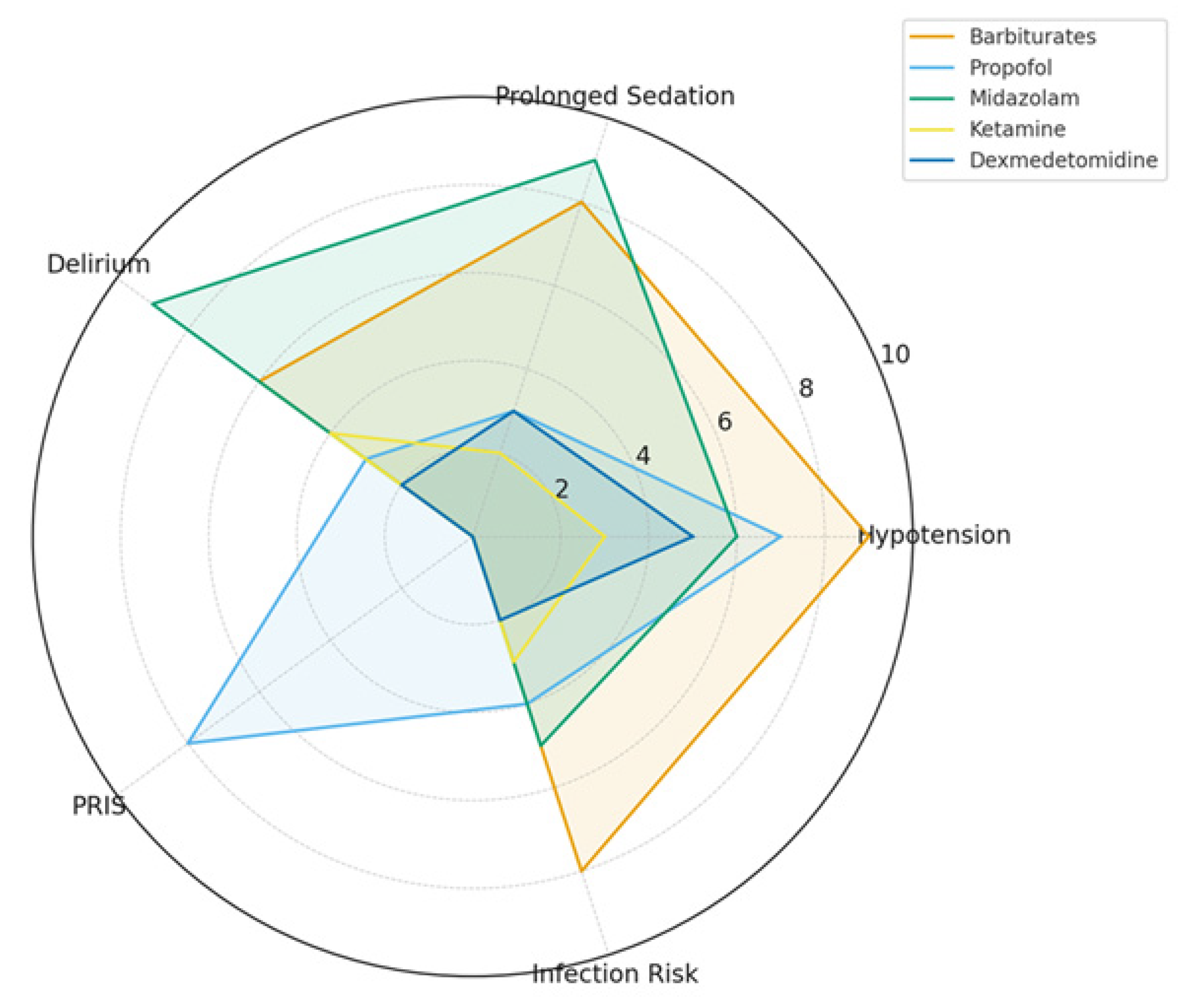
Figure 1. Adverse event risk profiles for sedatives in TBI.
Ctn 09 00053 g001
Figure 2. Guidelines for tiered ICP control measures.
Ctn 09 00053 g002
Table 1. Comparative sedative profiles of commonly used drugs.
AgentMechanism of ActionICP EffectBurst SuppressionSafety Profile/Adverse Effects
Barbiturates (Pentobarbital/Thiopental)GABA_A agonist, glutamate inhibitionStrong decrease, effective in refractory ICPYes, potent and reliableHypotension, immunosuppression, prolonged coma
PropofolGABA_A agonist, NMDA modulationStrong decrease, titratableYes, achievable at high dosesHypotension, risk of PRIS, hypertriglyceridemia
MidazolamGABA_A potentiator (benzodiazepine site)Moderate decreaseNot reliably, only at very high dosesAccumulation, delirium risk, prolonged sedation
KetamineNMDA antagonist, sympathomimeticNeutral to mild decrease (ICP safe)No (does not induce burst suppression)Tachycardia, hypertension, increased secretions
Dexmedetomidineα2-adrenergic agonistMinimal effect, ICP-neutralNoBradycardia, hypotension, arousable sedation
Table 2. Sedation protocols for commonly used drugs.
AgentLoading DoseMaintenance InfusionEEG Target
Pentobarbital10 mg/kg over 30 min, then 5 mg/kg q1h X31–3 mg/kg/hBurst suppression, 2–5 bursts/min
PropofolBolus 1–2 mg/kg (optional)2–5 mg/kg/h (up to 10 for burst suppression)Burst suppression, BIS < 20
Midazolam0.05–0.2 mg/kg IV bolus0.1–0.4 mg/kg/hSlowing, not reliable burst suppression
Ketamine0.5–1 mg/kg IV bolus0.5–3 mg/kg/hNo burst suppression, adjunct role
DexmedetomidineAvoid bolus (risk of brady/hypotension)0.2–0.7 mcg/kg/hLight sedation, cooperative state
Disclaimer/Publisher’s Note: The statements, opinions and data contained in all publications are solely those of the individual author(s) and contributor(s) and not of MDPI and/or the editor(s). MDPI and/or the editor(s) disclaim responsibility for any injury to people or property resulting from any ideas, methods, instructions or products referred to in the content.