Proceeding Paper

Catching the Sugars: Electrochemical Aptasensors for the Detection of Cancer-Related Glycosylation Changes in Prostate-Specific Antigen †

by
Ana Díaz-Fernández
1,2,*,
Rebeca Miranda-Castro
1,2,
Pedro Estrela
3,4,
Noemí de-los-Santos-Álvarez
1,2 and
María Jesús Lobo-Castañón
1,2
1
Departamento de Química Física y Analítica, Universidad de Oviedo, Av. Julián Clavería 8, 33006 Oviedo, Spain
2
Instituto de Investigación Sanitaria del Principado de Asturias, Av. de Roma, 33011 Oviedo, Spain
3
Centre for Biosensors, Bioelectronics and Biodevices (C3Bio), University of Bath, Bath BA2 7AY, UK
4
Department of Electronic and Electrical Engineering, University of Bath, Bath BA2 7AY, UK
*
Author to whom correspondence should be addressed.
Presented at the 1st International Electronic Conference on Biosensors, 2–17 November 2020; Available online: https://iecb2020.sciforum.net/.
Proceedings 2020, 60(1), 47; https://doi.org/10.3390/IECB2020-07023
Published: 2 November 2020
(This article belongs to the Proceedings of The 1st International Electronic Conference on Biosensors)

Abstract

:
Prostate-specific Antigen (PSA) is the biomarker that is used for prostate cancer (PCa) detection, although its lack of specificity results in a high rate of false-positives and many unnecessary biopsies. Therefore, there is a need for more specific cancer biomarkers for PCa. Recent studies have shown that the aberrant glycosylation of proteins is a common feature of the presence of cancer. In the case of prostate cancer, there are changes in core-fucose and sialic acids in the glycan structure of PSA. In this work, we describe two different strategies to direct the selection of aptamers toward the glycans of PSA. From these strategies, we identified two aptamers (PSA-1 and PSAG-1) that bind to the glycan structure of PSA with high affinity. Both aptamers were applied in the design of electrochemical aptasensors, in sandwich and direct formats, in order to detect the changes in the glycosylation of PSA. The sensors responded to different levels of PSA in serum, and they showed higher potential to discriminate clinically-meaningful PCa than the ELISA (Enzyme-linked immunosorbent assay) test used in hospitals (reducing the number of false positives), although validation on more samples is needed.

1. Introduction

Prostate cancer (PCa) is the most frequently diagnosed cancer, and the second most prevalent cause of cancer death in men. The approved tests to diagnose prostate cancer are digital rectal exam and the analysis of the levels of Prostate-specific Antigen (PSA) in the blood. However, because their low sensitivity and specificity, an invasive prostate biopsy is needed to confirm the presence of the tumor. PSA is the most prevalently used PCa biomarker in the clinical practice. The test consists in the measurement of the PSA levels in serum, with a cut-off of 4 ng/mL. However, this test is not specific in the gray zone, between 4 and 10 ng/mL, where the concentration of PSA is not only indicative of cancer but also of other benign prostate diseases. This results in a high number of false positives and leads to many unnecessary biopsies. Therefore, new tests for PCa diagnosis are needed [1,2].
The well-established association between aberrant glycosylation and cancer suggests that the analysis of tumor-linked glycan alterations could be a valuable tool for cancer diagnosis [3]. In prostate cancer, a variation in the content of the fucose and the sialic acids in the glycan linked to PSA was described [4]. The glycosylation profile is currently characterized by long and complicated protocols based on final mass spectrometry detection or the use of lectins. For routine analysis, however, the development of rapid, direct methods for the detection of aberrant glycosylated forms of PSA requires specific receptors with higher affinity than lectins. Aptamers can be tailored to specifically recognize the glycan moiety turning into promising synthetic receptors, in order to replace the current methods for glycosylation profiling [5].
In this work, we present a strategy to select aptamers toward the glycosylation site of the glycoprotein PSA. The aptamers were used in the design of electrochemical aptasensors for the detection of changes in the glycosylation pattern of PSA associated to PCa (Figure 1).

2. Materials and Methods

2.1. Materials and Apparatus

A detailed list of the materials and apparatus is available in references [6,7].

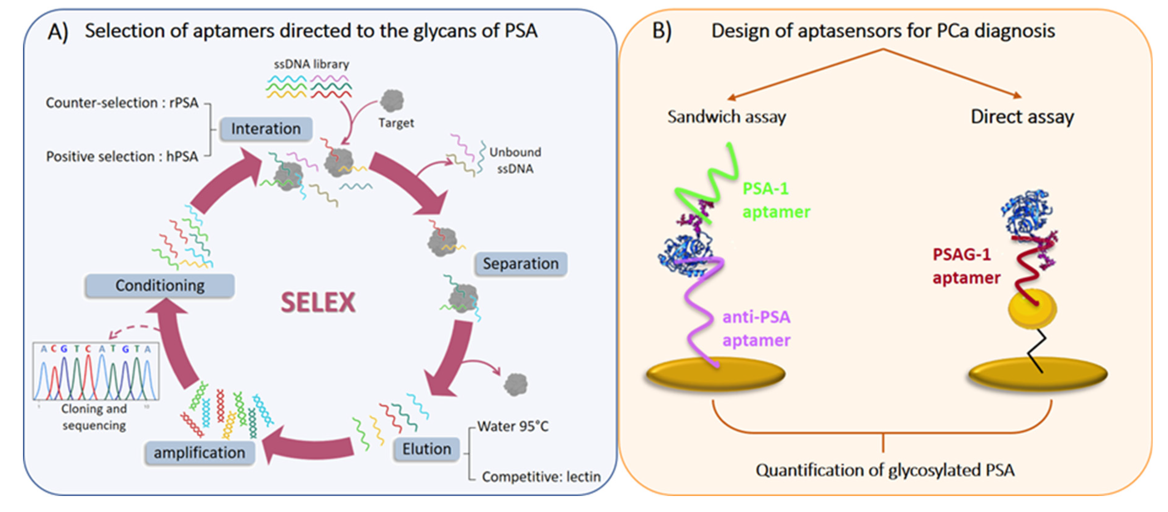
2.2. SELEX Procedure

The SELEX (Systematic Evolution of ligands by Exponential Enrichment) procedure is an iterative process, and each cycle consists of the following steps: the incubation of an ssDNA library with the target (PSA), the separation of the unbound sequences, the elution of the bound ones, the amplification of the eluted sequences by PCR (Polymerase chain reaction), and strand separation to start a new round of selection (Figure 1A).
In this work, we used an initial ssDNA library of 80 nucleotides (nt)-length with a 40 nt randomized central region, flanked at both ends by two constant sequences of 20 nucleotides each. To make the separation and elution steps easier, the protein PSA was immobilized on the surface of tosylactivated magnetic particles. In order to direct the selection toward the glycosylation site of the protein, we performed two strategies: the first was based on the introduction of counter-selection steps with recombinant PSA (rPSA), an unglycosylated form of the protein; and the second was based on counter-selection with rPSA and competitive elution with a lectin.
The counter-selection step consisted in the incubation of the ssDNA library with rPSA, and after the interaction, the unbound sequences were collected and incubated with PSA. The following steps of the SELEX were performed in the traditional way.
The competitive elution with the lectin consisted in the elution of the sequences bound to PSA by adding a high concentration of the lectin in solution, which displaced the sequences that bind to the lectin-binding site.
A comprehensive and detailed explanation of the protocols can be found in reference [6,7].

2.3. Electrochemical Binding Assays

The aptamers were screened by affinity assays with electrochemical detection. With this aim, PSA was covalently immobilized onto screen-printed gold electrodes. First, a self-assembled monolayer (SAM) of a mixture 1:3 11-mercaptoundecanoic acid and 6-mercaptohexanol (both 1 mM) in an NaAc buffer (10 mM pH 5.5) was prepared overnight at 4 °C. The carboxylic groups were activated with a solution of 100 mM EDC (N-(3-dimethylaminopropyl)-N′-ethylcarbodiimide hydrochloride) and 25 mM NHS (N-hydroxysuccinimide) in water for 30 min, and then PSA was covalently immobilized by incubating the electrodes in 50 µg/mL PSA for 30 min. Finally, the remaining active groups were blocked with ethanolamine 1 M in PBS (1 × pH 7.4) for 30 min.
Binding assays were performed by incubating the modified electrode with increasing concentrations of the aptamers labelled with fluorescein for 30 min in PBS. After the interaction, the conjugate Fab-antifluorescein-peroxidase (Fab-POD: 0.5 U/mL in PBS-0.5% casein) was added for 30 min. Finally, the electrochemical measurement was carried out with 35 µL TMB-H2O2, and the enzymatic product was measured after 30 s by chronoamperometry at −0.2 V for 1 min. The current obtained during the last 10 s was averaged and related with the aptamer concentration.

2.4. Deglycosyation Studies

The interaction of PSA with specific glycosidase enzymes was performed on screen-printed gold electrodes modified with PSA, as indicated above. The incubation order was as follows: neuraminidase-A, β-galactosidase, N-acetylhexosaminidase, mannosidase, fucosidase, and PNGaseF. The preparation of the enzymes (buffer and concentration) was performed following the manufacturer’s protocols. The incubations were performed at 37 °C in a volume of 10 µL in a wet atmosphere for 2 h, except for the PNGaseF enzyme, which had an incubation period of 24 h. Then, the binding capacity of the aptamers was tested by performing an electrochemical binding assay with PSAG-1 or PSA-1 as indicated above [7].

2.5. Sandwich Aptasensor

To fabricate the sensor, the capture biotinylated aptamer (anti-PSA) was immobilized onto screen-printed gold electrodes modified with streptavidin. For the PSA analysis, different concentrations of PSA prepared in TBS (10 mM Tris-HCl, 150 mM NaCl, 5 mM KCl, 5 mM MgCl2 pH 7.4) or diluted serum were incubated on the working electrode for 30 min, and then the detection aptamer (PSA-1) was added for 30 min. Next, the bound aptamer was labeled with Fab-POD for 30 min in TBS-0.5% casein. Finally, the electrochemical measurement was carried out as described above [6].

2.6. Impedimetric Aptasensor

A dual impedimetric platform for the direct measurement of PSA was developed by immobilizing a thiolated aptamer (anti-PSA or PSAG-1) onto nanostructured gold electrodes. Each aptamer layer was prepared on gold electrodes modified with 11-amino-1-undecanothiol and 6-mercapto-1-hexanol, and by attaching gold nanoparticles to the amine group in the SAM. For the PSA detection, the sensors were incubated with increasing concentrations of PSA in PBS for 30 min and, after the interaction, the impedance was measured in 5 mM Fe[(CN)6]3−/4− at 0.22 V vs. Ag|AgCl, KCl (3M)|KNO3 (3M).

3. Results and Discussion

3.1. Selection and Characterization of Aptamers

To direct the selection of aptamers toward the glycosylation site of the PSA, two counter-selection steps against the recombinant PSA (rPSA) were introduced into the six-round SELEX. One of the obtained aptamers (PSA-1) showed preferential binding for PSA over rPSA, and its affinity and selectivity was evaluated in detail. A dissociation constant of 357 ± 42 nM was estimated. However, this aptamer also recognized other glycoproteins (Lipocalin-2 or NGAL), meaning that PSA-1 binds to the glycan moiety and not to the peptide region of the protein. Consequently, PSA-1 might be a good receptor for human glycans [6].
In order to refine the selection of the aptamers, obtaining receptors that are able to differentiate glycoproteins with similar glycosylation patterns, we proposed a selection based on counter-selection steps against rPSA and competitive elution with the lectin PhoSL (specific binder of α1-6 core-fucose). After ten rounds of selection, one aptamer (PSAG-1) preferentially bound to PSA with a dissociation constant of 34 ± 9 nM. This aptamer showed selectivity against other glycoproteins: NGAL (lipocalin-2), AFP (α-fetoprotein), ovalbumin and transferrin. This indicates that the molecular interaction of PSAG-1 and PSA involved both the glycans and some of the amino acids of the protein [7].

3.2. Study of the Binding Site

To find out which sugars in the glycan structure are involved in the recognition of PSA by both of the aptamers, the sugars of PSA immobilized onto the gold electrodes were removed using selective glycosidases. After each sugar removal, the binding ability of the aptamers was tested. From this study, we observed that PSA-1 and PSAG-1 aptamers have a completely different binding pattern. PSA-1 recognizes the external sugars (sialic acid and galactose), while PSAG-1 recognizes the innermost sugars [7].

3.3. Detection of Glycosylated PSA Using Aptasensors

We employed the selected aptamers and a previously described aptamer (anti-PSA), which only recognizes the peptide moiety [8], for the development of the electrochemical aptasensors. Two different formats—sandwich and direct label-free approaches—were designed using gold electrodes as support.
The sandwich aptasensor, using anti-PSA as the capture aptamer and PSA-1 as the detection aptamer, has a working range between 0.66 ng/mL and 25 ng/mL in 1:1 diluted serum, and a detection limit of 0.66 ng/mL PSA. This sensor has an excellent selectivity against other proteins (rPSA and NGAL) and 92 analytes present in biological fluids [6].
The direct label-free assay was performed on a dual impedimetric platform with two sensors, one with anti-PSA and the other with the PSAG-1 aptamer. Both sensors respond to increasing concentrations of PSA in 1:5 diluted serum. The working range of the anti-PSA sensor is between 0.64 ng/mL and 62.5 ng/mL, and 0.26 ng/mL to 62.5 ng/mL for the PSAG-1 sensor [9].
Both aptasensors were applied to the analysis of serum samples from patients with benign or malignant prostate pathologies from the clinical analysis laboratory at Hospital de Cabueñes (Gijón, Spain). For samples with PSA concentrations in the grey zone or above, the aptasensors provided discordant values compared to those obtained by the ELISA, but which were in close agreement with the final diagnosis (no prostate cancer). Therefore, the results point to the detection of glycans as an alternative for the detection of prostate cancer, with potential to improve the clinical outcomes of the current PSA test, reducing the number of unnecessary biopsies [6,9].

Funding

This research was funded by the Spanish Government (project RTI-2018-095756-B-I00) and Principado de Asturias Government (IDI2018-000217), co-financed by FEDER funds. A.D-F. thanks Asociación Española Contra el Cáncer (AECC) for her pre-doctoral grant, and Banco Santander and University of Oviedo for the grant in the framework of the economic mobility of excellence for teachers and researchers.

Acknowledgments

We thank E. Fernández-Rodríguez and S. García-Alonso (Hospital Universitario de Cabueñes-Asturias) for providing the serum samples.

Conflicts of Interest

The authors declare no conflict of interest.

References

  1. Lilja, H.; Ulmert, D.; Vickers, A.J. Prostate-specific antigen and prostate cancer: Prediction, detection and monitoring. Nat. Rev. Cancer 2008, 8, 268–278. [Google Scholar] [CrossRef]
  2. Lomas, D.J.; Ahmed, H.U. All change in the prostate cancer diagnostic pathway. Nat. Rev. Clin. Oncol. 2020, 17, 372–381. [Google Scholar] [CrossRef] [PubMed]
  3. Pinho, S.S.; Reis, C.A. Glycosylation in cancer: Mechanisms and clinical implications. Nat. Rev. Cancer 2015, 15, 540–555. [Google Scholar] [CrossRef] [PubMed]
  4. Gilgunn, S.; Conroy, P.J.; Saldova, R.; Rudd, P.M.; O’Kennedy, R.J. Aberrant PSA glycosylation—A sweet predictor of prostate cancer. Nat. Rev. Urol. 2013, 10, 99–107. [Google Scholar] [CrossRef] [PubMed]
  5. Díaz-Fernández, A.; Miranda-Castro, R.; de-los-Santos-Álvarez, N.; Lobo-Castañón, M.J. Post-translational modifications in tumor biomarkers: The next challenge for aptamers? Anal. Bioanal. Chem. 2018, 410, 2059–2065. [Google Scholar] [CrossRef] [PubMed]
  6. Díaz-Fernández, A.; Miranda-Castro, R.; de-los-Santos-Álvarez, N.; Fernández-Rodríguez, E.; Lobo-Castañón, M.J. Focusing aptamer selection on the glycan structure of prostate-specific antigen: Toward more specific detection of prostate cancer. Biosens. Bioelectron. 2019, 128, 83–90. [Google Scholar] [CrossRef] [PubMed]
  7. Díaz-Fernández, A.; Miranda-Castro, R.; Díaz, N.; Suárez, D.; de-los-Santos-Álvarez, N.; Lobo-Castañón, M.J. Aptamers targeting protein-specific glycosylation in tumor biomarkers: General selection, characterization and structural modeling. Chem. Sci. 2020, 11, 9402–9413. [Google Scholar] [CrossRef] [PubMed]
  8. Savory, N.; Abe, K.; Sode, K.; Ikebukuro, K. Selection of DNA aptamer against prostate specific antigen using a genetic algorithm and application to sensing. Biosens. Bioelectron. 2010, 26, 1386–1391. [Google Scholar] [CrossRef] [PubMed]
  9. Díaz-Fernández, A.; Miranda-Castro, R.; de-los-Santos-Álvarez, N.; Lobo-Castañón, M.J.; Estrela, P. Impedimetric aptamer-based glycan PSA score for discrimination of prostate cancer from other prostate diseases. Biosens. Bioelectron. 2020. submitted. [Google Scholar] [CrossRef] [PubMed]
Figure 1. Overview of the work described in this conference proceeding paper: (A) the selection of aptamers directed to the glycan structure of PSA, and (B) the design of aptasensors for prostate cancer diagnosis.
Proceedings 60 00047 g001
Publisher’s Note: MDPI stays neutral with regard to jurisdictional claims in published maps and institutional affiliations.

