Abstract
Plasmonic heating finds multiple applications in cell manipulation and stimulation, where heat generated by metal nanoparticles can be used to modify cell adhesion, control membrane currents, and suppress neuronal action potentials among others. Metal nanoparticles can also be easily integrated in artificial extracellular matrices to provide tunable, thermal cueing functionalities with nanometer spatial resolution. In this contribution, we present a platform enabling the combination of plasmonic heating with localized temperature sensing using quantum dots (QDs). Specifically, a functional nanocomposite material was designed with gold nanorods (AuNRs) and QDs incorporated in a cell-permissive hydrogel (e.g., collagen) as well as an optical set-up combining optical heating and temperature imaging, respectively. Specific area stimulation/readout can be realized through structured illumination using digital micromirror device (DMD) projection.
1. Materials and Methods
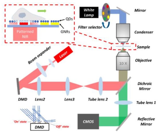
The setup is shown in Figure 1. A collimated beam was projected onto a digital micromirror device (DMD) to generate structured patterns on the sample plane for localized plasmonic heating [1]. To monitor cellular responses and local temperature changes, a rotating filter selector was placed in front of a white lamp [2]. Filtered blue light is used for QD excitation in temperature-related fluorescent image recording, while images of cell movement can be obtained by switching to a red filter.

Figure 1.
Experimental setup.
Gold nanorods (AuNRs) with local surface plasmon resonance (LSPR) in the near infrared (NIR) region were prepared by surfactant-mediated, seeded growth. CdSe/ZnS quantum dots with fluorescence emission around 630 nm were water-compatibilized through amphiphilic polymer Poly(styrene-co-maleic anhydride) (PSMA) encapsulation. 2D AuNR–QD assemblies with collagen matrices were prepared by first depositing pre-formed collagen fibers on glass coverslips. PSMA-QDs were then activated with 1-ethyl-3-(3-dimethylaminopropyl) carbodiimide together with N-hydroxysuccinimide (EDC-NHS) before being covalently bound to the surface-deposited collagen fibers. Finally, AuNRs were bound to the collagen-–QD matrices by incubation in an aqueous AuNR suspension containing 5 mM CTAB and 100 mM NaCl. Surfaces were rinsed in ultrapure water and dried in a flow of pure nitrogen gas between each deposition step.
2. Results
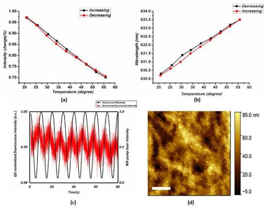
In this contribution we report the results on the temperature sensitivity of PSMA-coated CdSe/ZnS QDs [3]. Figure 2a, b illustrate the calibration of temperature sensitivity of QD fluorescence intensity and spectral shift, respectively. A linear and reversible variation is displayed with a thermal sensitivity of 0.7% peak value change and 0.09 nm peak wavelength shift per degree.

Figure 2.
Temperature calibration for (a) photoluminescence intensity and (b) temperature induced spectral shift of PSMA-coated CdSe/ZnS quantum dots (QDs) in PBS buffer (excitation at 532nm); (c) QDs PL intensity changes induced by modulated NIR illumination; (d) Collagen matrix with AuNRs; scale bar indicates a distance of 500 nm.
Furthermore, we demonstrate the possibility of using QDs to map temperature changes induced by NIR illumination of plasmonic AuNRs. Figure 2c illustrates the changes in QD (PL) intensity that occurred when a modulated NIR LED source (850 nm) was used for the AuNRs excitation.
Our contribution discusses the effects of the QD/AuNRs preparation method and experimental conditions on the temperature sensitivity in living cell preparation.
Author Contributions
Validation, W.Y., F.G. and P.Z. Sample preparation, O.D., S.J. and J.W. Writing—review and editing, O.D., C.G., and C.B.; Funding acquisition, C.G., and C.B. All authors have read and agreed to the published version of the manuscript.
Funding
This research was funded by KU Leuven C1 project, grant number C14/16/063 OPTIPROBE and FWO research grant, grant number G0947.17N.
Conflicts of Interest
The authors declare no conflict of interest.
References
- Min, Z.; Guillaume, B. Micropatterning Thermoplasmonic Gold Nanoarrays to Manipulate Cell Adhesion. ACS Nano 2012, 6, 7227–7233. [Google Scholar]
- Sangjin, Y.; Ji-Ho, P. Single-Cell Photothermal Neuromodulation for Functional Mapping of Neural Networks. ACS Nano 2019, 13, 544–551. [Google Scholar]
- Vasudevanpillai, B.; Yoji., M. Temperature-Sensitive Photoluminescence of CdSe Quantum Dot Clusters. J. Phys. Chem. B 2005, 109, 13899–13905. [Google Scholar]
Disclaimer/Publisher’s Note: The statements, opinions and data contained in all publications are solely those of the individual author(s) and contributor(s) and not of MDPI and/or the editor(s). MDPI and/or the editor(s) disclaim responsibility for any injury to people or property resulting from any ideas, methods, instructions or products referred to in the content. |
© 2021 by the authors. Licensee MDPI, Basel, Switzerland. This article is an open access article distributed under the terms and conditions of the Creative Commons Attribution (CC BY) license (https://creativecommons.org/licenses/by/4.0/).