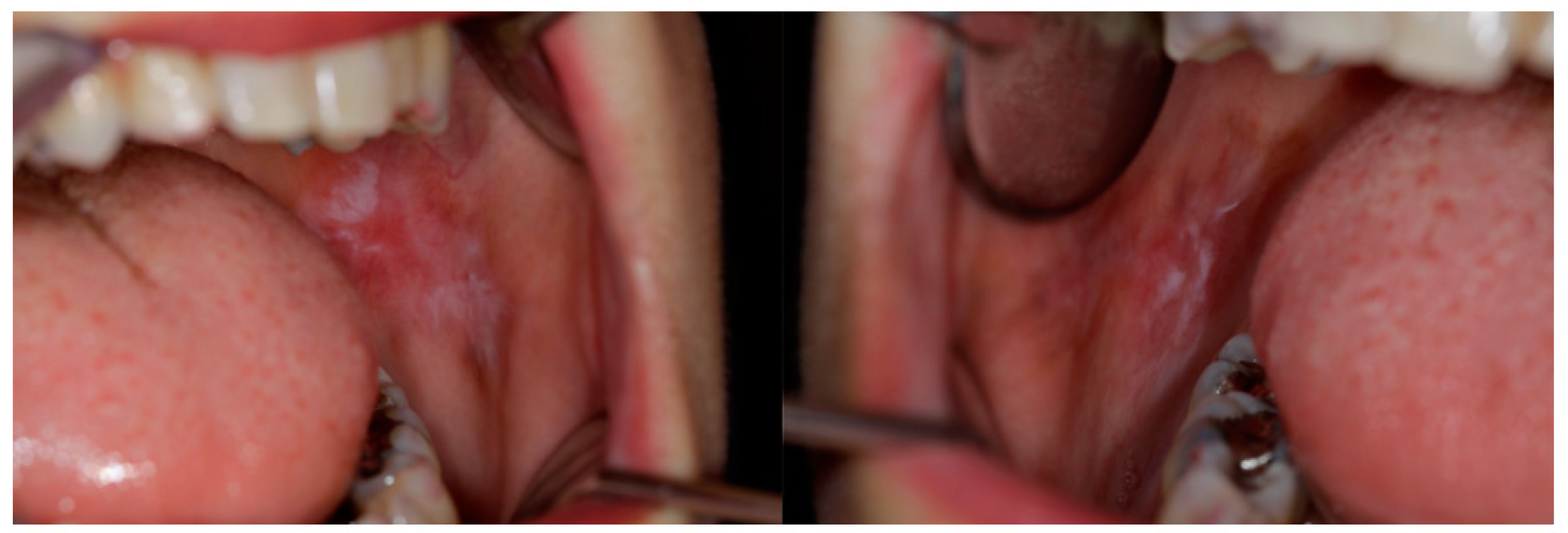
Unusual Manifestations of Oral Follicular Lymphoid Hyperplasia Mimicking Oral Lichen Planus †


Conflicts of Interest
References
- Menasce, L.P.; Shanks, J.H.; Banerjee, S.S.; Harris, M. Follicular lymphoid hyperplasia of the hard palate and oral mucosa: Report of three cases and a review of the literature. Histopathology 2001, 39, 353–358. [Google Scholar] [CrossRef]
- Kolokotronis, A.; Dimitrakopoulos, I.; Asimaki, A. Follicular lymphoid hyperplasia of the palate: Report of a case and review of the literature. Oral Surg. Oral Med. Oral Pathol. Oral Radiol. Endod. 2003, 96, 172–175. [Google Scholar] [CrossRef]
- Jham, B.C.; Binmadi, N.O.; Scheper, M.A.; Zhao, X.F.; Koterwas, G.E.; Kashyap, A.; Levy, B.A. Follicular lymphoid hyperplasia of the palate: Case report and literature review. J. Craniomaxillofac. Surg. 2009, 37, 79–82. [Google Scholar] [CrossRef]

© 2019 by the authors. Licensee MDPI, Basel, Switzerland. This article is an open access article distributed under the terms and conditions of the Creative Commons Attribution (CC BY) license (http://creativecommons.org/licenses/by/4.0/).
Share and Cite
Val, M.; Gobbo, M.; Rossi, M.; Ragazzo, M.; Nardini, L.G. Unusual Manifestations of Oral Follicular Lymphoid Hyperplasia Mimicking Oral Lichen Planus. Proceedings 2019, 35, 80. https://doi.org/10.3390/proceedings2019035080
Val M, Gobbo M, Rossi M, Ragazzo M, Nardini LG. Unusual Manifestations of Oral Follicular Lymphoid Hyperplasia Mimicking Oral Lichen Planus. Proceedings. 2019; 35(1):80. https://doi.org/10.3390/proceedings2019035080
Chicago/Turabian StyleVal, Matteo, Margherita Gobbo, Marco Rossi, Mirko Ragazzo, and Luca Guarda Nardini. 2019. "Unusual Manifestations of Oral Follicular Lymphoid Hyperplasia Mimicking Oral Lichen Planus" Proceedings 35, no. 1: 80. https://doi.org/10.3390/proceedings2019035080
APA StyleVal, M., Gobbo, M., Rossi, M., Ragazzo, M., & Nardini, L. G. (2019). Unusual Manifestations of Oral Follicular Lymphoid Hyperplasia Mimicking Oral Lichen Planus. Proceedings, 35(1), 80. https://doi.org/10.3390/proceedings2019035080