Treatment of Symptomatic Mandibular Tori: A Case Report †

1. Introduction
2. Case
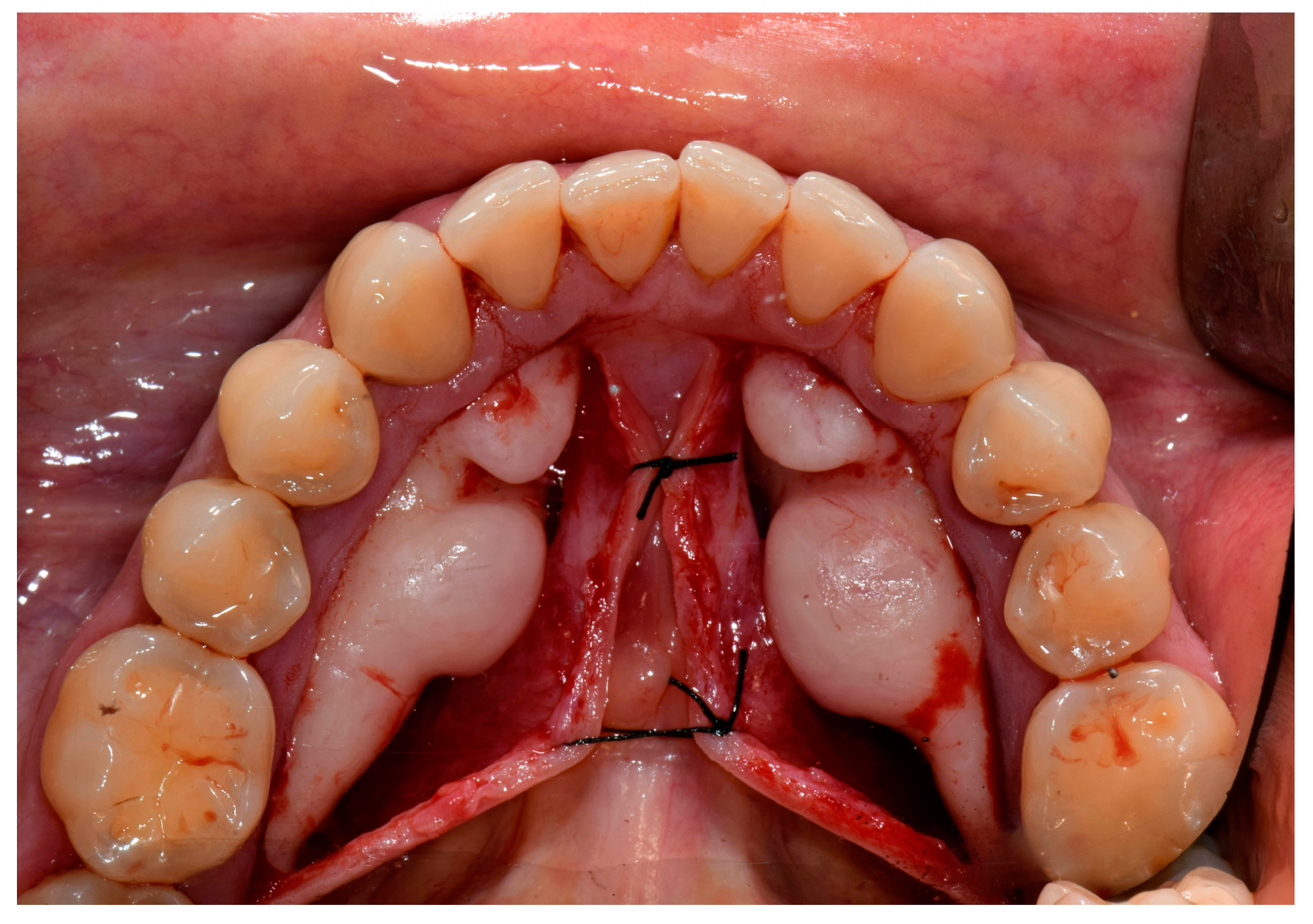
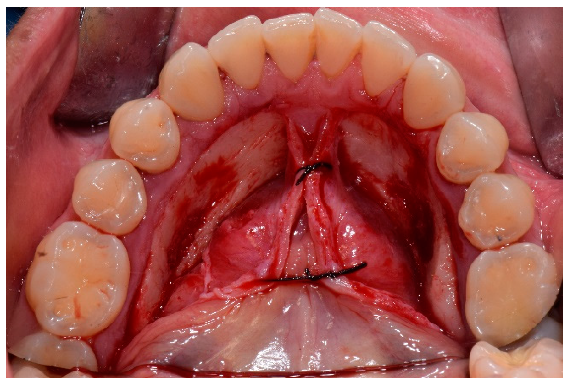
3. Treatment
4. Conclusions
Conflicts of Interest
References
- García-García, A.S.; Martínez-González, J.M.; Gómez-Font, R.; Soto-Rivadeneira, A.; Oviedo-Roldán, L. Current status of the torus palatinus and torus mandibularis. Med. Oral Patol. Oral Cir. Bucal 2010, 15, e353–e360. [Google Scholar] [CrossRef] [PubMed]
- Seah, Y.H. Torus palatinus and torus mandibularis: A review of the literature. Aust. Dental J. 1995, 40, 318–321. [Google Scholar] [CrossRef] [PubMed]
- Jainkittivong, A.; Langlais, R.P. Buccal and palatal exostoses: Pre-valence and concurrence with tori. Oral Surg. Oral Med. Oral Pathol. Oral Radiol. Endodontol. 2000, 90, 48–53. [Google Scholar] [CrossRef] [PubMed]


© 2019 by the authors. Licensee MDPI, Basel, Switzerland. This article is an open access article distributed under the terms and conditions of the Creative Commons Attribution (CC BY) license (http://creativecommons.org/licenses/by/4.0/).
Share and Cite
Sorrentino, D.; Lombardi, N.; Battilana, C.; Decani, S.; Henin, D.; Rossi, V. Treatment of Symptomatic Mandibular Tori: A Case Report. Proceedings 2019, 35, 75. https://doi.org/10.3390/proceedings2019035075
Sorrentino D, Lombardi N, Battilana C, Decani S, Henin D, Rossi V. Treatment of Symptomatic Mandibular Tori: A Case Report. Proceedings. 2019; 35(1):75. https://doi.org/10.3390/proceedings2019035075
Chicago/Turabian StyleSorrentino, Daniela, Niccolò Lombardi, Chiara Battilana, Sem Decani, Dolaji Henin, and Vincent Rossi. 2019. "Treatment of Symptomatic Mandibular Tori: A Case Report" Proceedings 35, no. 1: 75. https://doi.org/10.3390/proceedings2019035075
APA StyleSorrentino, D., Lombardi, N., Battilana, C., Decani, S., Henin, D., & Rossi, V. (2019). Treatment of Symptomatic Mandibular Tori: A Case Report. Proceedings, 35(1), 75. https://doi.org/10.3390/proceedings2019035075