Extended Abstract

Nevus in the Oral Cavity †

1
Dipartimento di Scienze Biomediche, Chirurgiche e Odontoiatriche, Università degli Studi di Milano, 20100 Milano, Italy
2
ASST Santi Paolo e Carlo, Ospedale San Paolo, UO Odontostomatologia II, 20141 Milano, Italy
*
Author to whom correspondence should be addressed.
Presented at the XV National and III International Congress of the Italian Society of Oral Pathology and Medicine (SIPMO), Bari, Italy, 17–19 October 2019.
Proceedings 2019, 35(1), 74; https://doi.org/10.3390/proceedings2019035074
Published: 12 December 2019

1. Introduction

The aim of this study is to describe a clinical case of a young female patient with oral nevus. Pigmented lesions of the oral cavity represent a variety of clinical entities, ranging from physiologic changes to manifestations of systemic illnesses and malignant neoplasms. Oral pigmentations have either a melanocytic (endogenous lesions, including racial pigmentations, melanotic macules, melanocytic nevi, malignant melanoma) or a nonmelanocytic origin (exogenous lesions: amalgam tattoos). Although they may show similar clinical presentations, different treatments apply. Therefore, differential diagnoses pose a challenge in some cases. Microscopic examination can be necessary to rule out an early-stage melanoma [1,2,3].

2. Case

A 20-years-old woman was referred to the San Paolo Hospital in Milan due to the diagnosis of a pigmented lesion in the oral cavity during a routine examination. Intraoral examination showed the presence of a painless, exophytic neoformation, covered whit dark spots, located on left mandibular gingiva, behind the wisdom tooth (Figure 1). The consistency was elastic-soft. The patient denied smoking and alcohol consumption.

3. Treatment

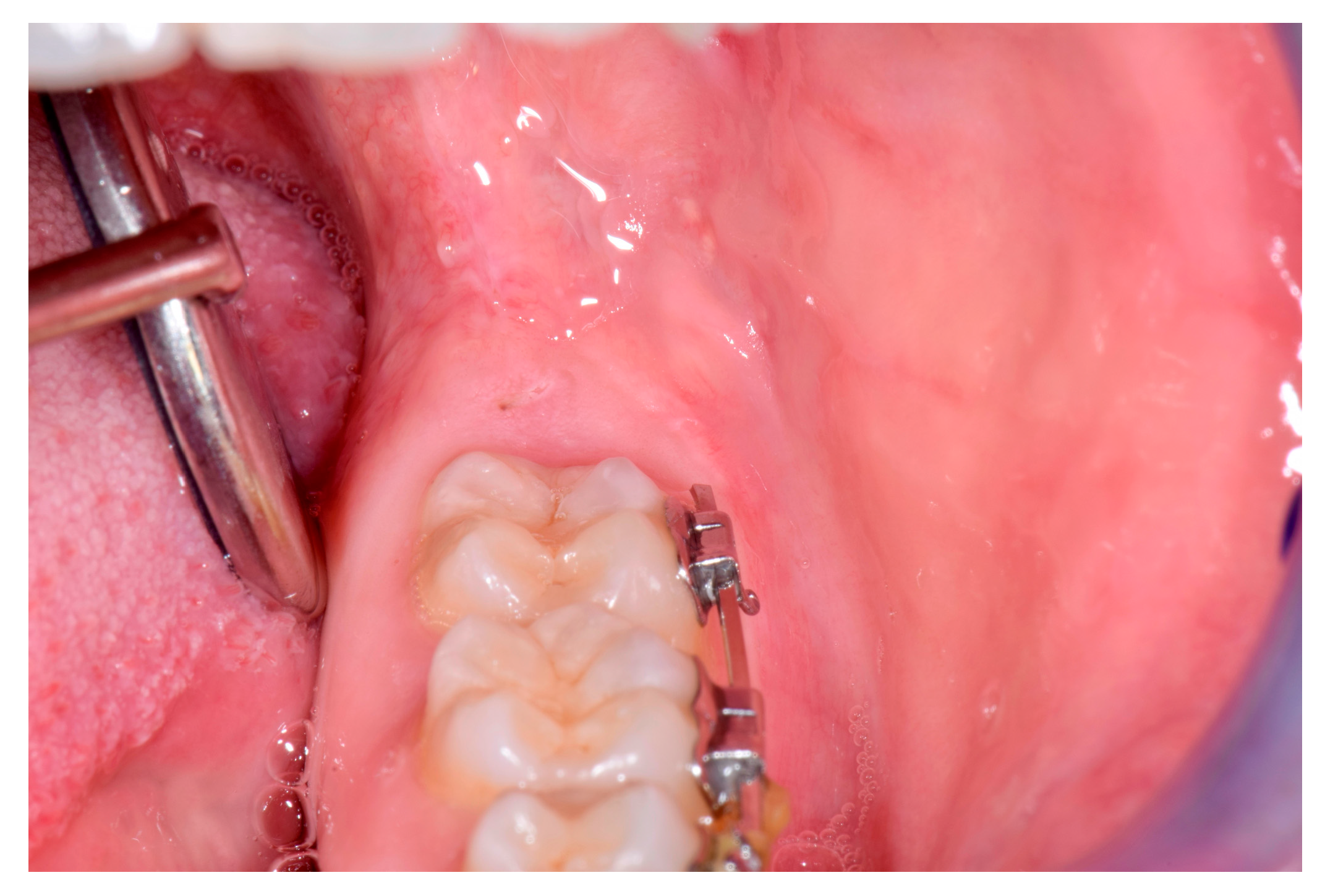
Therefore, an excisional biopsy was performed, under local anesthesia. The incision for biopsy was elliptic, and the lesion was completely removed, and the surgical area was sutured. The removed material was fixed in 10% formalin and sent for anatomohistopathological examination. The diagnosis of composit melanocityc nevus was confirmed. After a week the suture has been removed (Figure 2).

4. Conclusions

Pigmented lesions represent uncommon diagnoses at an oral pathology service. Often these lesions are found in routine examination. Nevi, while uncommon, can occur in the oral cavity, and should be differentiated from other pigmented lesions, including oral melanomas that, although also rare, have a high mortality rate. When the nevi occur, they are most often found in females, in the third and fourth decade of life and the most common histological type is located on the palate and in the buccal mucosa. In this light, visual inspection of the oral cavity should be done very carefully, and the ideal treatment is the removal of the pigmented lesion whit a safety margin of 2 mm.

Conflicts of Interest

The authors declare no conflict of interest.

References

  1. Popa, C.; Stelea, C.; Popa, R.; Popescu, E. Oral and perioral endogenous pigmented lesions. Rev. Med. Chir. Soc. Med. Nat. Iasi 2008, 112, 1054–1060. [Google Scholar] [PubMed]
  2. Tavares, T.S.; Meirelles, D.P.; de Aguiar MC, F.; Caldeira, P.C. Pigmented lesions of the oral mucosa: A cross-sectional study of 458 histopathological specimens. Oral Dis. 2018, 24, 1484–1491. [Google Scholar] [CrossRef] [PubMed]
  3. Freitas, D.A.; Bonan, P.R.; Sousa, A.A.; Pereira, M.M.; Oliveira, S.M.; Jones, K.M. Intramucosal nevus in the oral cavity. J. Contemp. Dent. Pract. 2015, 16, 74–76. [Google Scholar] [CrossRef] [PubMed]
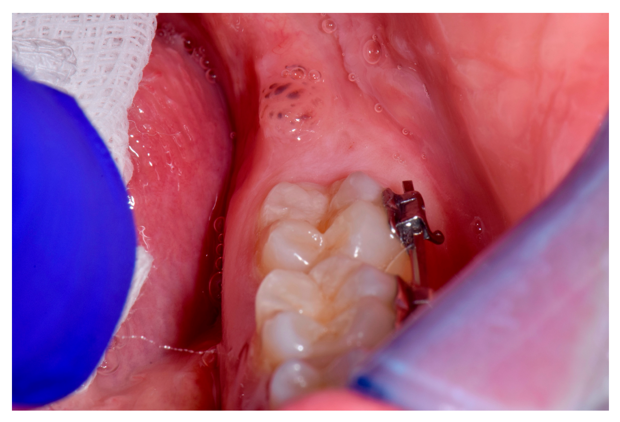
Figure 1. Clinical picture.
Proceedings 35 00074 g001
Figure 2. Postoperatory clinical picture.
Proceedings 35 00074 g002