Extended Abstract

Oral Granular Cell Tumour: A Case Report †

1
Department of Medicine and Surgery, University of Insubria, ASST Sette Laghi, Dental Clinic, Unit of Oral Medicine and Pathology, 21100 Varese, Italy
2
Department of Medicine and Surgery, University of Insubria, ASST Sette Laghi, Unit of Pathology, 21100 Varese, Italy
3
Department of Medicine and Surgery, University of Insubria, ASST Sette Laghi, Dental Clinic, Unit of Pediatric Dentistry, 21100 Varese, Italy
*
Author to whom correspondence should be addressed.
Presented at the XV National and III International Congress of the Italian Society of Oral Pathology and Medicine (SIPMO), Bari, Italy, 17–19 October 2019.
Proceedings 2019, 35(1), 55; https://doi.org/10.3390/proceedings2019035055
Published: 12 December 2019

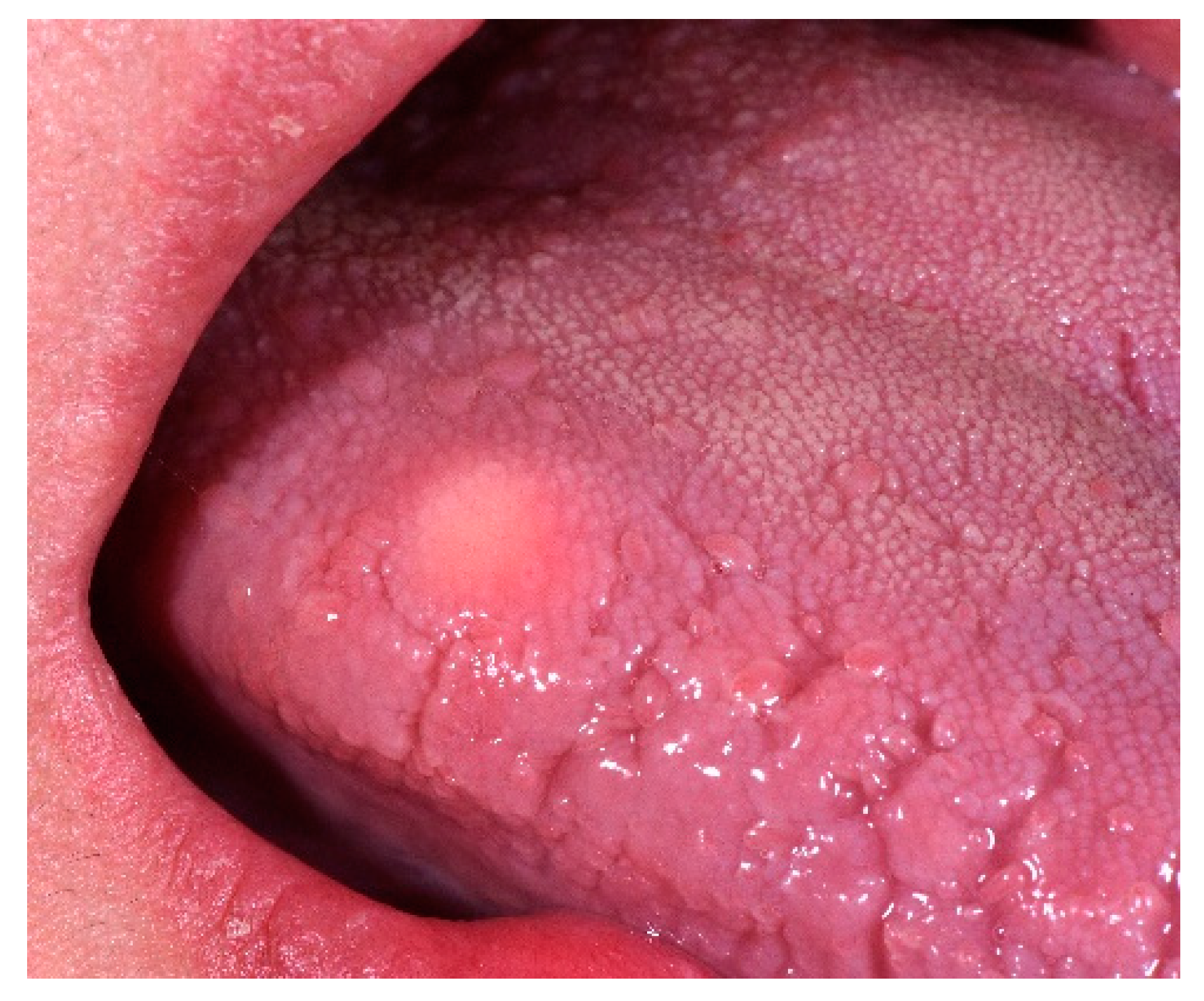
1. Case Presentation
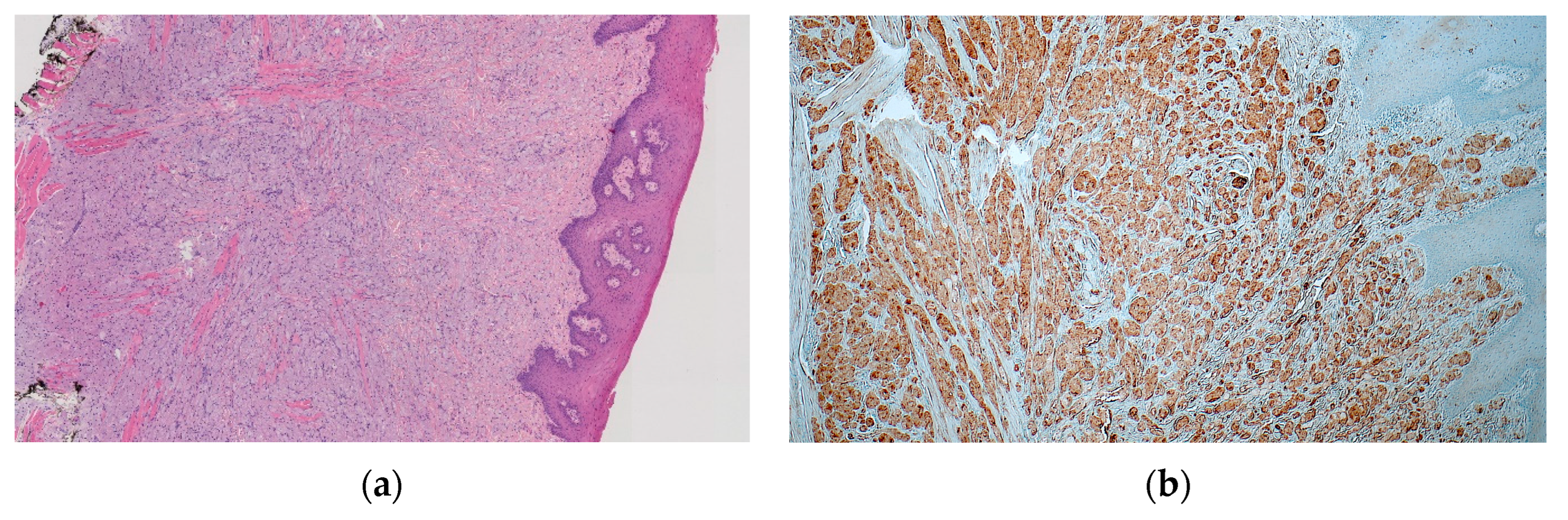
A 16-year-old girl presented at our attention with an asymptomatic nodular lesion on the tongue. Clinically, the lesion appeared as a submucosal smooth nodule, 5 mm in diameter, covered by a pink-yellowish looking mucosa (Figure 1). On palpation, it was well defined, but not encapsulated. The lesion was located on the right posterior dorsum of the tongue, it had been slowly growing for a few months, and it was completely asymptomatic. The patient’s anamnesis revealed atopic dermatitis and pityriasis rosea.
Clinically, the lesion was placed in differential diagnosis with other “soft tissue tumors”. Therefore, an excisional biopsy of the lesion was performed with subsequent histopathological analysis.

2. Histopathology

The connective tissue displayed a well-defined proliferation of cells with a characteristic granular cytoplasm. The cells had a characteristic granular eosinophilic cytoplasm positive to PAS staining. The neoplastic cells had a polygonal shape, large dimensions and indefinite margins (Figure 2). The nucleus was roundish and with free chromatin. There was no nuclear atypia and the Ki-67 proliferation index was low. A peculiar histological element of the granular cell tumor was the presence of pseudoepitheliomatous hyperplasia (PEH).

3. Conclusions

Granular cell tumor (GCT) is a benign soft tissue neoplasm. GCT can develop throughout the body but it shows a clear preference for the head-neck district. In particular, in 50% of cases it develops on the tongue, precisely on the lateral margins and back of the tongue [2].
PEH places this lesion in a differential diagnosis with a well-differentiated squamous cell tumor [3]. In order to avoid dangerous diagnostic errors, which can lead to unnecessary lingual mutilation, it is essential to perform a correct biopsy test. Bioptic sampling must include both the epithelial lamina and the lamina propria, where the granular cell tumor develops.

Conflicts of Interest

The authors declare no conflict of interest.

References

  1. Musha, A.; Ogawa, M.; Yokoo, S. Granular cell tumors of the tongue: Fibroma or schwannoma. Head Face Med. 2018, 14, 1. [Google Scholar] [CrossRef] [PubMed]
  2. Alotaibi, O.; Al Sheddi, M. Neurogenic tumors and tumor-like lesions of the oral and maxillofacial region: A clinicopathological study. Saudi Dent. J. 2016, 28, 76–79. [Google Scholar] [CrossRef] [PubMed]
  3. Al-Eryani, K.; Karasneh, J.; Sedghizadeh, P.P.; Ram, S.; Sawair, F. Lack of utility of cytokeratins in differentiating pseudocarcinomatous hyperplasia of granular cell tumors from oral squamous cell carcinoma. Asian Pac. J. Cancer Prev. 2016, 17, 1785–1787. [Google Scholar] [CrossRef] [PubMed]
Figure 1. Clinical aspect of the lesion. A pink-yellowish submucosal nodule was detected on the posterior lateral aspect of the tongue.
Proceedings 35 00055 g001
Figure 2. (a) Histopathological features of OGCT. The tumour was composed of large polygonal granular cells arranged in a pseudosyncitial pattern. The aspect is typical for GCT. (b) Immunohistochemical analysis revaled the tumour cells be positive for S-100, a marker that confirmed the origin from perineural tissues, like the Schwann cells [1].
Proceedings 35 00055 g002
Publisher’s Note: MDPI stays neutral with regard to jurisdictional claims in published maps and institutional affiliations.

