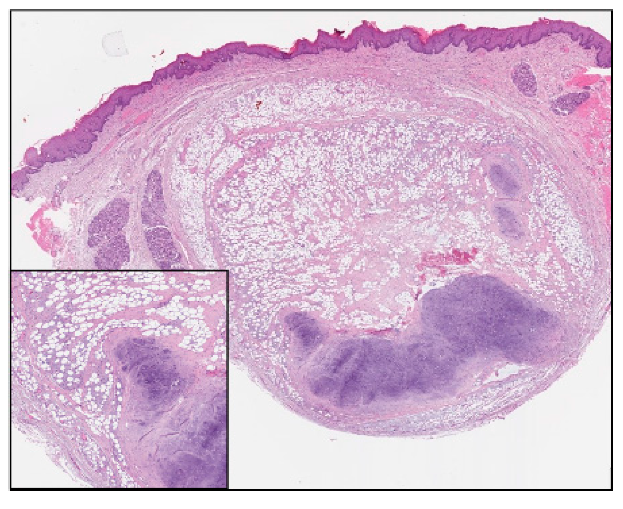
Cartilaginous Choristoma of the Lower Lip †

1. Introduction
2. Material and Methods
3. Discussion
4. Conclusions
Conflicts of Interest
References
- Halley, D.; Dargue, A.; Pring, M. Cartilaginous Choristoma of the lower lip: Report of a case and review of the literature. Oral Surg. 2014, 7, 48–50. [Google Scholar] [CrossRef]
- Batra, R. The pathogenesis of oral choristomas. J. Oral and Maxillofac. Surg. Med. Pathol. 2012, 24, 110–114. [Google Scholar] [CrossRef]

© 2019 by the authors. Licensee MDPI, Basel, Switzerland. This article is an open access article distributed under the terms and conditions of the Creative Commons Attribution (CC BY) license (http://creativecommons.org/licenses/by/4.0/).
Share and Cite
Bosotti, M.; Boggio, F.; Mascellaro, A.; Rossi, M.; Porrini, M.; Rosso, E.d.; Spadari, F. Cartilaginous Choristoma of the Lower Lip. Proceedings 2019, 35, 48. https://doi.org/10.3390/proceedings2019035048
Bosotti M, Boggio F, Mascellaro A, Rossi M, Porrini M, Rosso Ed, Spadari F. Cartilaginous Choristoma of the Lower Lip. Proceedings. 2019; 35(1):48. https://doi.org/10.3390/proceedings2019035048
Chicago/Turabian StyleBosotti, Moreno, Francesca Boggio, Anna Mascellaro, Margherita Rossi, Massimo Porrini, Ettore del Rosso, and Francesco Spadari. 2019. "Cartilaginous Choristoma of the Lower Lip" Proceedings 35, no. 1: 48. https://doi.org/10.3390/proceedings2019035048
APA StyleBosotti, M., Boggio, F., Mascellaro, A., Rossi, M., Porrini, M., Rosso, E. d., & Spadari, F. (2019). Cartilaginous Choristoma of the Lower Lip. Proceedings, 35(1), 48. https://doi.org/10.3390/proceedings2019035048