Use of Optical Coherence Tomography in Patients with Desquamative Gingivitis: A Case Series †

 ,
, 

 ,
, 
 and
and 
1. Introduction
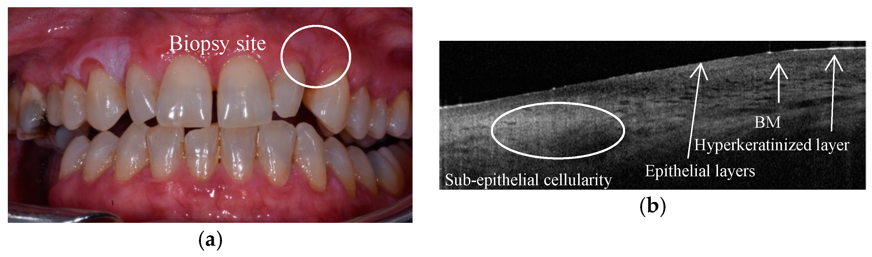
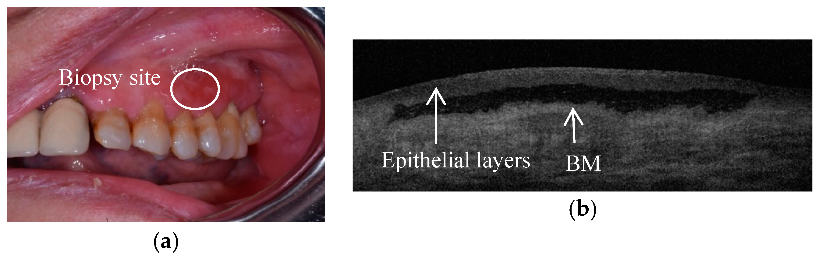
2. Case Series
3. Conclusions
Conflicts of Interest
References
- Lo Russo, L.; Fierro, G.; Guiglia, R.; Compilato, D.; Testa, N.F.; Lo Muzio, L.; Campisi, G. Epidemiology of desquamative gingivitis: Evaluation of 125 patients and review of the literature. Int. J. Dermatol. 2009, 48, 1049–1052. [Google Scholar] [CrossRef] [PubMed]
- Huang, D.; Swanson, E.A.; Lin, C.P.; Schuman, J.S.; Stinson, W.G.; Chang, W.; Hee, M.R.; Flotte, T.; Gregory, K.; Puliafito, C.A.; et al. Optical coherence tomography. Science 1991, 254, 1178–1181. [Google Scholar] [CrossRef] [PubMed]
- Capocasale, G.; Panzarella, V.; Rodolico, V.; Di Fede, O.; Campisi, G. In vivo optical coherence tomography imaging in a case of mucous membrane pemphigoid and a negative Nikolsky’s sign. J. Dermatol. 2018, 45, 603–605. [Google Scholar] [CrossRef] [PubMed]


© 2019 by the authors. Licensee MDPI, Basel, Switzerland. This article is an open access article distributed under the terms and conditions of the Creative Commons Attribution (CC BY) license (http://creativecommons.org/licenses/by/4.0/).
Share and Cite
Panzarella, V.; Bartolone, A.; Ciavarella, D.; Santarelli, A.; Fede, O.D.; Mauceri, R.; Campisi, G. Use of Optical Coherence Tomography in Patients with Desquamative Gingivitis: A Case Series. Proceedings 2019, 35, 47. https://doi.org/10.3390/proceedings2019035047
Panzarella V, Bartolone A, Ciavarella D, Santarelli A, Fede OD, Mauceri R, Campisi G. Use of Optical Coherence Tomography in Patients with Desquamative Gingivitis: A Case Series. Proceedings. 2019; 35(1):47. https://doi.org/10.3390/proceedings2019035047
Chicago/Turabian StylePanzarella, Vera, Alessia Bartolone, Domenico Ciavarella, Andrea Santarelli, Olga Di Fede, Rodolfo Mauceri, and Giuseppina Campisi. 2019. "Use of Optical Coherence Tomography in Patients with Desquamative Gingivitis: A Case Series" Proceedings 35, no. 1: 47. https://doi.org/10.3390/proceedings2019035047
APA StylePanzarella, V., Bartolone, A., Ciavarella, D., Santarelli, A., Fede, O. D., Mauceri, R., & Campisi, G. (2019). Use of Optical Coherence Tomography in Patients with Desquamative Gingivitis: A Case Series. Proceedings, 35(1), 47. https://doi.org/10.3390/proceedings2019035047