Extended Abstract

Hard Palate Hyperpigmentation Induced by Chloroquine: A Case Report †

1
Department of Dentistry, ASST-PG23: Ospedale Papa Giovanni XXIII, 24100 Bergamo, Italy
2
Department of Anatomical Pathology ASST-PG23: Ospedale Papa Giovanni XXIII, 24100 Bergamo, Italy
*
Author to whom correspondence should be addressed.
Presented at the XV National and III International Congress of the Italian Society of Oral Pathology and Medicine (SIPMO), Bari, Italy, 17–19 October 2019.
Proceedings 2019, 35(1), 45; https://doi.org/10.3390/proceedings2019035045
Published: 12 December 2019

1. Introduction

Pigmented oral mucosa lesions can have an exogenous or endogenous nature, depending on the determining causes. Among the exogenous pigmentations are included those related to heavy metal ingestion or intoxication, metal implantation, drug ingestion and “bad habits” like pencil chewing. A physiological and medical anamnesis is important to investigate the etiology of pigmentations [1].

2. Case Report

A 87-years old man was referred to the Oral Pathology division of Papa Giovanni XXIII hospital in Bergamo for xerostomia and temporomandibular joint (TMJ) disorders. The patient’s medical history revealed cardiopathy, uncompensated vestibulopathy and seronegative rheumatoid arthritis. He was receiving treatment with Chloroquine (250 mg/die since 2016), Vitamin “D”, Cardioaspirin, Atorvastatin and an antihypertensive drug. The patient stated he never smoked.
Intraoral examination showed a flat, homogeneous, grey discoloration regarding the whole hard palate mucosa. The pigmentated area was asymptomatic, symmetric and bilateral (Figure 1).
The rest of the oral mucosa appeared to be moistened and healthy, without leucoplasic or eritroplasic areas. Neither functional limitation nor muscular pain was noticed through clinical exam of TMJ.
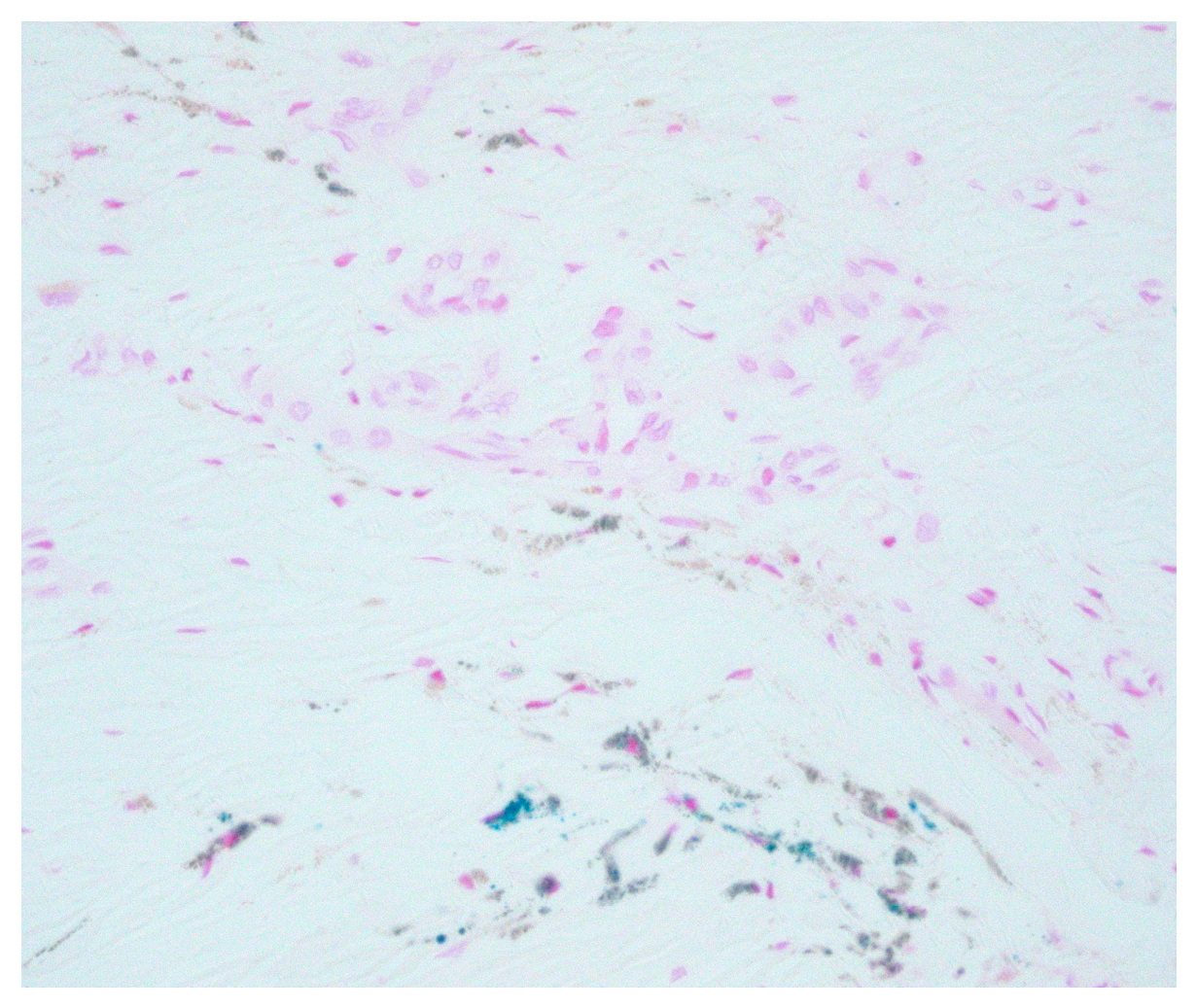
The patient reported to have never noticed the palate pigmentation before; 15 days later it was still present without any clinical change. After the patient’s written informed consent an incisional biopsy was performed in local anesthesia; beside standard histopathologic examination, Prussian blue (Perls’) reaction was used to investigate hemosiderin staining (Figure 2).
The clinical and histopathologic features confirmed the diagnosis of drug-induced oral pigmentation caused by chloroquine.

3. Discussion

Oral mucosa hyperpigmentation is a rare side effect of antimalarial agents like chloroquine, used also for management of rheumatoid arthritis.
The literature reported that prolonged systemic administration of this drug can determine the deposition of hemosiderin in the tissues of the oral cavity. Chloroquine chelates iron and/or melanin; moreover, melanocytes are stimulated in melanin production [2,3].
In most cases only hard palate is involved, but involvement of gingiva and labial or buccal mucosa has been reported too. For this type of oral pigmentation there is no a recommended treatment, the importance of an early diagnosis of chloroquine-induced hyperpigmentation of the oral mucosa is that it could be a marker of another adverse reaction: irreversible retinopathy. The drug binds pigmented ocular tissues and could lead to blindness. The patient should be referred to an ophthalmologist and, according to the rheumatologist, the drug should be discontinued or modified in the dosage.

Conflicts of Interest

The authors declare no conflict of interest.

References

  1. Joseph, A.; Regezi, J.S.; Richard, C.K.; Jordan, A. Oral Pathology: Clinical Pathologic Correlations, 7th ed.; Elsevier-Saunders: Amsterdam, The Netherlands, 2017. [Google Scholar]
  2. De Andrade, B.A.B.; Padron-Alvarado, N.A.; Muñoz-Campos, E.M.; Morais, T.M.L.; Martinez-Pedraza, R. Hyperpigmentation of hard palate induced by chloroquine therapy. J. Clin. Exp. Dent. 2017, 9, e1487–e1491. [Google Scholar] [CrossRef] [PubMed]
  3. Tosios, K.I.; Kalogirou, E.M.; Sklavounou, A. Drug-associated hyperpigmentation of the oral mucosa: report of four cases. Oral Surg. Oral Med. Oral Pathol. Oral Radiol. 2018, 125, e54–e66. [Google Scholar] [CrossRef] [PubMed]
Figure 1. Hard palate hyperpigmentation.
Proceedings 35 00045 g001
Figure 2. Perls’ Prussian blue stained section.
Proceedings 35 00045 g002