Primordial Odontogenic Tumour: A Systematic Review †

 , , , and
 1. Introduction
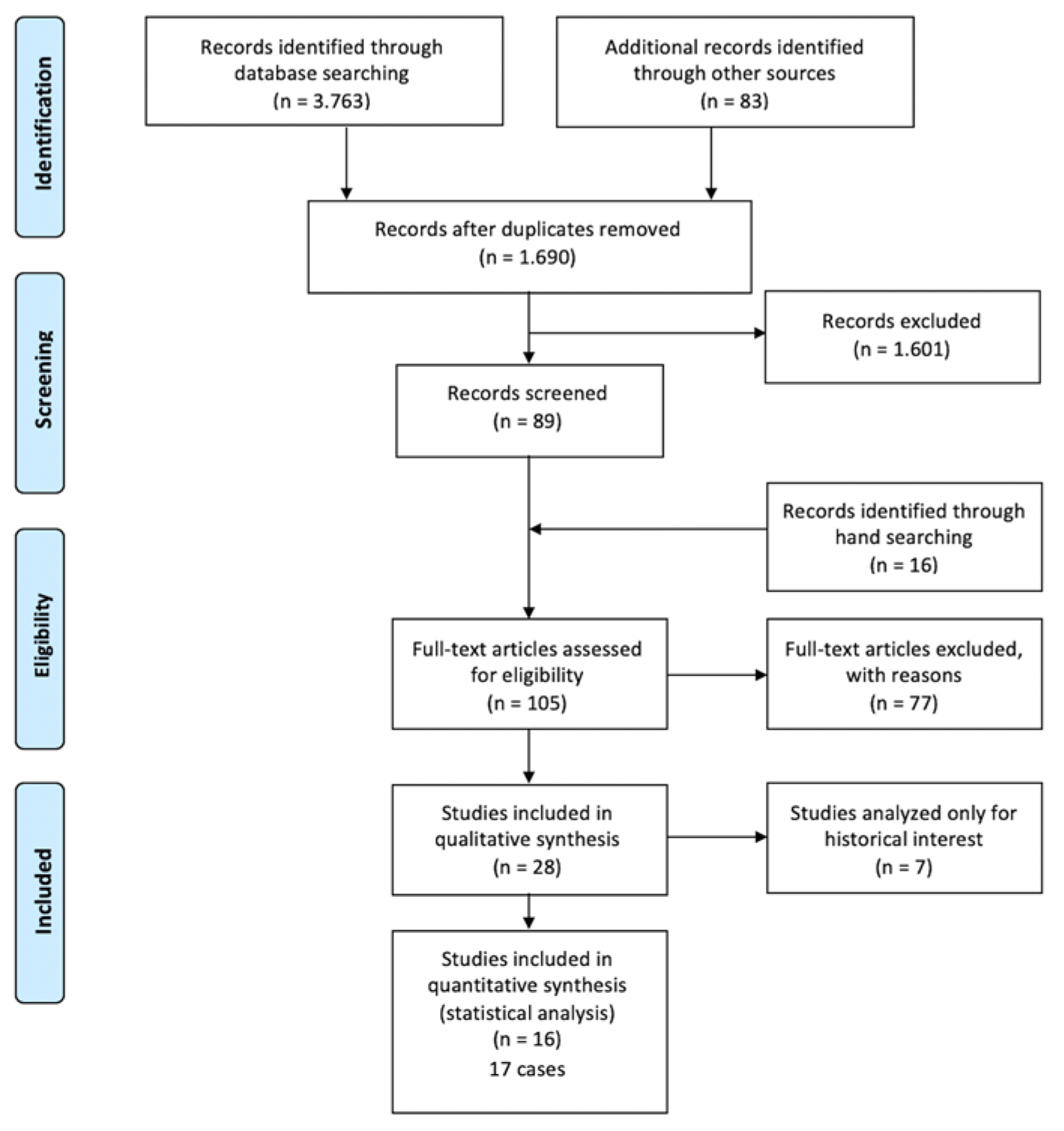
2. Materials and Methods
3. Results
4. Conclusions
Conflicts of Interest
References
- Mosqueda-Taylor, A.; Pires, F.R.; Aguirre-Urízar, J.M.; Carlos-Bregni, R.; de la Piedra-Garza, J.M.; Martínez-Conde, R.; Martínez-Mata, G.; Carreño-Álvarez, S.J.; da Silveira, H.M.; de Barros Dias, B.S.; et al. Primordial odontogenic tumour: Clinicopathological analysis of six cases of a previously undescribed entity. Histopathology 2014, 65, 606–612. [Google Scholar] [CrossRef] [PubMed]
- Liberati, A.; Altman, D.G.; Tetzlaff, J.; Mulrow, C.; Gøtzsche, P.C.; Ioannidis, J.P.; Clarke, M.; Devereaux, P.J.; Kleijnen, J.; Moher, D. The PRISMA statement for reporting systematic reviews and meta-analyses of studies that evaluate health care interventions: Explanation and elaboration. BMJ 2009, 339, b2700. [Google Scholar] [CrossRef] [PubMed]
- Almazyad, A.; Li, C.C.; Tapia, R.O.C.; Robertson, J.P.; Collette, D.; Woo, S.B. Primordial odontogenic tumour: Report of two cases. Histopathology 2018, 72, 1221–1227. [Google Scholar] [CrossRef] [PubMed]
- Bomfim, B.B.; Prado, R.; Sampaio, R.K.; Conde, D.C.; de Andrade, B.A.B.; Agostini, M.; Romañach, M.J. Primordial Odontogenic Tumor: Report of a New Case and Literature Review. Head Neck Pathol. 2019, 13, 125–130. [Google Scholar] [CrossRef] [PubMed]

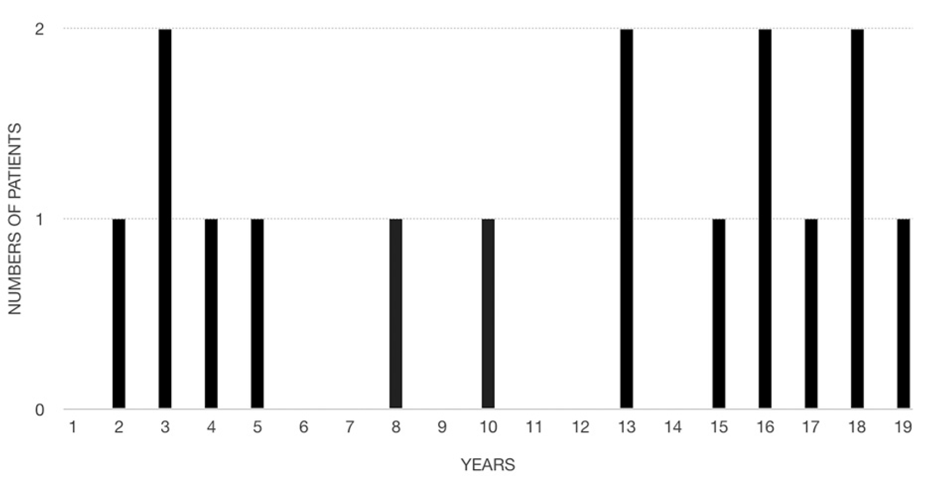
| EPIDEMIOLOGICAL DATA | CLINICAL FEATURES | RADIOLOGICAL ASPECTS | MACROSCOPIC APPEARANCE | THERAPY AND FOLLOW-UP | |||||||||||||||||
|---|---|---|---|---|---|---|---|---|---|---|---|---|---|---|---|---|---|---|---|---|---|
| CASE n° | Year | Author | AGE | SEX | REGION | PAIN | CLINICAL APPEARANCE | TIME BEFORE DIAGNOSIS | SITE | RX APPEARANCE | TOOTH INVOLVEMENT | ROOT RESORPTION | RX DIAMETERS | MACROSCOPIC DIMENSIONS | LOBULES | CYSTIC SPACES | TOOTH/NEOPLASM | DIFFERENTIAL DIAGNOSIS | SURGICAL APPROACH | RECURRENCE | FOLLOW-UP |
| 1 | 2014 | Mosqueda-Taylor A et al. | 18 | M | Latin America/Hispanic† | Asymptomatic | Buccal swelling | 6 months | Left posterior mandible; angle | RL, UL, well defined | enclosing the crown of the third molar | YES (second molar) | 45 X 40 mm | N/A | multilobated | NO | embedded | Dentigerous cyst, ameloblastoma | Enucleation with third molar | NO | 20 years |
| 2 | 2014 | Mosqueda-Taylor A et al. | 16 | M | Latin America/Hispanic† | Asymptomatic | Buccal and inferior mandibular cortical bone expansion | 4 months | Left posterior mandible; angle | RL, UL, well defined | enclosing the crown of the third molar | N/A | 55 x 50 mm | N/A | multilobated | NO | easily detatched | Ameloblastic fibroma | Enucleation with third molar | N/A | Lost to follow-up |
| 3 | 2014 | Mosqueda-Taylor A et al. | 16 | M | Latin America/Hispanic† | Asymptomatic | Buccal swelling | 12 months | Left posterior mandible; angle | RL, UL, well defined | enclosing the crown of the third molar | NO | 65 x 50 mm | N/A | multilobated | NO | embedded | Benign solid odontogenic tumour | Enucleation with third molar | NO | 10 years |
| 4 | 2014 | Mosqueda-Taylor A et al. | 3 | F | Latin America/Hispanic† | Asymptomatic | Buccal and lingual bony expansion | 22 months | Left posterior mandible; angle; ascending ramus | RL, BIL, well defined | enclosing the crowns of the second decidous and first permanent molars; absence of #20 tooth germ | YES (first decidous molar) | 90 x 70 mm | N/A | multilobated | NO | easily detached | Ameloblastic fibroma | Enucleation with involved teeth | NO | 9 years |
| 5 | 2014 | Mosqueda-Taylor A et al. | 13 | F | Latin America/Hispanic† | Asymptomatic | Buccal swelling | 4 months | Left posterior mandible; angle; ascending ramus | RL, BIL, well defined | enclosing entirely the third molar | YES (first and second molars) | 80 x 50 mm | N/A | multilobated | NO | embedded | Ameloblastoma; ameloblastic fibroma | Enucleation with third molar and 18-19 | NO | 3 years |
| 6 | 2014 | Mosqueda-Taylor A et al. | 3 | F | Latin America/Hispanic† | Asymptomatic | Buccal and palatal bony swelling | 3 months | Left posterior maxilla; sinus floor; tuberosity | RL, UL, well defined | second decidous and first permanent molar | N/A | 35 x 30 mm | N/A | multilobated | NO | easily detached | Benign solid odontogenic tumour | Enucleation with involved teeth | NO | 6 months |
| 7 | 2016 | Slater LJ et al. | 19 | M | USA | Asymptomatic | Buccal and lingual bony expansion (thinning and resorption of lingual and inferior cortical plates) | N/A | Right posterior mandible | RL, UL, well defined | enclosing the crown of the third molar | YES (second molar) | N/A | 25 x 19 x 15 mm | multilobated | NO | easily detached | N/A | Enucleation with third molar (no data on second molar) | NO | 7 months |
| 8 | 2016‡ | Almazyad A et al. | 15 | F | USA (Hispanic) | Asymptomatic | Buccal swelling | N/A | Right posterior mandible; angle | RL, ML, well defined | enclosing the crown of the third molar | YES (first and second molars) | 35 x 20 mm | N/A | N/A | NO | N/A | Dentigerous cyst | Enucleation with third molar | NO | 3 months |
| 9 | 2016‡ | Almazyad A et al. | 18 | M | Mexico (Hispanic) | Asymptomatic | None | N/A | Right posterior mandible | RL, UL, well defined | enclosing the crown of a third molar | NO | 17 x 12 mm | 17 x 12 mm | multilobated | NO | easily detached | Dentigerous cyst | Curettage and third molar extraction | NO | 20 months |
| 10 | 2017 | Ando T et al. | 8 | F | Japan | Asymptomatic | Buccal swelling | N/A | Left posterior maxilla; sinus floor | RL, UL, well defined | enclosing the crown of the first decidous molar | NO | N/A | N/A | multilobulated | NO | N/A | Dentigerous cyst, benign solid odontogenic tumour | Enucleation | NO | 16 months |
| 11 | 2017 | Mikami T et al. | 5 | M | Japan | Asymptomatic | Buccal swelling, 1st decidous molar mobility | N/A | Right posterior mandible | RL, UL, well defined | association with the second decidous molar | YES (first decidous molar) | N/A | 9 x 8 x 8 mm | nodule | NO | easily detached | N/A | Enucleation with involved teeth | NO | 7 months |
| 12 | 2018 | Pardhe N and Bajpai M | 17 | M | India | Asymptomatic | Buccal swelling | 6 months | Left posterior mandible; angle | RL, ML, well defined (scalloped upper margins) | enclosing the crown of the third molar | YES (second premolar, first and second molars) | N/A | N/A | N/A | N/A | N/A | Unicystic ameloblastoma | Enucleation with third molar; ID canal not preserved | NO | 6 months |
| 13 | 2018 | Amer H et al. | 2 | M | Egypt | Painful | Buccal swelling | 2 months | Right posterior mandible; angle; ramus | RL, ML, well defined | association with an impacted developing tooth | N/A | 30 x 40 mm | N/A | N/A | YES | N/A | N/A | Enucleation with the impacted tooth | NO | 2 years |
| 14 | 2018 | Bomfim BB et al. | 4 | M | Brazil (Black) | Asymptomatic | Buccal swelling; perforation of lingual cortical bone | 8 months | Left posterior mandible | RL, UL, well defined | enclosing the crown of the second decidous molar; absence of #20 tooth germ; displacement of IAN canal | YES (first decidous molar) | 30 x 20 mm | 40 X 30 mm | multilobulated | NO | easily detached | Immature complex odontoma | Enucleation with incolved teeth | N/A | Lost to follow-up |
| 15 | 2019 | Teixeira LN et al. | 13 | F | Brazil (Black) | Asymptomatic | Buccal swelling | 3 months | Left posterior mandible; angle; ascending ramus | RL, UL, well defined | enclosing the crown of the third molar | YES (at least, second molar) | N/A | N/A | N/A | N/A | N/A | Dentigerous cyst, odontogenic keratocyst, ameloblastoma | Enucleation with third molar (no data on other elements) | N/A | N/A |
| 16 | 2019 | Berdugo J and Bilodeau E | Preadolescent | M | N/A | Asymptomatic | Buccal swelling | N/A | Left posterior mandible; angle; ascending ramus | RL, UL, well defined | Absence of tooth 38 | NO | 22 x 20 mm | N/A | N/A | NO | Not applicable | Ameloblastic fibro-odontoma | Enucleation | N/A | N/A |
| 17 | 2019 | Sun Q et al. | 10 | M | South Korea | Asymptomatic | None | N/A | Left mandible, mesiolingual to the root of tooth 21 | RL, UL, well defined | In close proximity to the root of the tooth | NO | 5 x 5 x 5 mm | 5 x 5 x 5 mm | nodule | NO | Not applicable | Simple bone cyst, periapical cemental dysplasia, paradental cyst | Enucleation | NO | 12 months |
Publisher’s Note: MDPI stays neutral with regard to jurisdictional claims in published maps and institutional affiliations. |
© 2019 by the authors. Licensee MDPI, Basel, Switzerland. This article is an open access article distributed under the terms and conditions of the Creative Commons Attribution (CC BY) license (https://creativecommons.org/licenses/by/4.0/).
Share and Cite
Croveri, F.; Maurino, V.; d’Aiuto, A.; Dani, M.; Boggio, A.; Azzi, L. Primordial Odontogenic Tumour: A Systematic Review. Proceedings 2019, 35, 20. https://doi.org/10.3390/proceedings2019035020
Croveri F, Maurino V, d’Aiuto A, Dani M, Boggio A, Azzi L. Primordial Odontogenic Tumour: A Systematic Review. Proceedings. 2019; 35(1):20. https://doi.org/10.3390/proceedings2019035020
Chicago/Turabian StyleCroveri, Fabio, Vittorio Maurino, Alessandro d’Aiuto, Marta Dani, Andrea Boggio, and Lorenzo Azzi. 2019. "Primordial Odontogenic Tumour: A Systematic Review" Proceedings 35, no. 1: 20. https://doi.org/10.3390/proceedings2019035020
APA StyleCroveri, F., Maurino, V., d’Aiuto, A., Dani, M., Boggio, A., & Azzi, L. (2019). Primordial Odontogenic Tumour: A Systematic Review. Proceedings, 35(1), 20. https://doi.org/10.3390/proceedings2019035020