Automatic Identification of Diabetic Macular Edema Using a Transfer Learning-Based Approach †


Abstract
:1. Introduction
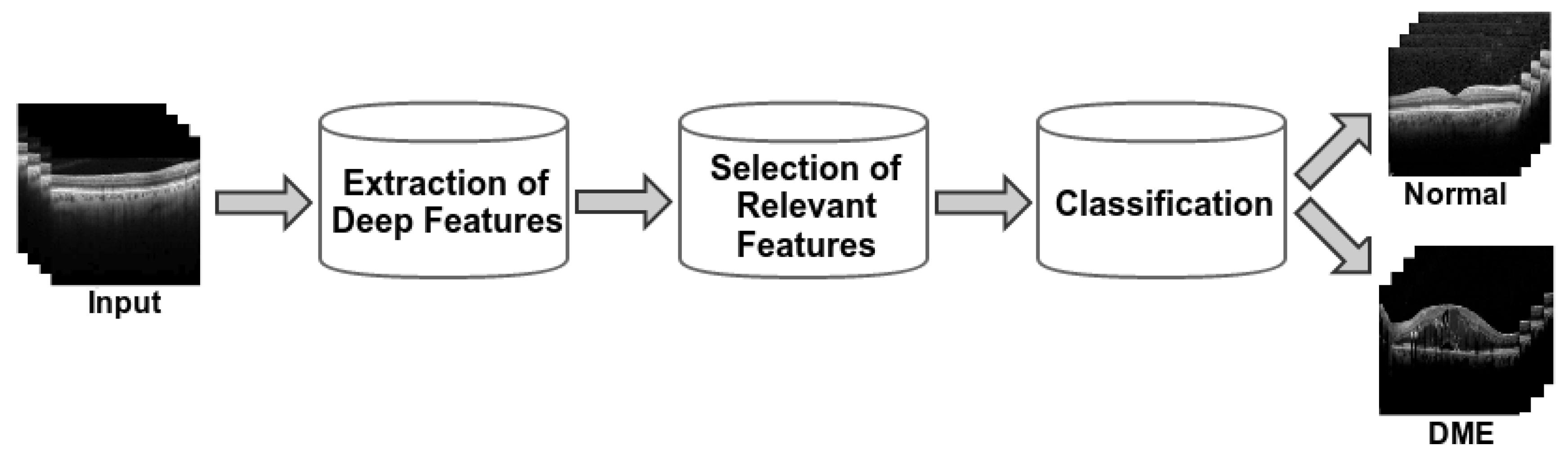
2. Methodology
3. Results and Conclusions
Author Contributions
Funding
Conflicts of Interest
References
- Samagaio, G.; Estévez, A.; de Moura, J.; Novo, J.; Fernández, M.; Ortega, M. ; Automatic macular edema identification and characterization using OCT images. Comput. Methods Programs Biomed. 2018, 163, 47–63. [Google Scholar] [CrossRef] [PubMed]
- Vidal, P.L.; de Moura, J.; Novo, J.; Penedo, M.G.; Ortega, M. Intraretinal fluid identification via enhanced maps using Optical Coherence Tomography images. Biomed. Opt. Express 2018, 9, 4730–4754. [Google Scholar] [CrossRef] [PubMed]
- De Moura, J.; Novo, J.; Ortega, M. Deep Feature Analysis in a Transfer Learning-based Approach for the Automatic Identification of Diabetic Macular Edema. In Proceedings of the 2019 International Joint Conference on Neural Networks (IJCNN), Budapest, Hungary, 14–19 July 2019; pp. 1–8. [Google Scholar]

Publisher’s Note: MDPI stays neutral with regard to jurisdictional claims in published maps and institutional affiliations. |
© 2019 by the authors. Licensee MDPI, Basel, Switzerland. This article is an open access article distributed under the terms and conditions of the Creative Commons Attribution (CC BY) license (https://creativecommons.org/licenses/by/4.0/).
Share and Cite
Moura, J.d.; Vidal, P.L.; Novo, J.; Ortega, M. Automatic Identification of Diabetic Macular Edema Using a Transfer Learning-Based Approach. Proceedings 2019, 21, 16. https://doi.org/10.3390/proceedings2019021016
Moura Jd, Vidal PL, Novo J, Ortega M. Automatic Identification of Diabetic Macular Edema Using a Transfer Learning-Based Approach. Proceedings. 2019; 21(1):16. https://doi.org/10.3390/proceedings2019021016
Chicago/Turabian StyleMoura, Joaquim de, Plácido L. Vidal, Jorge Novo, and Marcos Ortega. 2019. "Automatic Identification of Diabetic Macular Edema Using a Transfer Learning-Based Approach" Proceedings 21, no. 1: 16. https://doi.org/10.3390/proceedings2019021016
APA StyleMoura, J. d., Vidal, P. L., Novo, J., & Ortega, M. (2019). Automatic Identification of Diabetic Macular Edema Using a Transfer Learning-Based Approach. Proceedings, 21(1), 16. https://doi.org/10.3390/proceedings2019021016