1. Introduction
Sound is collected by the outer ear and propagates through the ear canal. These sound waves impinge on the eardrum, converting sound energy into mechanical energy. This mechanical energy is transferred to the inner ear by vibrations of three tiny ossicles: malleus, incus and stapes. This ossicular chain acts as a mechanical impedance matching system, reducing the impedance difference between the air and the fluid-filled cochlea.
The middle ear is prone to a number of pathologies, but as well to yourself! In literature, multiple cases of malleus fractures have been reported in which patients experienced such malleus fracture after retracting his/her finger out of the wet ear canal, during or shortly after a shower [
1]. In cases of malleus handle fracture, the sound transmission from eardrum to the cochlea is drastically reduced, resulting in severe hearing loss. Such cases are rare and can easily be missed if not specifically looked for.
It is not yet clear if reduction in sound transmission is solely due to the separation of the broken malleus fragments. Often, the eardrum is also affected by the malleus handle fracture. Deformation and sometimes wrinkling of the eardrum is observed, suggesting a reduction in tension.
In order to have a better understanding of the mechanics of malleus handle fractures on middle ear sound transmission, we have performed Laser Doppler vibrometry measurements and finite element simulations.
2. Materials and Methods
2.1. Laser Doppler Vibrometry Setup
In the experimental setup, a semi-closed cavity containing an earphone speaker and a condenser microphone was placed over the ear canal and was acoustically sealed using silicone paste. The condenser microphone was calibrated against a probe microphone (Bruel & Kjaer 4182).
The cochlea was removed and a reflective patch was placed on the medial side of the stapes footplate, allowing for velocity measurements using a Laser Doppler vibrometer (Polytec model 534 and controller OFV-5000). The vibrometer head was attached to an otology microscope. Piezo-actuated mirrors allowed precise positioning of the measurement beam within the field of view.
Single sine stimulation at 90 dB SPL was used at eight frequencies per octave between 250 Hz and 4 or 8 kHz. The stapes footplate velocity was measured for the intact middle ear and once more after a fracture in the malleus handle was introduced using a backbiting forceps. The velocity ratio of pre- to post-fracture reflects the hearing loss. The measurements shown in this paper originate from previous work in our lab and are used as a comparison with the finite element simulations [
2].
2.2. Finite Element Simulation
2.2.1. Intact Models
Two finite element models were constructed based on µCT data of two temporal bones. The full description of the construction and definition of the middle ear models (without fracture) can be found in De Greef et al. (2017) [
3] only the TM damping (
has been changed and a condensed description is provided here.
All materials except for the interior of the incudostapedial joint (ISJ) were modeled as solid materials. The tympanic membrane (TM) elasticity was non-isotropic. The TM was subdivided in an attempt to represent the arrangement of collagen fibers.
The periphery of the TM as well as the end surfaces of the anterior mallear ligament (AML), posterior incudal ligament (PIL), stapedial annular ligament (SAL), tensor tympani (TT) tendon and stapedius muscle (SM) tendon were fixed.
The stimulating load was a direct, uniform, harmonic pressure of 1 Pa peak amplitude on the lateral side of the TM. Since we will be comparing ratios before and after fracture, the stimulation levels actually do not matter.
All simulations were performed in COMSOL.
2.2.2. Malleus Fracture Models
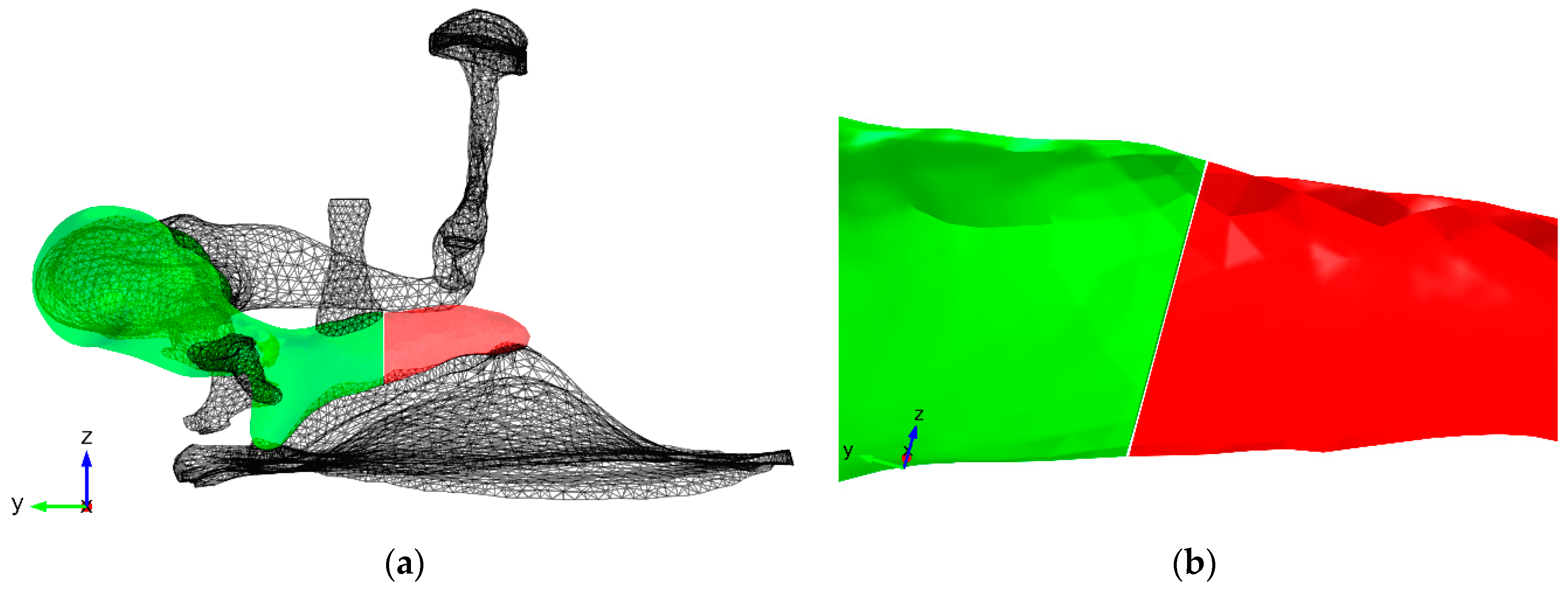
In order to simulate malleus fractures, the intact models were altered by removing a piece of the malleus handle (
Figure 1). This was done by partitioning the malleus geometry using two planes perpendicular to the malleus handle, spaced 10 micrometers apart, close to the TT. The exact width of malleus fractures is unknown. However, the fractures can be resolved on clinical CT [
4] indicating that the gap is large enough to neglect contact between the broken parts. For this reason, contact mechanics were not included in the model.
The deformation and occasional wrinkling of the TM observed in clinical cases was not taken into account directly. The geometry the tympanic membrane was unaltered. However, as mentioned in the introduction, such deformation and wrinkling suggests a release in tension. Additional models were made by crudely mimicking this tension release utilizing a lower TM Young’s modulus. The Young’s modulus of the TM was divided by three. The effect of this choice will be discussed further on.
3. Results
3.1. Measurements
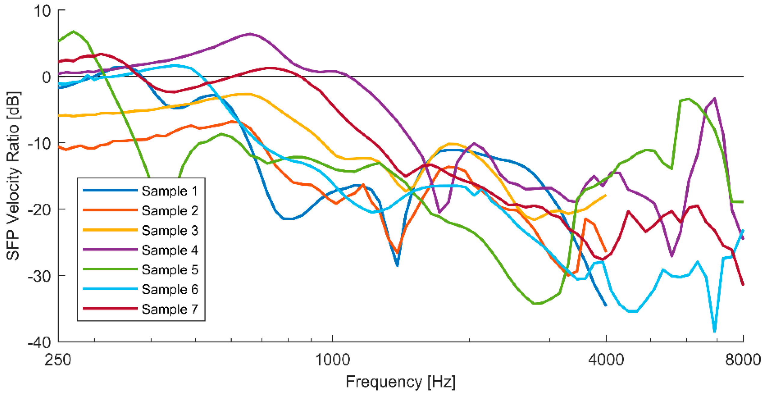
The ratio of the stapes velocity amplitude of the intact to that of the fractured middle ear shows a similar pattern for all samples (
Figure 2). For low frequencies (<500 Hz), the transmission loss is limited and for most samples it remains close to 0 dB. In some case the velocity ratio is positive, indicating an increase in transmission. Below 1 kHz, one or two local velocity ratio maxima are found, of which most occur at around 600–700 Hz with values ranging from +6 dB to −8 dB. At higher frequencies, the velocity ratio steeply decreases to values of at least −15 dB. Another local maximum is seen at around 1500–2000 Hz at about −13 to −10 dB for most samples. Another decrease occurs above 2000 Hz to values of around −20 dB or less and at around 4000 Hz another increase is present for most samples.
3.2. Simulations
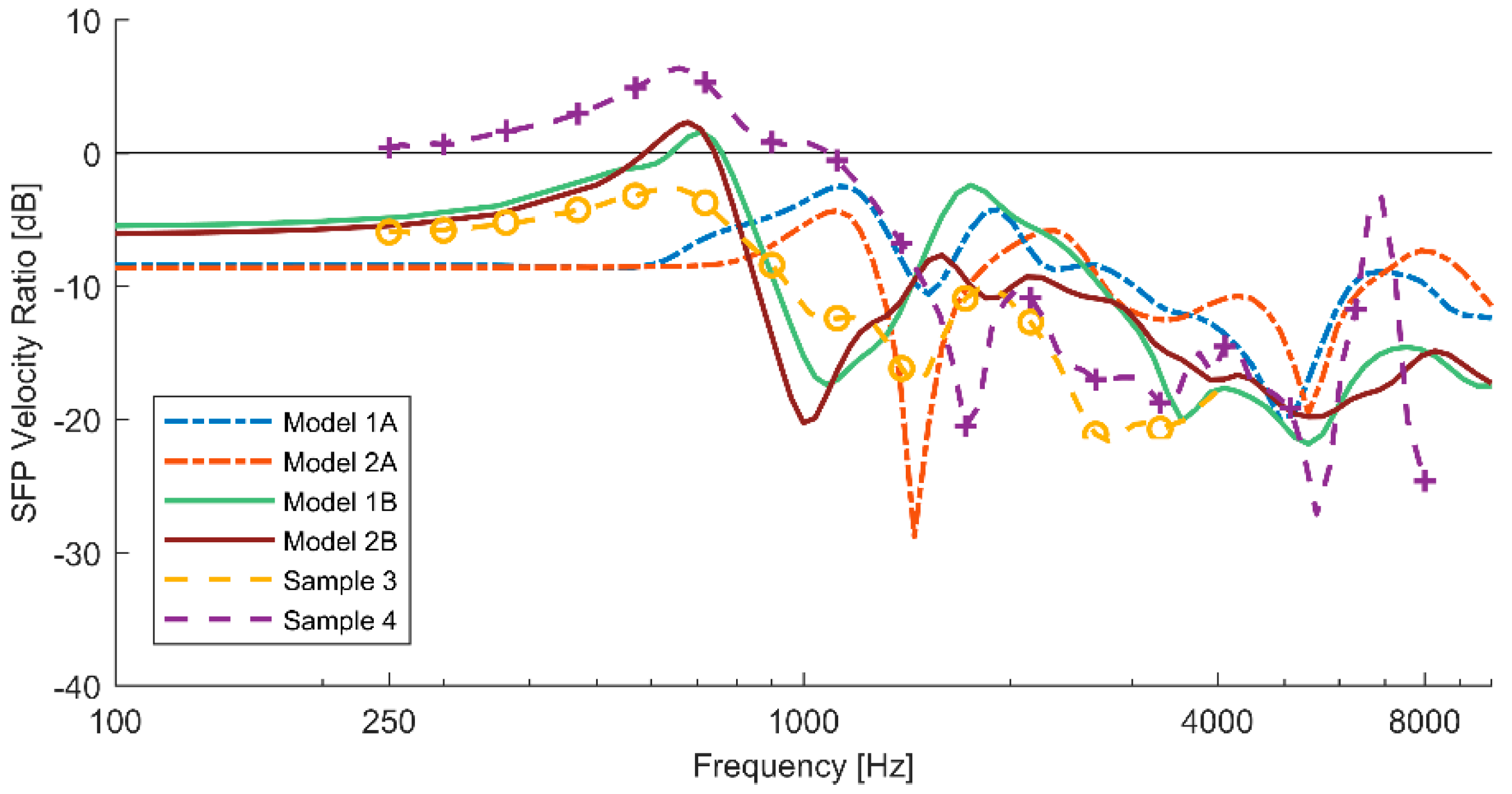
The fractured models without prestress reduction (1A & 2A) show an SFP velocity ratio of about −8.5 dB (
Figure 3). The first local maximum occurs at around 1100 Hz with ratio values of −2.5 dB and −4.3 dB for model 1A and 2A respectively. Above 1100 Hz, a steep decrease is observed. At around 2000 Hz, the velocity ratio increases again, almost reaching the same levels as the first local maximum. Between 2000 Hz and 4000 Hz, the ratio is about −10 dB. A dip occurs at around 5 kHz, after which the ratio increases again. The SFP velocity ratio is negative at all frequencies.
The fractured models with prestress reduction (1B & 2B) have a higher SFP velocity ratio at low frequencies of about −6 dB. The local maximum occurs at around 700 Hz. At this local maximum the velocity ratio becomes positive showing ratios of about +2 dB. At higher frequencies, a steep decrease is observed to values of −16 to −20 dB for model 1B and 2B respectively. After this dip, the ratio increases again to a new local maximum at around 1600–1750 Hz to ratio values of −2.5 and −7.5 dB (model 1B and 2B respectively). Above 1600–1750 Hz, the ratio drops further to values of about −20 dB.
4. Discussion
4.1. Measurements
The measurements presented in
Figure 1 have shown that malleus fractures can cause tremendous hearing losses almost up to −40 dB at some frequencies. The losses are minimal at low frequencies and mainly increase with increasing frequency. Two things stand out:
The global maximum usually occurs at a few hundred Hertz below the middle ear resonance frequency which is typically around 1–1.2 kHz.
The SFP velocity ratio can be positive and is close to 0 dB for most samples at low frequencies.
Both of these observations seem to be due to a shift of the middle ear resonance frequency to lower frequencies after fracture. If the resonance frequency is lowered, the local velocity ratio maximum will be present at this new resonance frequency and a steep decrease will occur at higher frequencies. The velocity ratio will also be boosted at frequencies below this new resonance frequency. The lack of contact between the broken malleus parts will thus produce an overall loss in transmission which can be (partly) countered at low frequencies by this resonance shift.
4.2. Simulations
Simulations without the mimicking of prestress reduction do not show such properties. The maximal velocity ratio occurs at the original resonance frequency of the model. The velocity ratio at low frequencies is also comparable to the lowest observed in the experimental study. No resonance shift is present for this model. This suggests that the resonance shift present in the experimental data is a consequence of something else than just the separation of the two malleus handle pieces. The most likely possibilities are then the change in eardrum prestress and or shape change. Additionally, the local maximum at 2 kHz is too large and decrease at higher frequencies is too small.
Simulations with prestress reduction mimicking show an increased SFP velocity ratio at lower frequencies and a global maximum below the original resonance frequency due to a resonance shift to lower frequencies. However, the first local minimum occurs at a too low frequency and the local maximum at around 2 kHz is still too large. At frequencies higher than 2 kHz, the achieved velocity ratios resemble a bit more the experimental data, reaching levels of −15 and −20 dB before 4 kHz.
It is clear that the models are not yet in good agreement with the experimental data. However, it has been shown that effect of a malleus fracture on sound transmission is caused by more than just the lack of contact between the two broken parts of the malleus handle. A slightly better agreement was achieved by crudely mimicking release of TM pretension. It is possible that the change in shape after fracture also plays an important role.
In the future we plan to perform experiments to measure the change in strain and shape of the tympanic membrane after malleus fracture and to incorporate such measurements in the models.
5. Conclusions
The ratios of stapes footplate velocity of intact middle ears to that of middle ears containing a malleus fracture have shown that hearing losses almost up to −40 dB can occur. It has been shown that for low frequencies the sound transmission can even increase. This is possibly due to a shift in middle ear resonance frequency.
The finite elements simulations have shown that the effect of a malleus fracture on sound transmission is caused by more than just the lack of contact between the two broken parts of the malleus handle. Other important effects of malleus fractures on sound transmission are assumed to be due to release in pretension and the change in shape of the tympanic membrane.
Author Contributions
K.G. has performed the simulation, analysis of the simulation data and has written the paper. P.M. has performed the experimental work and analysis of the experimental data. J.D. has cowritten and proofread the paper.
Acknowledgments
Pieter Muyshondt was supported by an FWO grant. Other researchers of this paper did not receive any grants.
Conflicts of Interest
The authors declare no conflict of interest. The founding sponsors had no role in the design of the study; in the collection, analyses, or interpretation of data; in the writing of the manuscript, and in the decision to publish the results.
References
- Niklasson, A.; Tano, K. Self-inflicted negative pressure of the external ear canal: A common cause of isolated malleus fractures. Acta Otolaryngol. 2010, 130, 410–416. [Google Scholar] [CrossRef] [PubMed]
- Niklasson, A.; Rönnblom, A.; Muyshondt, P.G.G.; Dirckx, J.; von Unge, M.; Tano, K. Ossiculoplasty on Isolated Malleus Fractures. Otol Neurotol. 2016, 37, 895–901. [Google Scholar] [CrossRef] [PubMed]
- De Greef, D.; Pires, F.; Dirckx, J.J.J. Effects of model definitions and parameter values in finite element modeling of human middle ear mechanics. Hear. Res. 2017. [Google Scholar] [CrossRef] [PubMed]
- Blanchard, M.; Abergel, A.; Vérillaud, B.; Williams, M.T.; Ayache, D. Isolated malleus-handle fracture. Auris Nasus Larynx 2011, 38, 439–443. [Google Scholar] [CrossRef] [PubMed]
| Publisher’s Note: MDPI stays neutral with regard to jurisdictional claims in published maps and institutional affiliations. |
© 2018 by the authors. Licensee MDPI, Basel, Switzerland. This article is an open access article distributed under the terms and conditions of the Creative Commons Attribution (CC BY) license (https://creativecommons.org/licenses/by/4.0/).


