1. Introduction
With advances in medical technology and sensors, interests have been piqued about using these advanced technologies to better understand diseases, disorders and therapeutic interactions. West Virginia University’s ambulatory microdose positron emission tomography (AMPET) helmet project involves a collaboration of WVU (West Virginia University) with multiple institutions, including GE Medical, for their groundbreaking technical advances [
1]. Standard PET scanners are most commonly used to detect cancer, heart problems, brain disorders, and problems with the central nervous system, but require patients to be still for an extended time [
2]. AMPET is a portable, lightweight PET scanner that allows a patient to complete various tasks to get real time images of the brain. Furthermore, the novel system allows the use of significantly lower dosage of the radioactive tracers compared to full size scanner systems [
3]. The original AMPET Helmet was created by WVU Medicine made of a plexiglass helmet with 12 photo-detector modules positioned in a ring formation around the head. This prototype confirms the proof of concept for AMPET and delivers real-time, high-definition images of the brain as a patient is preforming a simple task. AMPET solves issues that arise in the current state of the art brain scanning and imaging equipment, combining motion tolerance and coverage of deep brain structures. Standard PET scanners in hospitals are heavy, mostly stationary, and expensive. The AMPET breaks the mold with a light-weight solution that is mobile and inexpensive in comparison to regular PET scanners [
4].
Many challenges can be derived from this helmet with one of the main obstacles being the weight of the 12 photo-detector modules; the modules weigh a total of 5.5 pounds, thus an additional structure to alleviate the weight of the helmet was required. The WVU team created a structure that would ease some of the weight of the helmet using a simple bungee cord design which works when the patient is in a sitting position but can move his/her head in various directions. However, the structure of the original AMPET Helmet’s shifting modules with relation to the patient’s head is higher than it should ideally be, resulting in skewed images and inaccurate readings. The challenge was brought to WVU’s Benjamin M. Statler College of Engineering and Mineral Resources (CEMR) to advance the original plexiglass structure and create a Generation 1 prototype. The criteria being high definition images where the modules move with the head and the creation of a support system that alleviates the weight of the helmet for the reduction of pressure, stress, and weight felt by the patient. And thus the purpose of this paper, the design of a mechanical system to collect high quality imaging with WVU’s portable AMPET helmet with consideration of a patient’s comfort and safety.
The scanning resolution and results have proved the concept of mobile brain imaging with the ability to move and complete simple tasks, i.e., tapping your foot. Issues with limited movement and possible fatigue from the weight of the AMPET helmet upon the subjects’ head has been identified as possible points of intervention by the engineering team.
The current (Generation 1) prototype is affixed to a football helmet for stability and uses a new modular ring in order to allow for more degrees of freedom for the subject wearing the helmet. A Biodex system will be used as the mechanical support system for the PET scanning mobile helmet design, providing an unweighing system for the subjects’ ease of use and safety [
5]. The Biodex will allow the patient to walk on a treadmill and therefore the capturing of images of brain activity during the complex task of walking is possible.
The future of AMPET is full of possibilities. A vision is to make this PET scanner available, completely mobile and able to be used by medical research professionals in the emergency room or onto the field [
6]. For example, a rural town in Africa has a problem with a certain brain disease, but there are no medical facilities in the area equipped with the right systems that are capable of producing the needed insights into the brain activity. This is a situation in which the mobile AMPET might come into play, allowing for highly dynamic brain images to be captured locally, in a small village with mobile technology. In addition, with concussions as an ever-growing issue (especially in sports such as football and soccer), the AMPET scanner could be used to assist the medical staff at the game in making life saving decisions. “Imagine imaging a savant while painting or a chess master in action: We might be able to tap into the mechanisms behind these super abilities,” as Dr. Brefczynski-Lewis explains potential applications for imaging parts of the brain that have not been scanned in this style before [
7].
2. Methods
The methods section will discuss the original AMPET Helmet designed by neuroscientists (OPNS) at West Virginia University (WVU) and the current (Generation 1) design of the AMPET Helmet.
Multiple limitations for these models include the following:
The module must have zero light emitted
The modules must stay at a (near) constant temperature
The modules must not move with relation to the head
The modules must be close in proximity to one another
The modules must be arranged between 28 and 34 cm in diameter
The helmet must be supported for a patient for the duration of a test run (~30 min)
2.1. Original AMPET Helmet
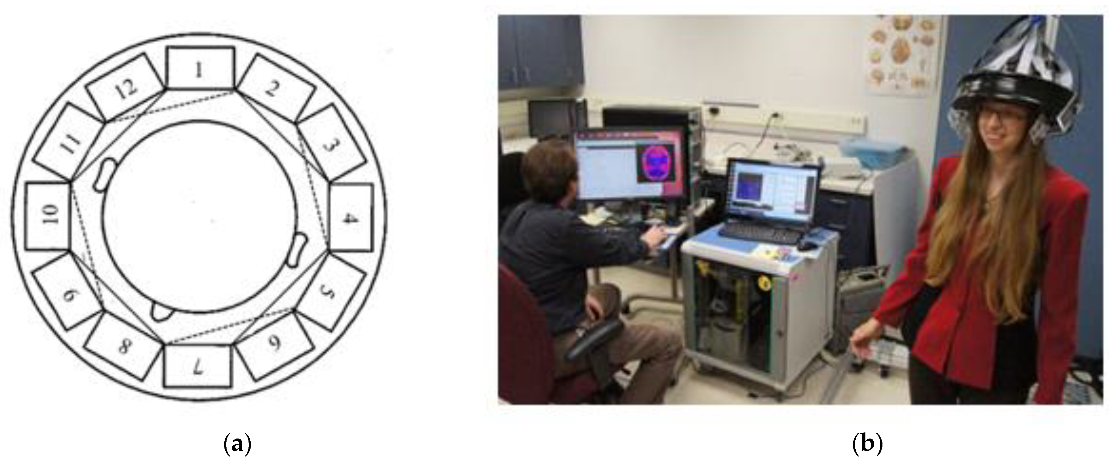
The AMPET Helmet was developed to create a portable PET scanner for more individualized brain imaging as compared to the commonly known PET/CT scanner where the patient is required to remain still in an isolated environment. The original AMPET Helmet is composed of twelve photo-detector modules evenly spaced around a patient’s head. This high-resolution brain imager is capable of fixed head-to-imager geometry, high-efficiency and high-resolution in a compact device, early detection of brain diseases, real time tomographic imaging, 3D reconstruction and fast continuous dynamic scans.
Figure 1 displays the twelve photo-detector modules orientation with relation to the patient’s head. The original AMPET Helmet’s modules dimensions’ measure 40 mm × 40 mm × 25 mm and weigh 200 g each. The twelve modules currently weigh a total of 2.4 km (almost 5.5 pounds) which is too heavy for a patient to support independently over the testing period, thus an apparatus must be constructed to alleviate this weight [
4].
One reason the system has high quality imaging is due to the close proximity of the photo-detector modules to the brain. These proximities not only alleviate the distance between the modules and the head but the distance from module to module, which should be kept to a minimum. The distance between the modules is critical due to the image overlapping that must be done to scan the entire brain without imagery gaps.
As mentioned previously, the original AMPET Helmet’s prototype is made of cut plexiglass pieced together in a circular form to accommodate the twelve photo-detector modules as indicated in
Figure 1. Black electrical tape fastens them around the plexiglass structure to block light interference from the modules. The original AMPET Hemet uses a compressive fitting for the helmet with the use of an adjustable inner ring, similar to that of a construction hat, with an adjustable knob in back to loosen or tighten the helmet and accommodate multiple head sizes. This original prototype allows a patient to sit in a chair to do various activities such as tap a foot, or clap one’s hands, however many obstacles have occurred with the plexiglass structure such as the moving of the original AMPET helmet with relation to the patient’s head orientation, loss of module maintenance time and lack of heat shield from the patient’s head resulting in, among other issues, increased noise present in the collected data.
Due to the weight of the modules and the plexiglass structure the original AMPET Helmet weighs around eight pounds and must have a support system to alleviate the weight on the patient head to reduce the risk of injury, e.g., neck. The original AMPET Helmet design uses a bungee cord attached to a sturdy structure to ease the weight of the helmet on the patient. At the center of the helmet, the bungie cord is attached and does not restrict the degrees of freedom that is associated with normal head movements (i.e., rotation about the sagittal, coronal and transverse planes). The helmet design that provides the ideal conditions to capture the most accurate readings from the photo-detector modules, translates to virtually no relative movement of photo-detector modules to the patient’s head. To accomplish this objective, a helmet with a tight fit on the patient’s head is crucial. If the helmet, and thus the modules, move with relation to the patient's head, the readings will not give the high-quality image of the patient’s brain.
2.2. Generation 1 AMPET Helmet
Building on these concepts from the OPNS, the next prototype (Generation 1) aims to advance this design and tackle these challenges. Generation 1’s main objective is to improve the support system of the helmet, by creating a support structure that can free the patient of major force due to the weight of the helmet. This design employs a standard American football helmet to create a better, in terms of tightness and comfort, fit for the patient. This design took advantage of the air bladder system on the inside of modern football helmets that is designed to adjust the helmet’s fit to individual head forms. The built in air bladder goes around the head with a port in the back of the helmet. The port allows to pump up or deflate the bladder as needed to provide a comfortable, secure fit to the patient’s head. With the use of the football helmet, the weight of the helmet system increases. However, the advantage of having a better fit and thus reduced relative movement of sensors to head justifies the trade off in the perspective of the design team.
Generation 1’s support structure will employ a novel counterweight and pulley system to create a virtually weightless experience for the patient. This system will attach to the helmet in an attempt to reduce the weight of the helmet experienced by the patient. When the patient’s head is moving in a positive vertical direction, the counterweight will retract the helmet up with a weight approximately the same as the helmet. This system will also keep tension on the helmet so when the patient moves in the negative vertical direction, the helmet won’t push down on their head. The rotation of the head is still accounted for in this design. Generation 1 is using a ball-in-socket joint to allow the patient to rotate their head in all directions, a patient’s head might move during the expected routines. This mount will attach directly to the center of the helmet and provide the user the ability to move their head around without major restriction. The photo-detector modules on the Generation 1 helmet will be contained in a 3D-printed (modular) ring arrangement. A modular design of the ring will allow for the flexibility to add and subtract the number of sensors used on the helmet and also significantly improve the accessibility and thus maintenance activities.
3. Results and Discussion
Generation 1, through a preliminary visual inspection, has improved the original AMPET Helmet, developed by the OPNS in terms of reducing the observed relative movement of head to sensor. A measurable confirmation will be made in the coming weeks with the use of a Vicon-infrared, marker-tracking system to compare the two systems in realistic scenarios.
3.1. Biodex Unweighing System
The Biodex system is meant to be used for physical therapy treatments to help those who have difficulties in walking (or need to build up strength), have the ability to do so with the help of a harness that is attached to the Biodex as the structural support. The AMPET team is using the harness provided by the Biodex for the safety of the patient and modules where the structure is to support the patient in case of a fall and to support the AMPET helmet. This system can do so with a 300-pound capacity. The design of the Biodex system was to meet the goal of allowing the helmet to have all degrees of freedom and feel nearly weightless on the patient. Another advantage of the use of the Biodex is it is meant to be used to walk freely on a treadmill, but also has wheels on the bottom to allow the patient to walk around for future designs. The design was able to fulfill the needs of Generation 1 in many different ways. The counter weight system preformed as intended to move with the helmet up and down while taking the weight of the helmet off of the patient. Instead of the use of a common counterweight as seen used on exercise equipment, the AMPET design team used a counterbalance where the weight can be manually changed from nine to thirteen pounds. The counterbalance was placed on the side of the Biodex system with its cable threaded through a system of pulleys. The pulleys were used to guide the cable to be directed over the center of the helmet. The group made minor modifications to the Biodex system. The system has a bar above the harness. This bar serves the purpose of a single point suspension. This permits functional pelvic rotation and versatility when walking. The bar’s length was too short compared to the diameter of the helmet in use. Here, the group lengthened the bar. The attachment rings are now further outside of the helmet allowing for the correct functional use.
3.2. Module and Helmet Design
As mentioned before, an American football helmet is the main structure for the next generation ring of photo-detector modules. The football helmet has many advantages including a tight fit with the use of an air bladder to adapt and adjust to each individual’s head shape, a radius that accommodates the 28–34 cm diameter limitation and the helmets sturdiness material to support the weight of each module and distribute it more evenly. These modules form a ring around the helmet that can be tilted in various orientations to get a better picture of specific areas of interest of the subject’s brain, for example motor control of deep brain structures like the basal ganglia. A swivel mount is attached to the top of the helmet to allow for movement of the patient’s head in all directions; thus, where the helmet is connected to the Biodex system. This is mounted at the center of the helmet’s weight; therefore, it does not affect the patients head movements. The modules themselves can be taken out of the ring easily by removing the screws around the “lid” and taking the module right out of the 3D printed model. This allows for easy maintenance of the modules. Future manufacturing designs will involve a plastic mold with an easy to use “door” with a clip so there are no screws that can be dropped, and thus improved associability.
3.3. Future for AMPET
The current system has certain limitations, such as the inability to walk through a normal doorway, non-uniform support when patients head is tilted (due to the upwards force only), and the modules themselves heat up over the duration of a measurement with a patient. The original design allowed for a patient to sit in a chair, the design currently under development will allow a patient to walk on a treadmill and future envisioned designs to be explored intend to be completely portable and able for social interaction and behaviors in a virtual reality environment. Part of the possible options for a completely portable design is using a robotic arm with the use of sensors, such as accelerometers, to move along with the head for a virtual weightless system. The next helmet (Generation 2) will be made with 16 higher sensitivity module’s weighing 500 g each and placed in a ring formation similar to that of Generation 1.
Another area in which the Generation 1 design aims to improve upon is keeping the sensor modules at a constant temperature during a measurement as with changing temperature inside the module, especially rising temperatures, the sensors lose accuracy. As of today, three potential sources for increasing temperature at the sensor modules are identified:
- (i)
the patient’s head (body temperature) (external)
- (ii)
changing room (environment) temperature (external)
- (iii)
emerging from the sensor modules themselves when processing data (internal)
Therefore, the temperature control is not trivial, especially as the target delta (temperature change) during a measurement cycle is lower than 0.5 degrees Celsius, ideally even lower. This might require an active temperature control system within each individual module casing.
AMPET could allow researchers to study the neuro-correlations of active behavior, and discover new mechanisms and targets for brain disorders and how other aspects of brain research are conducted. The AMPET helmet will change lives as well as save lives with the ability for early detection of cancer and other diseases in the brain.
4. Conclusions
The AM-PET helmet allows measuring a patient’s brain activities using (i) far reduced exposure to radioligand (ii) relevant activities like, e.g., movement or social interaction. In this paper, the current work and future vision of an engineering re-design of the helmet and support structure focusing on identified challenges of the current system are described. From a visual inspection of the helmet there is no slipping of the modules with relation to the head and thus the objective has been achieved. This modular design is proven to be the best form (with the current use of a 3D printer) due to the ease of access of the modules. Generation 2’s design will involve a 16 photo-detector module ring with each module weighing a pound, thus the helmet will weigh upwards of 20 pounds. The current system will be improved upon to accommodate the increased weight.
Author Contributions
Samantha Melroy, Mathew McHugh, Garret Carden and Thorsten Wuest are part of the engineering team who designed the Generation 1 support and helmet and built the support structure; Julie Brefczynski-Lewis is a neuroscientist who performed testing with the original AMPET helmet and analyzed results.
Acknowledgments
The authors thank West Virginia University, University of Virginia, University of California, Davis, University of Washington, and GE Global Research for technical assistance and NIH grant #R24 MH106057 for the funding assistance. We also thank the individuals representing these fine institutions, Stan Majewski, Jinyi Qi, Kuang Gong, Paul Kinahan, Robert Harrison, Brian Elston, Sergei Dolinsky, Michael Rishel, Ravi Manjeshwar for the unwavering support and collaboration for innovation.
Conflicts of Interest
The authors declare no conflict of interest.
References
- Kinahan, P.; Majewski, S.; Elston, B.; Harrison, R.; Qi, J.; Manjeshwar, R.; Dolinsky, S.; Stolin, A.; Brefczynski-Lewis, J. Design Considerations for AMPET: The Ambulatory Micro-Dose, Wearable PET Brain Imager. J. Nuclear Med. 2015, 3, 1540–1540. [Google Scholar]
- Krans, B. PET Scan, 2 November 2015. Available online: http://www.healthline.com/health/pet-scan#Overview1 (accessed on 16 October 2016).
- MLN Matters Articles. Fluorodeoxyglucose (FDG) Positron Emission Tomography (PET) for Solid Tumors, 6 February 2014. Available online: https://www.cms.gov/Outreach-and-Education/Medicare-Learning-Network-MLN/MLNMattersArticles/downloads/MM8739.pdf (accessed on 16 October 2016).
- Majewski, S.; Proffitt, J. Compact and Mobile High Resolution PET Brain Imager. U.S. Patent 7,884,331, 8 February 2011. [Google Scholar]
- Biodex Medical Systems, “Unweighing System”. 2016. Available online: http://www.biodex.com/physical-medicine/products/pbws/unweighing-system (accessed on 17 October 2016).
- Lewis, T. “Wearable Brain Scanner Measures Activity on the Go”, 3 January 2015. Available online: http://www.livescience.com/49548-portable-brain-scanner.html (accessed on 17 October 2016).
- Freeman, T. “Portable Brain Scanner Allows PET in Motion”, 28 October 2015. Available online: http://medicalphysicsweb.org/cws/article/research/63031 (accessed on 17 October 2016).
| Publisher’s Note: MDPI stays neutral with regard to jurisdictional claims in published maps and institutional affiliations. |
© 2016 by the authors. Licensee MDPI, Basel, Switzerland. This article is an open access article distributed under the terms and conditions of the Creative Commons Attribution (CC BY) license (https://creativecommons.org/licenses/by/4.0/).


