Article

Investigation of Upper Respiratory Carriage of Bacterial Pathogens among University Students in Kampar, Malaysia

1
Department of Biological Science, Faculty of Science, Kampar Campus, Universiti Tunku Abdul Rahman, Kampar 31900, Malaysia
2
Department of Allied Health Sciences, Faculty of Science, Kampar Campus, Universiti Tunku Abdul Rahman, Kampar 31900, Malaysia
3
Centre for Biomedical and Nutrition Research, Kampar Campus, Universiti Tunku Abdul Rahman, Kampar 31900, Malaysia
4
Faculty of Medicine and Institute for Life Sciences, University of Southampton, Southampton SO16 6YD, UK
5
NIHR Southampton Biomedical Research Centre, University Hospital Southampton NHS Trust, Southampton SO16 6YD, UK
6
Global Health Research Institute, University of Southampton, Southampton SO17 1BJ, UK
7
School of Postgraduate Studies, International Medical University, Kuala Lumpur 57000, Malaysia
8
Centre for Translational Research, Institute for Research, Development, and Innovation, International Medical University, Kuala Lumpur 57000, Malaysia
*
Author to whom correspondence should be addressed.
Trop. Med. Infect. Dis. 2023, 8(5), 269; https://doi.org/10.3390/tropicalmed8050269
Submission received: 27 March 2023 / Revised: 29 April 2023 / Accepted: 2 May 2023 / Published: 8 May 2023
(This article belongs to the Special Issue Advances in Emerging and Re-emerging Infectious Diseases)

Abstract

:
The carriage of bacterial pathogens in the human upper respiratory tract (URT) is associated with a risk of invasive respiratory tract infections, but the related epidemiological information on this at the population level is scarce in Malaysia. This study aimed to investigate the URT carriage of Streptococcus pneumoniae, Haemophilus influenzae, Neisseria meningitidis, Staphylococcus aureus, Klebsiella pneumoniae and Pseudomonas aeruginosa among 100 university students by nasal and oropharyngeal swabbing. The presence of S. aureus, K. pneumoniae and P. aeruginosa was assessed via swab culture on selective media and PCR on the resulting isolates. For S. pneumoniae, H. influenzae and N. meningitidis, their presence was assessed via multiplex PCR on the total DNA extracts from chocolate agar cultures. The carriage prevalence of H. influenzae, S. aureus, S. pneumoniae, K. pneumoniae, N. meningitidis and P. aeruginosa among the subjects was 36%, 27%, 15%, 11%, 5% and 1%, respectively, by these approaches. Their carriage was significantly higher in males compared to females overall. The S. aureus, K. pneumoniae and P. aeruginosa isolates were also screened by the Kirby-Bauer assay, in which 51.6% of S. aureus were penicillin-resistant. The outcomes from carriage studies are expected to contribute to informing infectious disease control policies and guidelines.

1. Introduction

Streptococcus pneumoniae, Haemophilus influenzae, Neisseria meningitidis, Staphylococcus aureus, Klebsiella pneumoniae and Pseudomonas aeruginosa are among the bacterial pathogens that colonize the human upper respiratory tract (URT) [1,2,3]. Their pathogenicity is mediated by some major virulence factors involved in host cell adherence and invasion, tissue damage and immune evasion [4,5,6,7]. Despite being pathogenic, most individuals do not show any symptoms when these bacteria colonize the URT. Under certain conditions, some of them (e.g., S. pneumoniae) can translocate to other URT sites causing local infections, such as otitis media and sinusitis, or to the lungs, bloodstream and brain, causing potentially life-threatening diseases, such as pneumonia, septicemia and meningitis, respectively [8,9,10,11,12]. One study reported that high-density colonization of the URT by S. pneumoniae, H. influenzae and Klebsiella spp. was associated with increased risks of lower respiratory tract infections [11]. This indicates that asymptomatic colonization (i.e., carriage) is the first step in developing respiratory tract infections [13].
The microbial communities in the URT vary significantly among different sites [2,11]. With regard to the target bacteria in this study, the microbiome of anterior nares is predominated by S. aureus [14], while the oropharynx commonly shows the presence of S. pneumoniae and H. influenzae [15]. Some bacterial pathogens carried in the URT, for instance S. pneumoniae and N. meningitidis, can be spread via airborne respiratory droplets, leading to colonization and possibly infection in the new hosts. This is especially apparent in crowded environments and among household contacts [15,16,17]. Hence, a higher prevalence of carriage might increase the risk of infection or outbreak within a population, particularly among the age extremes [18,19].
Respiratory carriage studies are a pragmatic solution to getting real-time epidemiological data on the carriage of pathogens at the population level [19]. Several carriage studies have been conducted among university students (representing young adults). A study of this type in Nepal reported that 35% and 12.5% of nasal and pharyngeal isolates were of S. aureus and H. influenzae, respectively [20]. A review of N. meningitidis carriage studies among university students in various countries revealed the highest carriage in the Americas (4–71.1%) and European region (10.4–61.9%), followed by Nigeria (5.1%), Chile (4%) and India (1.5%) [21]. In Malaysia, existing studies among university students mainly focused on S. aureus, with carriage prevalence from 10% to as high as 93% recorded [22,23,24]. Other carriage studies emphasized on pneumococcal carriage in children, in which the prevalence of about 10% was documented based on nasopharyngeal swab assessment [25,26]. Recent carriage studies were carried out among the indigenous communities (locally known as Orang Asli) in certain parts of Malaysia, in which high carriage of S. aureus (47.2%) and K. pneumoniae (30%) was observed [27,28].
To date, respiratory carriage studies are still limited in Malaysia, in which those reported previously are outdated and were skewed toward certain age and ethnic groups [27,28,29]. Further epidemiological studies are needed to increase the data on pathogenic bacterial carriage, especially among young adults and ethnic minority groups. Therefore, this study set out to investigate the URT carriage of S. pneumoniae, H. influenzae, N. meningitidis, S. aureus, K. pneumoniae and P. aeruginosa, focusing on students at a private university. The objectives were to determine the carriage prevalence of these bacteria and preliminarily assess their association with several host factors to provide a better understanding of the risk of disease acquisition from their carriage. The antibiotic susceptibility of isolates of the three ESKAPE pathogens (S. aureus, K. pneumoniae and P. aeruginosa) was also evaluated; these pathogens are commonly associated with high antibiotic resistance.

2. Materials and Methods

2.1. Study Population, Design and Procedures

The total number of subjects (n = 100) were randomly recruited from the student population at Universiti Tunku Abdul Rahman (UTAR), Kampar, located in the state of Perak, which lies on the west coast of Peninsular Malaysia. Ethical approval for the study was obtained from the UTAR Scientific and Ethical Review Committee (approval no. U/SERC/49/2018). The sample collection was carried out during the period of 1 May to 31 August 2018.
The sample size required was calculated using the Epi Info™ 7.2.2.6 software [30] available at the Centers for Disease Control and Prevention (CDC) website. The UTAR Kampar Campus (study location) saw the enrolment of approximately 13,280 students during the study period. Based on this population size and the expected frequency of 10% reported for pneumococcal carriage in a Malaysian study [28], the sample size of n = 95 was determined to be sufficient to attain a statistical power of at least 95% confidence level, with a margin of error of 6%.
Informed consent was obtained from all participants prior to sample collection. The information sheet provided to participants includes guidelines on confidentiality and anonymity of the data obtained. A questionnaire to collect socio-demographic and health-related data was completed for each subject, including the subject’s gender, age, ethnicity, vaccination status, self-reported respiratory symptoms and antibiotic consumption in the previous month. A nasal (anterior nares) swab and an oropharyngeal swab were taken from each subject by using sterile cotton swabs of the DRYSWAB™ range (MWE, Wiltshire, UK) in accordance with the WHO Pneumococcal Carriage Working Group guidelines [31]. The swab was placed in the skim milk, tryptone, glucose and glycerol (STGG) medium for laboratory analysis and long-term storage.

2.2. Swab Culture for Target Bacteria

The swab suspensions obtained were cultured onto general chocolate agar, mannitol salt agar (for isolation of S. aureus), MacConkey agar (K. pneumoniae) and Pseudomonas agar supplemented with cetrimide and sodium nalidixate (C-N) (P. aeruginosa). All the components for preparing these media were sourced from Oxoid (Hampshire, UK). The chocolate agar plates were incubated at 37 °C in a candle jar, while the other culture media were incubated at 37 °C under aerobic conditions. All the plates were incubated for 1–3 d. Preliminary identification of the resulting isolates was by their growth characteristics on the respective culture media. Potential S. aureus and K. pneumoniae isolates were also subjected to the coagulase and oxidase tests, respectively. The former was performed with the Bactident® EDTA-rabbit plasma (Merck, Rahway, NJ, USA) according to the manufacturer’s instructions, while the latter was performed with the BactiDrop™ oxidase test reagent (Thermo Fisher Scientific, Lenexa, KS, USA). The chocolate agar cultures were transferred to the STGG medium for long-term storage.

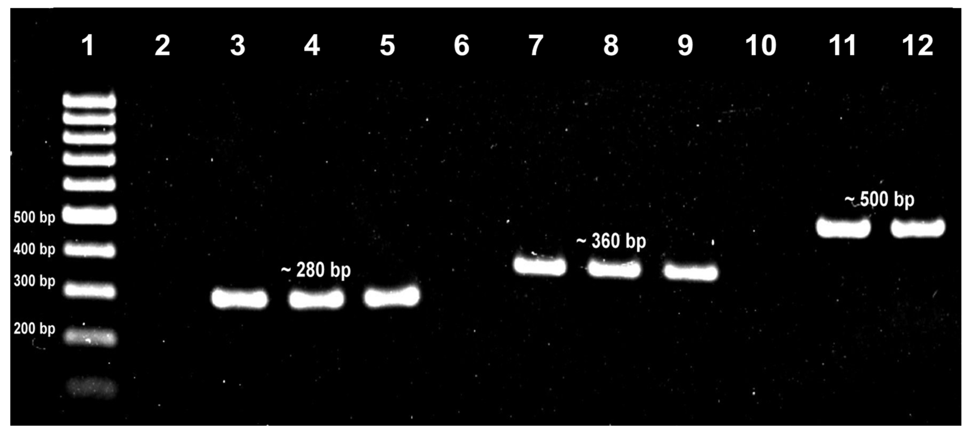
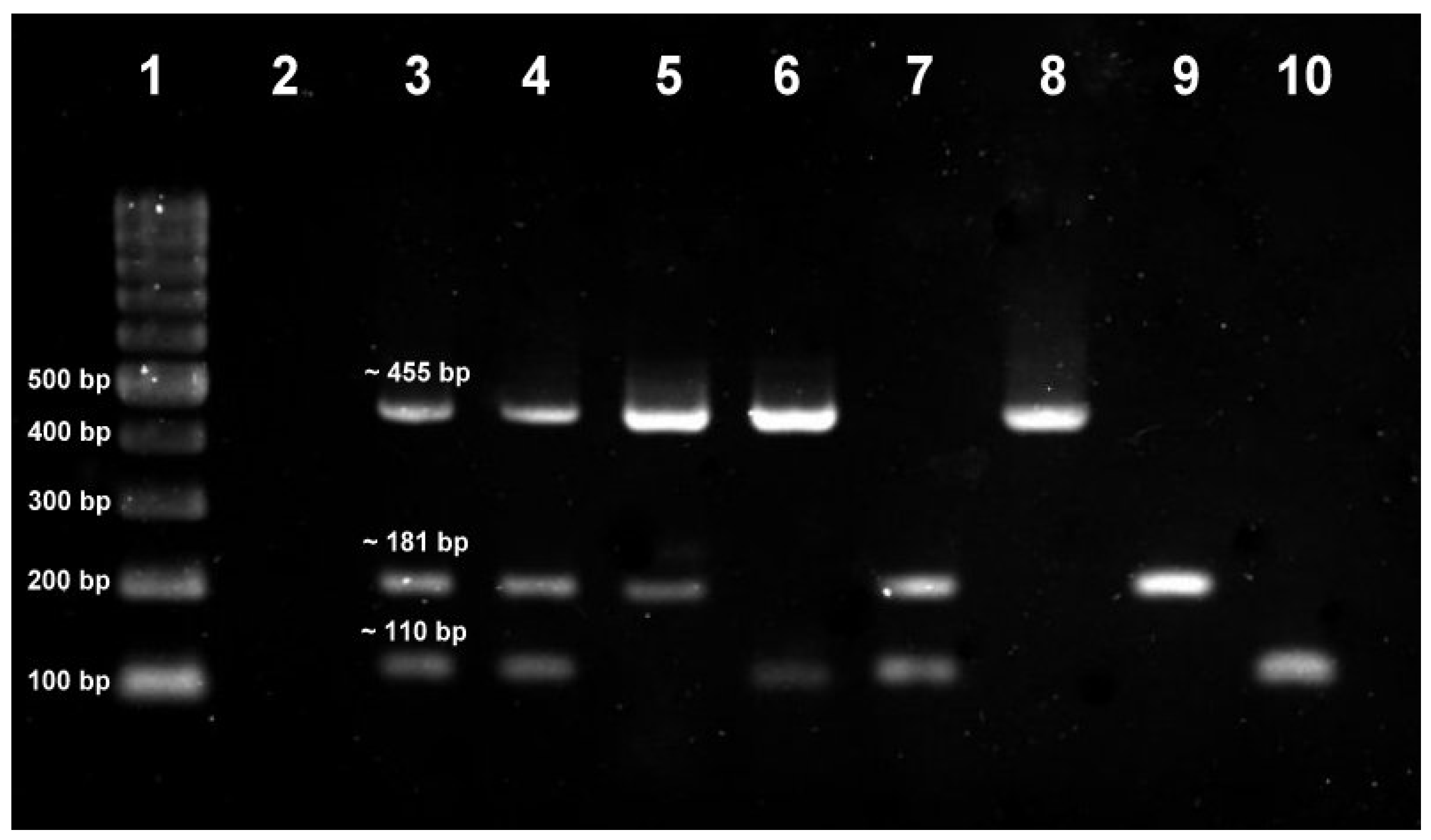
2.3. PCR Identification of Target Bacteria from Swab Cultures

For the detection of S. aureus, K. pneumoniae and P. aeruginosa, the potential isolates were subcultured onto LB agar and the resulting cultures were subjected to boiling DNA extraction. The respective DNA extracts were subjected to specific PCR assays targeting the S. aureus nuc, K. pneumoniae mdh and P. aeruginosa oprL genes. For the detection of S. pneumoniae, H. influenzae and N. meningitidis, the suspensions of chocolate agar cultures were subjected to DNA extraction with the GF-1 Tissue DNA Extraction Kit (Vivantis, Selangor, Malaysia) according to the manufacturer’s instructions. The culture DNA extracts were subjected to a multiplex PCR assay targeting the S. pneumoniae lytA, H. influenzae bex and N. meningitidis ctrA genes. The sequences of the primer pairs used in these PCR assays and their respective amplicon sizes are shown in Table 1.
The PCR conditions were adapted from the respective studies [32,33,34,35,36], with some optimizations performed (data not shown). The final conditions used in this study are as follows. Each PCR reaction consisted of 1× PCR buffer (Invitrogen, Carlsbad, CA, USA), 0.3 μM each for forward and reverse primers (Integrated DNA Technologies, Singapore), 0.3 mM dNTPs (RBC Bioscience, New Taipei City, Taiwan), 2.5 mM MgCl2 (Invitrogen), 2.5 U of Taq DNA polymerase (Invitrogen) and 80-100 ng of template DNA in a total volume of 20 μL. The PCR conditions were 95 °C for 5 min, followed by 35 cycles of 95 °C for 30 s, 57 °C for 40 s and 72 °C for 1 min. A no-template control (NTC) and positive control were included in each run. PCR amplicons were subjected to electrophoresis on 2% (w/v) agarose gel, which was then stained in RedSafe™ Nucleic Acid Staining Solution (iNtRON Biotechnology, Gyeonggi-do, Korea) for visualization under the ultraviolet transilluminator.

2.4. Antibiotic Susceptibility Assessment of ESKAPE Target Bacteria

The Kirby-Bauer antibiotic susceptibility test was performed on the S. aureus, K. pneumoniae and P. aeruginosa isolates, which are three species of the ESKAPE pathogens that are commonly associated with high antibiotic resistance. The bacterial test suspension, with turbidity equivalent to that of the 0.5 McFarland standard, was inoculated over the Mueller–Hinton agar (Oxoid) with a sterile cotton swab to create a bacterial lawn. Antibiotic discs (Oxoid) were placed over the agar and the plates were incubated at 37 °C for 18 h. The results were interpreted by comparing the diameter of the zones of inhibition measured to the Clinical and Laboratory Standards Institute (CLSI) standards [37] to determine the susceptibility of the isolates. S. aureus was tested with chloramphenicol (30 μg), ciprofloxacin (5 μg) doxycycline (30 μg), gentamicin (10 μg), methicillin (5 μg), penicillin G (10 U), quinupristin-dalfopristin (15 μg), trimethoprim-sulfamethoxazole (1.25/23.75 μg) and tetracycline (30 μg). K. pneumoniae was tested with ampicillin (10 μg), cefpodoxime (10 μg), ceftazidime (30 μg), ceftriaxone (30 μg), ciprofloxacin (5 μg), gentamicin (10 μg), imipenem (10 μg) and tetracycline (30 μg). P. aeruginosa was tested with ceftazidime (30 μg), ciprofloxacin (5 μg), gentamicin (10 μg) and imipenem (10 μg). These antibiotics were selected based on the CLSI guidelines, in which many are of the following classes: beta-lactams, aminoglycosides, carbapenems, macrolides, quinolones and tetracyclines.

2.5. Data Analysis

Statistical analyses were conducted using the SPSS software version 25.0 (IBM, Armonk, NY, USA). A univariate analysis was performed to determine the distribution of values for all the variables. Fisher’s exact test was used to analyze the associations between demographic characteristics or host factors and bacterial carriage. A p-value of less than 0.05 would indicate statistical significance at a 95% confidence level.

3. Results

3.1. Subject Demographic and Health-Related Profiles

A total of 100 subjects participated in this study, in which 100 nasal and 100 oropharyngeal swabs were collected and tested. The median age of the subjects was 21 years old, with an equal number in gender distribution. The ethnicity distribution is as follows: 92 Chinese, 5 Indian and 3 Malay. This skewed distribution mirrored the ethnic composition of the student population at the university’s main campus (the study location) during the study period: 92% Chinese (n = 12,184), 6.2% Indian (n = 828), 0.6% Malay (n = 75) and 1.5% Others (n = 193). Based on eligibility for receiving vaccines under the Malaysian National Immunisation Programme (NIP), only 27% of the subjects were aware of their vaccination status, while 55% were unsure and the remaining thought they did not receive any of the vaccines. Thirty-five subjects reported having URT symptoms that could be those of respiratory infections (e.g., cold, flu, ear infection, etc.) in the month prior to sample collection. Five subjects had taken antibiotics in this period.

3.2. Carriage Prevalence of Target Bacteria

Potential isolates of S. aureus, K. pneumoniae and P. aeruginosa from swab cultures on mannitol salt agar (yellow colonies with yellow zones), MacConkey agar (pink mucoid colonies) and Pseudomonas agar supplemented with C-N (green colonies), respectively, were subjected to their respective PCR assays. Based on the PCR results, the carriage prevalence among the 100 subjects was 27%, 11% and 1%, respectively. Figure 1 shows the representative gel analysis for the S. aureus nuc, K. pneumoniae mdh and P. aeruginosa oprL PCR assays. All the potential S. aureus isolates were coagulase-positive, while all the K. pneumoniae isolates were oxidase-negative. The detection of S. pneumoniae, H. influenzae and N. meningitidis in the swab cultures on chocolate agar was by a multiplex PCR assay on their total DNA extracts. Based on the PCR results, the carriage prevalence among the 100 subjects was 15%, 36% and 5%, respectively. Figure 2 shows the representative gel analysis for the multiplex PCR assay.

3.3. Bacterial Carriage by URT Sites

The distribution of target bacterial carriage between the two URT sites surveyed, the anterior nares and the oropharynx, was also determined. From a total of 31 S. aureus isolates obtained from 27 subjects, 21 (67.7%) were from the nasal swabs and 10 (32.3%) were from the oropharyngeal swabs. Four subjects were positive for S. aureus by both swabs. For the 12 K. pneumoniae isolates obtained from 11 subjects, 5 (41.7%) were of nasal origin and 7 (58.3%) were from the oropharynx. The sole P. aeruginosa in this study was isolated from the oropharyngeal swab. For S. pneumoniae, 13 of 15 subjects (86.7%) carried it in the oropharynx while the remaining two subjects (13.3%) showed nasal carriage. All the H. influenzae (n = 36) and N. meningitidis (n = 5) carriage was in the oropharynx.

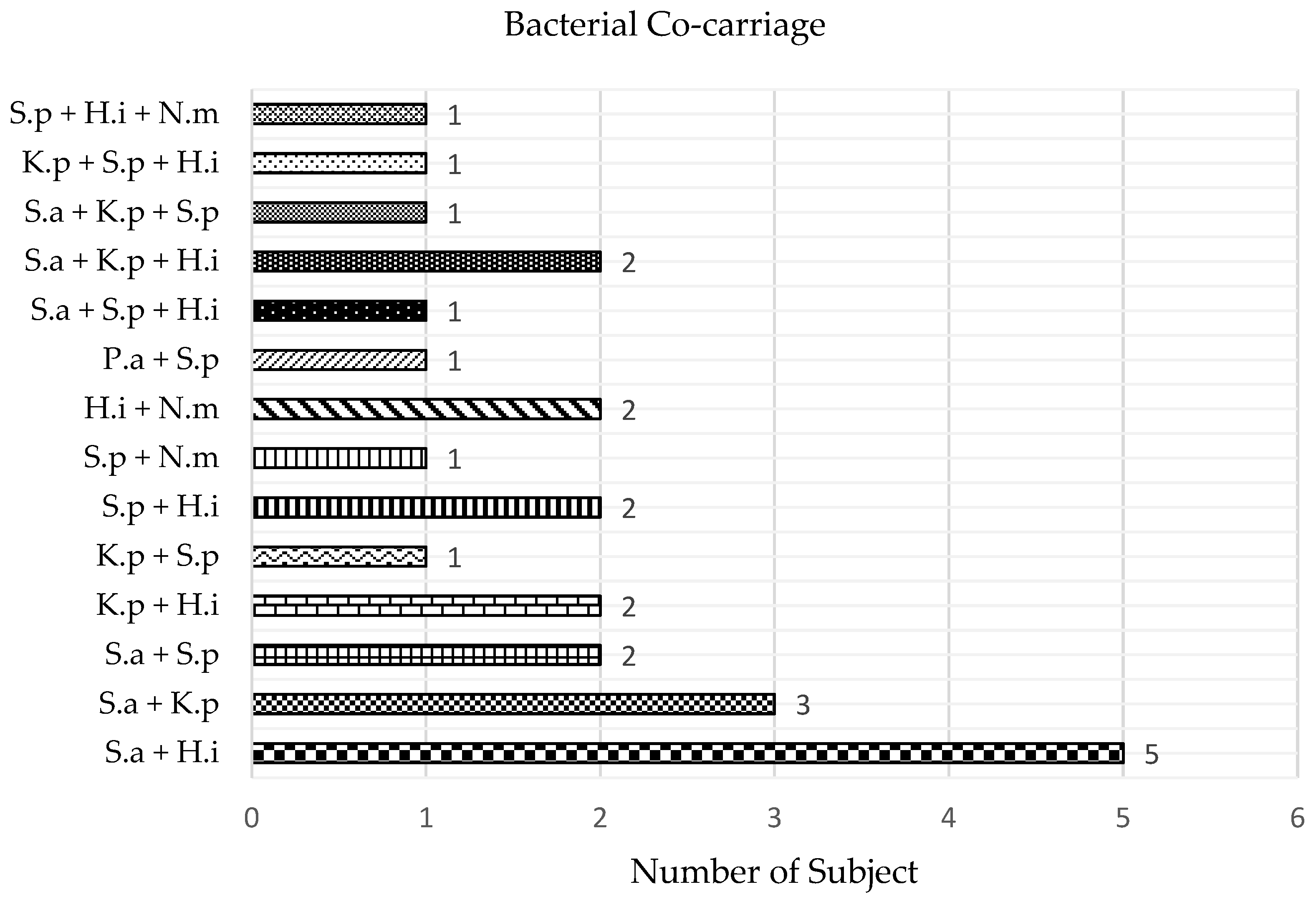
3.4. Bacterial Co-Carriage

Co-carriage prevalence was determined by the proportions of subjects carrying more than one target bacterium. Of the 64 carriers in this study, 25 belonged to this category, of which 19 subjects (29.7%) carried two target bacteria and 6 subjects (9.4%) carried three target bacteria. Figure 3 shows that the co-carriage of S. aureus and H. influenzae was the highest (ո = 5), followed by that of S. aureus and K. pneumoniae (ո = 3). Other co-carriage combinations were observed in 1–2 subjects (Figure 3). For the 39 subjects with single bacterial carriage, there were 20 (31.3%) H. influenzae carriers, 13 (20.3%) S. aureus carriers, 4 (6.3%) S. pneumoniae carriers and one carrier (1.6%) each for K. pneumoniae and N. meningitidis.

3.5. Association between Bacterial Carriage and Host Factors

As shown in Table 2, the overall carriage of target bacteria was significantly higher in males (n = 39; 78%) as compared to in females (n = 25; 50%) (p < 0.01). Additionally, males showed a higher frequency of multiple bacterial carriage (≥2 target bacteria) as compared to females (p < 0.01). No significant associations were demonstrated between the bacterial carriage and the other factors (ethnicity, NIP vaccination, previous URT symptoms and antibiotic intake). When comparing the URT sites among the target bacterial carriers, there are no significant differences for all the factors (data not shown). It is noteworthy that the assessment for some of these factors was limited by skewed distribution in their variables, which is especially apparent for ethnicity and antibiotic intake. The reliability of responses to NIP vaccination is another concern.

3.6. Antibiotic Susceptibility of ESKAPE Target Bacteria

A high level of penicillin resistance (51.6%, n = 16) was observed among the S. aureus isolates (Table 3). For quinupristin-dalfopristin, 2 (6.5%) isolates were resistant to it, but 8 (25.8%) isolates fall into the category of intermediate resistance. Resistance or intermediate resistance to chloramphenicol, doxycycline, gentamicin, methicillin and tetracycline was also occasionally observed in some isolates. All the S. aureus isolates were susceptible to ciprofloxacin and trimethoprim/sulfamethoxazole. For K. pneumoniae, all the isolates were resistant to ampicillin but susceptible to the other seven antibiotics (Table 3). The only P. aeruginosa isolate from this study was susceptible to all four antibiotics tested, which were ceftazidime, ciprofloxacin, gentamicin and imipenem. None of the bacterial isolates showed multidrug resistance, which is commonly defined as resistance to antibiotics of three or more classes [38].

4. Discussion

Based on the results obtained, the carriage prevalence of the target bacteria among the university students (n = 100) was as follows: S. pneumoniae (15%), H. influenzae (36%), N. meningitidis (5%), S. aureus (27%), K. pneumoniae (11%) and P. aeruginosa (1%). In two Nepalese studies on undergraduate medical students and healthcare workers, the S. pneumoniae carriage prevalence was 4% and 21%, respectively [20,39]. The pneumococcal carriage was also documented to be relatively low (3.2%) among medical students in a Turkish study [40]. A recent study among the indigenous community (70.7% adults) in Malaysian Borneo and another on adults in Indonesia recorded carriage prevalence of 10% and 11%, respectively [28,41]. With regard to S. aureus carriage, several studies reported a prevalence of 20–30% [28,42,43], which is consistent with that observed in our study. Nevertheless, two studies among students of two Malaysian universities recorded a lower prevalence of 10% and 16%, respectively [22,23]. For K. pneumoniae carriage, the prevalence in our study (11%) is identical to that documented among adults in an Indonesian study [41]. The Turkish study on medical students reported a significantly lower carriage prevalence of 0.8% [40], while the Borneo study reported a much higher prevalence of 30% among their target indigenous community in rural East Malaysia [28].
Meningococcal carriage is relatively more common in late adolescents and young adults [44]. A study on South Australian university students showed a carriage prevalence of 6.2% [44], which is comparable to the prevalence (5%) in our study. The Borneo study also recorded a prevalence of 5% [28]. However, another study reported a higher carriage prevalence of 12.7–14.6% among undergraduate students in a university in the USA [45]. In two carriage studies in Turkey, a low prevalence of N. meningitidis carriage was observed among medical students and the community, both at 0.6% [40,46]. For P. aeruginosa carriage, only one subject (1%) in our study was positive, consistent with the low carriage prevalence reported among medical students in the Turkish study [40]. P. aeruginosa is a nosocomial pathogen that seldom circulates in the healthy population, which might explain its low carriage in the community. However, a significantly higher prevalence of 6.4% was reported among the indigenous population in the Borneo study [28].
Intriguingly, the H. influenzae carriage was unexpectedly high in our study as compared to that reported by studies in other countries. The Nepalese studies documented carriage of 12.5% and 8% for H. influenzae among undergraduate medical students and healthcare workers, respectively [20,39]. The H. influenzae carriage among medical students in the Turkish study was also observed less frequently, with a prevalence of 3.2% [40], while the Borneo study recorded a prevalence of 9.3% among the indigenous community [28]. The Haemophilus type b (Hib) conjugate vaccine was added to the Malaysian NIP in 2002; it is given to children at 2, 3 and 5 months of age [47]. Despite its implementation, the effects of Hib vaccination have yet to be sufficiently evaluated and monitored. It is noteworthy that all our subjects were born before 2002 and might not benefit from certain protective effects of the Hib vaccine. The possibility of cross-detection of closely related Haemophilus species or strains by the PCR used cannot be excluded at this stage, though in silico analysis has demonstrated its specificity for capsulated H. influenzae [48].
The distribution of target bacterial carriage in the URT sites surveyed was also determined. Most of the S. aureus isolates in this study (21 of 31 isolates, 67.7%) and other carriage studies were recovered from the anterior nares in line with its predominance in the nasal microbiome [14]. For S. pneumoniae and H. influenzae, 13 of 15 subjects (86.7%) and all 36 subjects (100%) carried them in the oropharynx, respectively. Similar to H. influenzae, all 5 subjects (100%) that were positive for N. meningitidis carried it in the oropharynx. The common presence of these three bacteria in the oropharynx is often reported [10,15]. Though K. pneumoniae carriage is commonly reported in the oropharynx [28], our findings show significant nasal carriage among the subjects. This illustrates the difference in microbial profile among the URT sites that would influence target bacterial detection by the sampling methods used. Individual variation in this is another factor to be considered.
Interactions between different species (also known as polymicrobial interactions) in URT carriage may be key to understanding their role in the occurrence of lower respiratory tract infection [11,18]. One of the most common interactions documented is between S. pneumoniae and H. influenzae [11,13,49]. In our study, the highest co-carriage was that between S. aureus and H. influenzae. Similarly, a study demonstrated that levels of H. influenzae were higher when S. aureus was the initial colonizer in the murine nasopharynx [50]. However, one study reported a negative association between S. aureus and H. influenzae for carriage in children, thus suggesting their competition for URT colonization [18]. Though some subjects in this study showed S. aureus and S. pneumoniae co-carriage, several studies reported their negative association due to the inflammatory and humoral immune responses triggered by the latter [11,18]. The low co-carriage prevalence in this study is expected to limit the interpretation of their significance.
The male subjects in this study showed significantly higher bacterial carriage relative to the female subjects. Several carriage studies have reported this observation, in which two demonstrated higher carriage of S. aureus and methicillin-resistant S. aureus (MRSA) among males [20,42]. Available data also point to males being more susceptible to respiratory and other infections and likely to develop more severe infections than females [51,52,53]. These could be supported by the effects of hormonal differences, with the female hormone estrogen assumed to have immune-stimulating properties at physiological concentrations, while the male hormone testosterone is often associated with immune suppression [51,52,54]. There is strong evidence that postmenopausal women with undetectable plasma estrogen have a reduced adaptive immune response [55]. Despite their asymptomatic presence, the interplay between mucosal immunity and URT colonization by pathogens is generally known [15]. One study showed the production of salivary antibodies against the capsular polysaccharides and surface-associated proteins of S. pneumoniae in response to its URT colonization [56]. Another consideration for the gender factor is how differences in behavioral practices, such as hand hygiene, would influence bacterial colonization and infection rates [53,57,58]. Nevertheless, one study on the indigenous population and another on university students in Malaysia showed that S. aureus carriage was more prevalent among females [23,27].
No significant associations were demonstrated for the other host factors (ethnicity, NIP vaccination, previous URT symptoms and antibiotic intake) in this study. The NIP aims to increase national immunization coverage to help reduce vaccine-preventable diseases, in which it provides free vaccines to protect those eligible against these diseases. Under the Malaysian NIP, 13 vaccines are provided for protection against major childhood diseases, with the pneumococcal conjugate vaccine added in December 2020 [59,60]. The latter highlights the need for respiratory carriage studies as surveillance and to evaluate its success. In the questionnaire survey, more than half of our subjects (55%) were unsure if they received any of the vaccines provided under the NIP, while 18% thought they never received any. This is of concern and reflects a lack of awareness and knowledge in this health aspect, even among the educated segment of the population. A KAP (knowledge, attitude and practice) survey on this and other health aspects among the target population is warranted to complement the existing carriage studies.
The antibiotic susceptibility of S. aureus, K. pneumoniae and P. aeruginosa was emphasized in this study due to them being members of the ESKAPE family, which is commonly associated with increased ability to acquire antibiotic resistance and the emergence of multidrug resistance [61]. The term “ESKAPE” is an acronym for scientific names of six bacterial pathogens, which include Enterococcus faecium, S. aureus, K. pneumoniae, Acinetobacter baumannii, P. aeruginosa and Enterobacter spp. Their presence is of concern to community spread of multidrug-resistant strains and the use of antibiotic prophylaxis to reduce URT bacterial carriage [62]. The high rate of penicillin resistance among the S. aureus isolates in our study (51.6%) was in concordance with another study performed on students in another local university (49%) [43]. Widespread penicillin usage rapidly resulted in the emergence of 𝛽-lactamase-producing Staphylococcus strains, with 65–85% of circulating S. aureus showing resistance to penicillin G [63]. All the K. pneumoniae isolates and the sole P. aeruginosa isolate obtained were susceptible to all the antibiotics tested, except for ampicillin for the former. Klebsiella species are known to be intrinsically resistant to ampicillin due to the production of SHV-1 beta-lactamase; this can be used as a marker for the identification of K. pneumoniae [64,65]. The inclusion of some third-generation cephalosporins in the test was to screen for the potential presence of extended-spectrum beta-lactamase (ESBL) K. pneumoniae strains. Even though published clinical data for antimicrobial resistance has increased, there is a scarcity of this information among carriage isolates and their surveillance [28]. Bacterial carriage has been recognized as a potential source of antibiotic-resistant pathogens, especially among those that practice self-medication with antibiotics from local retail pharmacies. One study reported this practice among students at a Malaysian university in which penicillin was one of the most commonly abused antibiotics [66].

5. Conclusions

The findings of this study are of value in providing important insights into the epidemiology and drug susceptibility profiles of the bacterial pathogens commonly associated with URT carriage, studies of which are currently still limited in the Malaysian population. Continued surveillance of URT carriage in the community will provide a better understanding of the risk of disease acquisition from bacterial carriage and help to evaluate the effectiveness of current and future infection control practices at the population level.

Author Contributions

H.H.O.—methodology, validation, investigation, formal analysis, writing—original draft, writing—review and editing; W.K.T.—validation, investigation, formal analysis; L.Y.T.—validation, investigation; L.Q.P.—supervision, writing—review and editing; S.C.C.—conceptualization, supervision, writing—review and editing; E.S.G.C.—conceptualization, resources, funding acquisition, project administration, supervision, writing—original draft, writing—review and editing. All authors have read and agreed to the published version of the manuscript.

Funding

The research was supported by the Ministry of Higher Education (MoHE), through the Fundamental Research Grant Scheme (FRGS/1/2018/SKK06/UTAR/02/4), and the Universiti Tunku Abdul Rahman (UTAR) Research Fund (IPSR/RMC/UTARRF/2017-C2/E01).

Institutional Review Board Statement

The study was approved by the UTAR Scientific and Ethical Review Committee (approval no. U/SERC/49/2018).

Informed Consent Statement

Informed consent was obtained from all subjects involved in the study.

Data Availability Statement

All data generated or analyzed during this study are included in this published article.

Acknowledgments

The authors are sincerely grateful to all the subjects who gave their consent and participated in the study.

Conflicts of Interest

S.C.C. acts as principal investigator on studies conducted on behalf of University Hospital Southampton NHS Foundation Trust and the University of Southampton that are sponsored by vaccine manufacturers but receives no personal payments. S.C.C. has received financial assistance from vaccine manufacturers to attend conferences. S.C.C. has participated in advisory boards for vaccine manufacturers but receives no personal payments. All other authors declare no conflict of interest.

References

  1. García-Rodríguez, J.Á.; Martínez, M.J.F. Dynamics of nasopharyngeal colonization by potential respiratory pathogens. J. Antimicrob. Chemother. 2002, 50, 59–74. [Google Scholar] [CrossRef]
  2. Kumpitsch, C.; Koskinen, K.; Schöpf, V.; Moissl-Eichinger, C. The microbiome of the upper respiratory tract in health and disease. BMC Biol. 2019, 17, 87. [Google Scholar] [CrossRef]
  3. Cleary, D.W.; Clarke, S.C. The nasopharyngeal microbiome. Emerg. Top. Life Sci. 2017, 1, 297–312. [Google Scholar] [PubMed]
  4. Ghssein, G.; Ezzeddine, Z. The key element role of metallophores in the pathogenicity and virulence of Staphylococcus aureus: A Review. Biology 2022, 11, 1525. [Google Scholar] [CrossRef] [PubMed]
  5. Ghssein, G.; Ezzeddine, Z. A review of Pseudomonas aeruginosa metallophores: Pyoverdine, pyochelin and pseudopaline. Biology 2022, 11, 1711. [Google Scholar] [CrossRef]
  6. Tsang, R.S.W. A narrative review of the molecular epidemiology and laboratory surveillance of vaccine preventable bacterial meningitis agents: Streptococcus pneumoniae, Neisseria meningitidis, Haemophilus influenzae and Streptococcus agalactiae. Microorganisms 2021, 9, 449. [Google Scholar] [CrossRef]
  7. Martin, R.M.; Bachman, M.A. Colonization, infection, and the accessory genome of Klebsiella pneumoniae. Front. Cell Infect. Microbiol. 2018, 8. [Google Scholar] [CrossRef] [PubMed]
  8. Clark, S.C. Commensal bacteria in the upper respiratory tract regulate susceptibility to infection. Curr. Opin. Immunol. 2020, 66, 42–49. [Google Scholar] [CrossRef]
  9. Haak, B.W.; Brands, X.; Davids, M.; Peters-Sengers, H.; Kullberg, R.F.J.; van Houdt, R.; Hugenholtz, F.; Faber, D.R.; Zaaijer, H.L.; Scicluna, B.P.; et al. Bacterial and viral respiratory tract microbiota and host characteristics in adults with lower respiratory tract infections: A case-control study. Clin. Infect. Dis. 2022, 74, 776–784. [Google Scholar] [CrossRef]
  10. Feldman, C.; Anderson, R. Meningococcal pneumonia: A review. Pneumonia 2019, 11, 3. [Google Scholar] [CrossRef]
  11. Claassen-Weitz, S.; Lim, K.Y.L.; Mullally, C.; Zar, H.J.; Nicol, M.P. The association between bacteria colonizing the upper respiratory tract and lower respiratory tract infection in young children: A systematic review and meta-analysis. Clin. Microbiol. Infect. 2021, 27, 1262–1270. [Google Scholar] [CrossRef]
  12. Subramanian, K.; Henriques-Normark, B.; Normark, S. Emerging concepts in the pathogenesis of the Streptococcus pneumoniae: From nasopharyngeal colonizer to intracellular pathogen. Cell Microbiol. 2019, 21, e13077. [Google Scholar] [CrossRef]
  13. Coughtrie, A.L.; Whittaker, R.N.; Begum, N.; Anderson, R.; Tuck, A.; Faust, S.; Jefferies, J.M.; Yuen, H.M.; Roderick, P.J.; A Mullee, M.; et al. Evaluation of swabbing methods for estimating the prevalence of bacterial carriage in the upper respiratory tract: A cross sectional study. BMJ Open 2014, 4, e005341. [Google Scholar] [CrossRef] [PubMed]
  14. Brown, A.F.; Leech, J.M.; Rogers, T.R.; McLoughlin, R.M. Staphylococcus aureus colonization: Modulation of host immune response and impact on human vaccine design. Front. Immunol. 2014, 8, 507. [Google Scholar] [CrossRef]
  15. Bogaert, D.; de Groot, R.; Hermans, P.W.M. Streptococcus pneumoniae colonisation: The key to pneumococcal disease. Lancet Infect. Dis. 2004, 4, 144–154. [Google Scholar] [CrossRef] [PubMed]
  16. Christensen, H.; May, M.; Bowen, L.; Hickman, M.; Trotter, C. Meningococcal carriage by age: A systematic review and meta-analysis. Lancet Infect. Dis. 2010, 10, 853–861. [Google Scholar] [CrossRef]
  17. Schenck, L.P.; Surette, M.G.; Bowdish, D.M. Composition and immunological significance of the upper respiratory tract microbiota. FEBS Lett. 2016, 590, 3705–3720. [Google Scholar] [CrossRef]
  18. Pettigrew, M.M.; Gent, J.F.; Revai, K.; Patel, J.A.; Chonmaitree, T. Microbial interactions during upper respiratory tract infections. Emerg. Infect. Dis. 2008, 14, 1584–1591. [Google Scholar] [CrossRef]
  19. McNeil, H.C.; Clarke, S.C. Serotype prevalence of Streptococcus pneumoniae in Malaysia—The need for carriage studies. Med. J. Malays. 2016, 71, 134–138. [Google Scholar]
  20. Bhatta, D.R.; Hamal, D.; Shrestha, R.; Parajuli, R.; Baral, N.; Subramanya, S.H.; Nayak, N.; Gokhale, S. Nasal and pharyngeal colonization by bacterial pathogens: A comparative study between preclinical and clinical sciences medical students. Can. J. Infect. Dis. Med. Microbiol. 2018, 2018, 7258672. [Google Scholar] [CrossRef] [PubMed]
  21. Peterson, M.E.; Mile, R.; Li, Y.; Nair, H.; Kyaw, M.H. Meningococcal carriage in high-risk settings: A systematic review. Int. J. Infect. Dis. 2018, 73, 109–117. [Google Scholar] [CrossRef]
  22. Nordin, S.A.; Za’im, N.A.N.; Sahari, N.N.; Jamaluddin, S.F.; Ahmadi, S.; Desa, M.N.M. Staphylococcus aureus nasal carriers among medical students in a medical school. Med. J. Malays. 2012, 67, 636–638. [Google Scholar]
  23. Zuridah, H.; Wirdatul, N.M.K.; Rashidah, S.; Evana, K.; Ahmad, N.M.R.; Siti, A.B.; Zakri, A.H.Z.; Izham, N.S.A.S. Carriage patterns and susceptibility testing of Staphylococcus aureus in healthy nasal carriers in UiTM. Biohealth Sci. Bull. 2011, 3, 29–36. [Google Scholar]
  24. Fong, I.L.; Abdul, R.E.E.; Tham, J.J.M.; Safian, N.A.; Ong, S.T.; Ng, P.P.; Hazmi, H. Prevalence and antibiotic sensitivity profiles of Staphylococcus aureus nasal carriage among preclinical and clinical medical students in a Malaysian university. Malays. J. Microbiol. 2018, 14, 351–355. [Google Scholar]
  25. Le, C.F.; Jefferies, J.M.; Yusof, M.Y.M.; Sekaran, S.D.; Clarke, S. The epidemiology of pneumococcal carriage and infections in Malaysia. Expert Rev. Anti-Infect. Ther. 2012, 10, 707–719. [Google Scholar] [CrossRef]
  26. Malik, A.S.; Ismail, A.; Pennie, R.A.; Naidu, J.V. Susceptibility pattern of Streptococcus pneumoniae among pre-school children in Kota Bharu, Malaysia. J. Trop. Pediatr. 1998, 44, 10–14. [Google Scholar] [CrossRef]
  27. Cleary, D.W.; Morris, D.E.; Anderson, R.A.; Jones, J.; Alattraqchi, A.G.; Rahman, N.I.A.; Ismail, S.; Razali, M.S.; Amin, R.M.; Aziz, A.A.; et al. The upper respiratory tract microbiome of indigenous Orang Asli in north-eastern Peninsular Malaysia. npj Biofilms Microbiomes 2021, 7, 1. [Google Scholar] [CrossRef]
  28. Morris, D.E.; on behalf of the MYCarriage group; McNeil, H.; Hocknell, R.E.; Anderson, R.; Tuck, A.C.; Tricarico, S.; Norazmi, M.N.; Lim, V.; Siang, T.C.; et al. Carriage of upper respiratory tract pathogens in rural communities of Sarawak, Malaysian Borneo. Pneumonia 2021, 13, 6. [Google Scholar] [CrossRef] [PubMed]
  29. Lister, A.J.J.; Le, C.F.; Cheah, E.S.G.; Desa, M.N.M.; Cleary, D.W.; Clarke, S.C. Serotype distribution of invasive, non-invasive and carried Streptococcus pneumoniae in Malaysia: A meta-analysis. Pneumonia 2021, 13, 9. [Google Scholar] [CrossRef]
  30. Dean, A.G.; Arner, T.G.; Sunki, G.G.; Friedman, R.; Lantinga, M.; Sangam, S.; Zubieta, J.C.; Sullivan, K.M.; Brendel, K.A.; Gao, Z.; et al. Epi Info™, Version 7.2.2.6; A Database and Statistics Program for Public Health Professionals; CDC: Atlanta, GA, USA, 2011.
  31. Satzke, C.; Turner, P.; Virolainen-Julkunen, A.; Adrian, P.V.; Antonio, M.; Hare, K.M.; Henao-Restrepo, A.M.; Leach, A.J.; Klugman, K.P.; Porter, B.D.; et al. Standard method for detecting upper respiratory carriage of Streptococcus pneumoniae: Updated recommendations from the World Health Organization Pneumococcal Carriage Working Group. Vaccine 2013, 32, 165–179. [Google Scholar] [CrossRef]
  32. Xirogianni, A.; Tzanakaki, G.; Karagianni, E.; Markoulatos, P.; Kourea-Kremastinou, J. Development of a single-tube polymerase chain reaction assay for the simultaneous detection of Haemophilus influenzae, Pseudomonas aeruginosa, Staphylococcus aureus, and Streptococcus spp. directly in clinical samples. Diagn. Microbiol. Infect. Dis. 2009, 63, 121–126. [Google Scholar] [CrossRef] [PubMed]
  33. Thong, K.L.; Lai, M.Y.; Teh, C.S.J.; Chua, K.H. Simultaneous detection of methicillin-resistant Staphylococcus aureus, Acinetobacter baumannii, Escherichia coli, Klebsiella pneumoniae and Pseudomonas aeruginosa by multiplex PCR. Trop. Biomed. 2011, 28, 21–31. [Google Scholar] [PubMed]
  34. Jiang, L.X.; Ren, H.Y.; Zhou, H.J.; Zhao, S.H.; Hou, B.Y.; Yan, J.P.; Qin, T.; Chen, Y. Simultaneous detection of 13 key bacterial respiratory pathogens by combination of multiplex PCR and capillary electrophoresis. Biomed. Environ. Sci. 2017, 30, 549–561. [Google Scholar] [PubMed]
  35. Lee, C.-T.; Hsiao, K.-M.; Chen, J.-C.; Su, C.-C. Multiplex polymerase chain reaction assay developed to diagnose adult bacterial meningitis in Taiwan. APMIS 2015, 123, 945–950. [Google Scholar] [CrossRef] [PubMed]
  36. Tzanakaki, G.; Tsopanomichalou, M.; Kesanopoulos, K.; Matzourani, R.; Sioumala, M.; Tabaki, A.; Kremastinou, J. Simultaneous single-tube PCR assay for the detection of Neisseria meningitidis, Haemophilus influenzae type b and Streptococcus pneumoniae. Clin. Microbiol. Infect. 2005, 11, 386–390. [Google Scholar] [CrossRef]
  37. CLSI. Performance Standards for Antimicrobial Susceptibility Testing. In CLSI Supplement M100, 30th ed.; Clinical and Laboratory Standards Institute: Wayne, PA, USA, 2020. [Google Scholar]
  38. Magiorakos, A.-P.; Srinivasan, A.; Carey, R.B.; Carmeli, Y.; Falagas, M.E.; Giske, C.G.; Harbarth, S.; Hindler, J.F.; Kahlmeter, G.; Olsson-Liljequist, B.; et al. Multidrug-resistant, extensively drug-resistant and pandrug-resistant bacteria: An international expert proposal for interim standard definitions for acquired resistance. Clin. Microbiol. Infect. 2012, 18, 268–281. [Google Scholar] [CrossRef]
  39. Hosuru Subramanya, S.; Thapa, S.; Dwedi, S.K.; Gokhale, S.; Sathian, B.; Nayak, N.; Bairy, I. Streptococcus pneumoniae and Haemophilus species colonization in health care workers: The launch of invasive infections? BMC Res. Notes. 2016, 9, 66. [Google Scholar] [CrossRef]
  40. Karapinar, B.A.; Yürüyen, C.; Gürler, N.; Kayacan, C. Nasopharyngeal carriage of Neisseria meningitidis among medical school students in Turkey. Biomed Res. 2019, 30, 1–5. [Google Scholar] [CrossRef]
  41. Farida, H.; Severin, J.A.; Gasem, M.H.; Keuter, M.; van den Broek, P.; Hermans, P.W.; Wahyono, H.; Verbrugh, H.A. Nasopharyngeal carriage of Klebsiella pneumoniae and other Gram-negative bacilli in pneumonia-prone age groups in Semarang, Indonesia. J. Clin. Microbiol. 2013, 51, 1614–1616. [Google Scholar] [CrossRef]
  42. Humphreys, H.; Fitzpatick, F.; Harvey, B.J. Gender differences in rates of carriage and bloodstream infection caused by methicillin-resistant Staphylococcus aureus: Are they real, do they matter and why? Clin. Infect. Dis. 2015, 61, 1708–1714. [Google Scholar]
  43. Suhaili, Z.; Rafee, P.; Mat Azis, N.; Yeo, C.C.; Nordin, S.A.; Abdul Rahim, A.R.; Al-Obaidi, M.M.J.; Mohd Desa, M.N. Characterization of resistance to selected antibiotics and Panton-Valentine leukocidin-positive Staphylococcus aureus in a healthy student population at a Malaysian University. Germs 2018, 8, 21–30. [Google Scholar] [CrossRef]
  44. McMillan, M.; Walters, L.; Mark, T.; Lawrence, A.; Leong, L.E.X.; Sullivan, T.; Rogers, G.B.; Andrews, R.M.; Marshall, H.S. B part of it study: A longitudinal study to assess carriage of Neisseria meningitidis in first year university students in South Australia. Hum. Vaccin Immunother. 2018, 15, 987–994. [Google Scholar] [CrossRef] [PubMed]
  45. Breakwell, L.; Whaley, M.; Khan, U.I.; Bandy, U.; Alexander-Scott, N.; Dupont, L.; Vanner, C.; Chang, H.-Y.; Vuong, J.T.; Martin, S.; et al. Meningococcal carriage among a university student population–United States, 2015. Vaccine 2018, 36, 29–35. [Google Scholar] [CrossRef] [PubMed]
  46. Kepenekli Kadayifci, E.; Güneşer Merdan, D.; Soysal, A.; Karaaslan, A.; Atıcı, S.; Durmaz, R.; Boran, P.; Turan, İ.; Söyletir, G.; Bakır, M. Prevalence of Neisseria meningitidis carriage: A small-scale survey in Istanbul, Turkey. J. Infect. Dev. Ctries. 2016, 10, 413–417. [Google Scholar] [CrossRef]
  47. Ministry of Health Malaysia, Health Facts. 2019. Available online: http://www.moh.gov.my/moh/resources/Penerbitan/Penerbitan%20Utama/HEALTH%20FACTS/Healh%20Facts%202019_Booklet.pdf (accessed on 3 January 2023).
  48. Torigoe, H.; Seki, M.; Yamashita, Y.; Sugaya, A.; Maeno, M. Detection of Haemophilus influenzae by loop-mediated isothermal amplification (LAMP) of the outer membrane protein P6 gene. Jpn. J. Infect. Dis. 2007, 60, 55–58. [Google Scholar]
  49. Cleary, D.; Devine, V.; Morris, D.; Osman, K.; Gladstone, R.; Bentley, S.; Faust, S.; Clarke, S. Pneumococcal vaccine impacts on the population genomics of non-typeable Haemophilus influenzae. Microb. Genom. 2018, 4, e000209. [Google Scholar] [CrossRef]
  50. Margolis, E.; Yates, A.; Levin, B.R. The ecology of nasal colonization of Streptococcus pneumoniae, Haemophilus influenzae and Staphylococcus aureus: The role of competition and interactions with host’s immune response. BMC Microbiol. 2010, 10, 59. [Google Scholar] [CrossRef]
  51. Falagas, M.E.; Mourtzoukou, E.G.; Vardakas, K.Z. Sex differences in the incidence and severity of respiratory tract infections. Respir. Med. 2007, 101, 1845–1863. [Google Scholar] [CrossRef] [PubMed]
  52. Vázquez-Martínez, E.R.; García-Gómez, E.; Camacho-Arroyo, I.; González-Pedrajo, B. Sexual dimorphism in bacterial infections. Biol. Sex Differ. 2018, 9, 27. [Google Scholar] [CrossRef]
  53. Ibrahim, J.N.; Eghnatios, E.; El Roz, A.; Fardoun, T.; Ghssein, G. Prevalence, antimicrobial resistance and risk factors for campylobacteriosis in Lebanon. J. Infect. Dev. Ctries. 2019, 13, 11–20. [Google Scholar] [CrossRef]
  54. Trigunaite, A.; Dimo, J.; Jørgensen, T.N. Suppressive effects of androgens on the immune system. Cell. Immunol. 2015, 294, 87–94. [Google Scholar] [CrossRef] [PubMed]
  55. Kamada, M.; Irahara, M.; Maegawa, M.; Yasui, T.; Takeji, T.; Yamada, M.; Tezuka, M.; Kasai, Y.; Aono, T. Effect of hormone replacement therapy on post-menopausal changes of lymphocytes and T cell subsets. J. Endocrinol. Investig. 2000, 23, 376–382. [Google Scholar] [CrossRef] [PubMed]
  56. Simell, B.; Kilpi, T.M.; Käyhty, H. Pneumococcal carriage and otitis media induce salivary antibodies to pneumococcal capsular polysaccharides in children. J. Infect. Dis. 2002, 186, 1106–1114. [Google Scholar] [CrossRef] [PubMed]
  57. Garbutt, C.; Simmons, G.; Patrick, D.; Miller, T. The public hand hygiene practices of New Zealanders: A national survey. New Zealand Med. J. 2007, 120, U2810. [Google Scholar]
  58. Mackert, M.; Liang, M.C.; Champlin, S. “Think the sink:” Preliminary evaluation of a handwashing promotion campaign. Am. J. Infect. Control 2013, 41, 275–277. [Google Scholar] [CrossRef] [PubMed]
  59. Immunise4Life. The Malaysian National Immunisation Programme (NIP). Available online: https://immunise4life.my/the-malaysian-national-immunisation-programme-nip/ (accessed on 12 March 2023).
  60. CodeBlue. Malaysia Chooses PCV10 Pneumococcal Vaccine, Immunisation Starts December. Available online: https://codeblue.galencentre.org/2020/11/24/malaysia-chooses-pcv10-pneumococcal-vaccine-immunisation-starts-december/ (accessed on 12 March 2023).
  61. WHO. Prioritization of Pathogens to Guide Discovery, Research and Development of New Antibiotics for Drug-Resistant Bacterial Infections, Including Tuberculosis; World Health Organization: Geneva, Switzerland, 2017. [Google Scholar]
  62. Tacconelli, E.; Carrara, E.; Savoldi, A.; Harbarth, S.; Mendelson, M.; Monnet, D.L.; Pulcini, C.; Kahlmeter, G.; Kluytmans, J.; Carmeli, Y.; et al. Discovery, research, and development of new antibiotics: The WHO priority list of antibiotic-resistant bacteria and tuberculosis. Lancet Infect. Dis. 2018, 18, 318–327. [Google Scholar] [CrossRef] [PubMed]
  63. Cheng, M.P.; René, P.; Cheng, A.P.; Lee, T.C. Back to the future: Penicillin-susceptible Staphylococcus aureus. Am. J. Med. 2016, 129, 1331–1333. [Google Scholar] [CrossRef]
  64. Wyres, K.L.; Holt, K.E. Klebsiella pneumoniae as a key trafficker of drug resistance genes from environmental to clinically important bacteria. Curr. Opin. Microbiol. 2018, 45, 131–139. [Google Scholar] [CrossRef]
  65. Shaikh, S.; Fatima, J.; Shakil, S.; Rizvi, S.M.D.; Kamal, M.A. Antibiotic resistance and extended spectrum beta-lactamases: Types, epidemiology and treatment. Saudi J. Biol. Sci. 2015, 22, 90–101. [Google Scholar] [CrossRef]
  66. Haque, M.; A Rahman, N.A.; McKimm, J.; Kibria, G.M.; Majumder, A.A.; Haque, S.Z.; Islam, Z.; Abdullah, S.L.B.; Daher, A.M.; Zulkifli, Z.; et al. Self-medication of antibiotics: Investigating practice among university students at the Malaysian National Defence University. Infect. Drug Resist. 2019, 12, 1333–1351. [Google Scholar] [CrossRef]
Figure 1. Gel analysis for singleplex PCR assays. Lane 1: 100-bp DNA ladder; Lane 2: nuc PCR NTC; Lane 3: nuc PCR positive control (S. aureus ATCC 6538), Lanes 4–5: nuc PCR test isolates; Lane 6: mdh PCR NTC; Lane 7: mdh PCR positive control (K. pneumoniae ATCC 13883), Lanes 8–9: mdh PCR test isolates; Lane 10: oprL PCR NTC; Lane 11: oprL PCR positive control (P. aeruginosa ATCC 9027), Lane 12: oprL PCR test isolate.
Tropicalmed 08 00269 g001
Figure 2. Gel analysis for multiplex PCR assay targeting S. pneumoniae, H. influenzae and N. meningitidis. Lane 1: 100-bp DNA ladder; Lane 2: NTC; Lane 3: positive control: S. pneumoniae ATCC 6303 lytA (455 bp), H. influenzae ATCC 10211 bex (181 bp) and N. meningitidis ATCC 10377 ctrA (110 bp); Lane 4: sample with all three target bacteria; Lane 5: sample with S. pneumoniae and H. influenzae; Lane 6: sample with S. pneumoniae and N. meningitidis; Lane 7: sample with H. influenzae and N. meningitidis; Lane 8: sample with only S. pneumoniae; Lane 9: sample with only H. influenzae; Lane 10: sample with only N. meningitidis.
Tropicalmed 08 00269 g002
Figure 3. Bacterial co-carriage among the subjects. S.a—S. aureus; K.p—K. pneumoniae; P.a—P. aeruginosa; S.p—S. pneumoniae; H.i—H. influenzae; N.m—N. meningitidis.
Tropicalmed 08 00269 g003
Table 1. Sequences of primer pairs used in the PCR assays.
Target
Bacterium
Target GenePrimer Sequences (5′ → 3′)Amplicon SizeReference
S. aureusnucForward: GCGATTGATGGTGATACGGTT
Reverse: AGCCAAGCCTTGGAACTAAAGC
279 bp[32]
K. pneumoniaemdhForward: GCGTGGCGGTAGATCTAAGTCATA
Reverse: TTCAGCTCCGCCACAAAGGTA
364 bp[33]
P. aeruginosaoprLForward: ATGGAAATGCTGAAATTCGGC
Reverse: CTTCTTCAGCTCGACGCGACG
504 bp[34]
S. pneumoniaelytAForward: TGCCTCAAGTCGGCGTGCAA
Reverse: CTGCTCACGGCTAATGCCCCAT
455 bp[35]
H. influenzaebexForward: TATCACACAAATAGCGGTTGG
Reverse: GGCCAAGAGATACTCATAGAACG
181 bp[36]
N. meningitidisctrAForward: GCTGCGGTAGGTGGTTCAA
Reverse: TTGTCGCGGATTTGCAACTA
110 bp[36]
Table 2. Bacterial carriage and its association with host factors.
Host FactorOverall Bacterial CarriageBacterial Carriage, n (%)
n (%)p-value aSingle sp.Multiple spp.p-value b
Gender
Male39 (78)0.006 *21 (42)18 (36)0.005 *
Female25 (50)18 (36)7 (14)
Ethnicity
Malay2 (66.7)0.992 (66.7)0 (0)0.96
Chinese59 (64.1)35 (38)24 (26.1)
Indian3 (60)2 (40)1 (20)
NIP Vaccination
Yes18 (66.7)0.8811 (40.7)7 (25.9)0.99
No12 (66.7)7 (38.9)5 (27.8)
Unsure34 (61.8)21 (38.2)13 (23.6)
Previous URT Symptoms
Yes24 (68.6)0.5213 (37.1)11 (31.4)0.56
No40 (61.5)26 (40)14 (21.5)
Previous Antibiotic Intake
Yes2 (40)0.351 (20)1 (20)0.53
No62 (65.3)38 (40)24 (25.3)
a Between group comparison for overall bacterial carriage by Fisher’s exact test. b Between group comparison for multiple bacterial carriage by Fisher’s exact test. * p < 0.05.
Table 3. Antibiotic susceptibility profiles of the ESKAPE target isolates.
AntibioticsSusceptibility
SusceptibleIntermediateResistant
S. aureus (n = 31)
Chloramphenicol29 (93.5%)2 (6.5%)0 (0%)
Ciprofloxacin31 (100%)0 (0%)0 (0%)
Doxycycline30 (96.8%)0 (0%)1 (3.2%)
Gentamicin30 (96.8%)1 (3.2%)0 (0%)
Methicillin30 (96.8%)0 (0%)1 (3.2%)
Penicillin G15 (48.4%)0 (0%)16 (51.6%)
Quinupristin-dalfopristin21 (67.7%)8 (25.8%)2 (6.5%)
Trimethoprim/sulfamethoxazole31 (100%)0 (0%)0 (0%)
Tetracycline30 (96.8%)0 (0%)1 (3.2%)
K. pneumoniae (n = 12)
Ampicillin0 (0%)0 (0%)12 (100%)
Chloramphenicol12 (100%)0 (0%)0 (0%)
Cefpodoxime12 (100%)0 (0%)0 (0%)
Ceftazidine12 (100%)0 (0%)0 (0%)
Ceftriaxone12 (100%)0 (0%)0 (0%)
Gentamicin12 (100%)0 (0%)0 (0%)
Imipenem12 (100%)0 (0%)0 (0%)
Tetracycline12 (100%)0 (0%)0 (0%)
P. aeruginosa (n = 1)
Ceftazidime1 (100%)0 (0%)0 (0%)
Ciprofloxacin1 (100%)0 (0%)0 (0%)
Gentamicin1 (100%)0 (0%)0 (0%)
Imipenem1 (100%)0 (0%)0 (0%)
Disclaimer/Publisher’s Note: The statements, opinions and data contained in all publications are solely those of the individual author(s) and contributor(s) and not of MDPI and/or the editor(s). MDPI and/or the editor(s) disclaim responsibility for any injury to people or property resulting from any ideas, methods, instructions or products referred to in the content.

