

Optimized Protocol for Preservation of Human Platelet Samples for Fluorometric Polyphosphate Quantification

 ,
, 
Abstract
1. Introduction
2. Experimental Design
3. Procedure
3.1. Preparation of Pure-Platelet-Rich Plasma and Platelet Suspension in PBS
3.2. Fixation and Preservation Conditions
3.3. Quantification of Platelet PolyP Levels
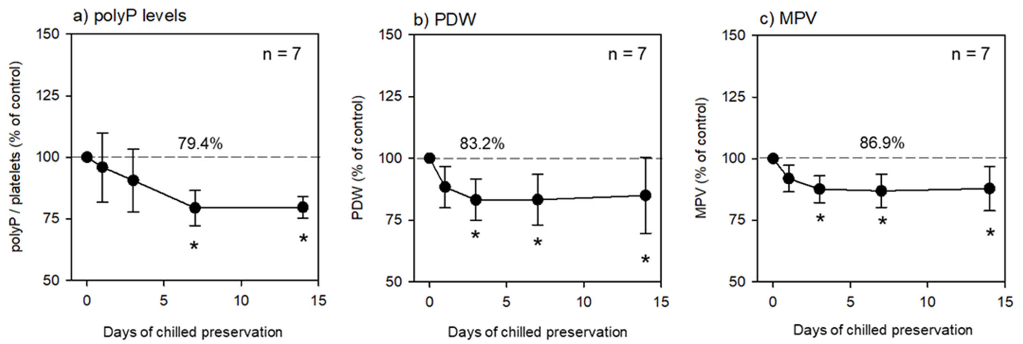
3.4. Preservation at 4 °C
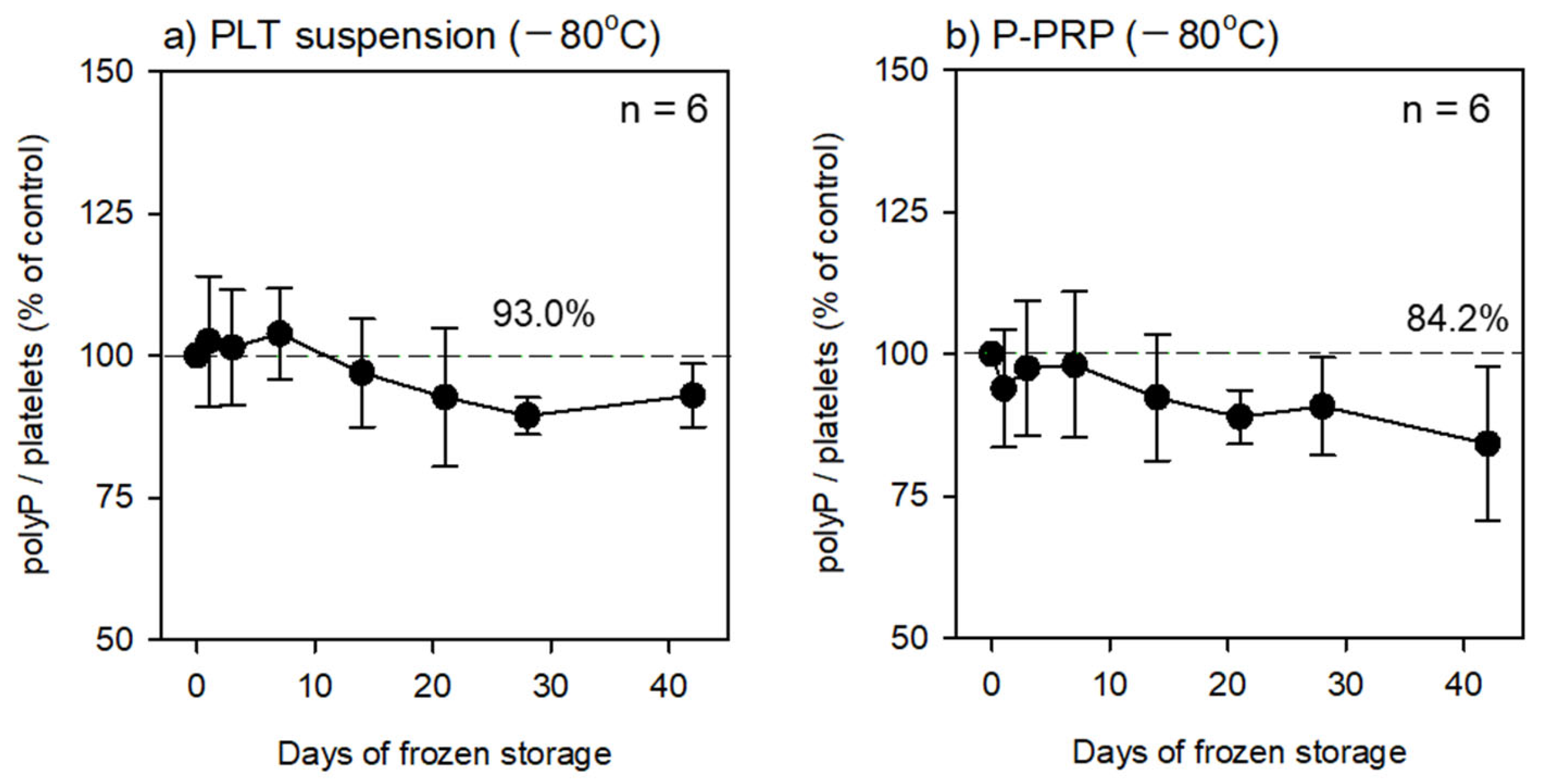
3.5. Preservation at −80 °C
3.6. Statistical Analysis
4. Expected Results and Discussion
Author Contributions
Funding
Institutional Review Board Statement
Informed Consent Statement
Data Availability Statement
Conflicts of Interest
References
- Kus, F.; Smolenski, R.T.; Tomczyk, M. Inorganic Polyphosphate-Regulator of Cellular Metabolism in Homeostasis and Disease. Biomedicines 2022, 10, 913. [Google Scholar] [CrossRef] [PubMed]
- Omelon, S.; Georgiou, J.; Habraken, W. A cautionary (spectral) tail: Red-shifted fluorescence by DAPI-DAPI interactions. Biochem. Soc. Trans. 2016, 44, 46–49. [Google Scholar] [CrossRef] [PubMed]
- Aschar-Sobbi, R.; Abramov, A.Y.; Diao, C.; Kargacin, M.E.; Kargacin, G.J.; French, R.J.; Pavlov, E. High sensitivity, quantitative measurements of polyphosphate using a new DAPI-based approach. J. Fluoresc. 2008, 18, 859–866. [Google Scholar] [CrossRef] [PubMed]
- Martin, R.M.; Leonhardt, H.; Cardoso, M.C. DNA labeling in living cells. Cytometry Part A 2005, 67, 45–52. [Google Scholar] [CrossRef] [PubMed]
- Tanious, F.A.; Veal, J.M.; Buczak, H.; Ratmeyer, L.S.; Wilson, W.D. DAPI (4′,6-diamidino-2-phenylindole) binds differently to DNA and RNA: Minor-groove binding at AT sites and intercalation at AU sites. Biochemistry 1992, 31, 3103–3112. [Google Scholar] [CrossRef] [PubMed]
- Grossgebauer, K.; Küpper, D. Interactions between DNA-binding fluorochromes and mucopolysaccharides in agarose-diffusion test. Klin Wochenschr. 1981, 59, 1065–1066. [Google Scholar] [CrossRef] [PubMed]
- Watanabe, T.; Kitamura, Y.; Aizawa, H.; Masuki, H.; Tsujino, T.; Sato, A.; Kawabata, H.; Isobe, K.; Nakata, K.; Kawase, T. Fluorometric Quantification of Human Platelet Polyphosphate Using 4’,6-Diamidine-2-phenylindole Dihydrochloride: Applications in the Japanese Population. Int. J. Mol. Sci. 2021, 22, 7257. [Google Scholar] [CrossRef] [PubMed]
- Ushiki, T.; Mochizuki, T.; Suzuki, K.; Kamimura, M.; Ishiguro, H.; Suwabe, T.; Kawase, T. Modulation of ATP Production Influences Inorganic Polyphosphate Levels in Non-Athletes’ Platelets at the Resting State. Int. J. Mol. Sci. 2022, 23, 11293. [Google Scholar] [PubMed]
- Ushiki, T.; Mochizuki, T.; Suzuki, K.; Kamimura, M.; Ishiguro, H.; Watanabe, S.; Omori, G.; Yamamoto, N.; Kawase, T. Platelet polyphosphate and energy metabolism in professional male athletes (soccer players): A cross-sectional pilot study. Physiol. Rep. 2022, 10, e15409. [Google Scholar] [CrossRef] [PubMed]
- Sato, A.; Aizawa, H.; Tsujino, T.; Isobe, K.; Watanabe, T.; Kitamura, Y.; Kawase, T. Fluorescent Cytochemical Detection of Polyphosphates Associated with Human Platelets. Int. J. Mol. Sci. 2021, 22, 1040. [Google Scholar] [CrossRef] [PubMed]
- Bru, S.; Jiménez, J.; Canadell, D.; Ariño, J.; Clotet, J. Improvement of biochemical methods of polyP quantification. Microb. Cell 2016, 4, 6–15. [Google Scholar] [CrossRef] [PubMed]


| Fixation (1 d, 4 °C) | Suspension | Replacement | Preservation Temperature (1 d–6 w) | To be Tested in This Study |
|---|---|---|---|---|
| no | Plasma | no | 4 °C | Not tested * |
| no | PBS | no | 4 °C | Not applicable ** |
| no | Plasma | no | −80 °C | Not applicable |
| no | PBS | no | −80 °C | Not applicable |
| ThromboFix | PBS | no | 4 °C | As control |
| ThromboFix | PBS | pure water | 4 °C | Not tested |
| ThromboFix | PBS | no | −80 °C | Not applicable |
| ThromboFix | PBS | pure water | −80 °C | As a candidate |
| ThromboFix | Plasma | no | 4 °C | Not tested |
| ThromboFix | Plasma | pure water | 4 °C | Not tested |
| ThromboFix | Plasma | no | −80 °C | Not applicable |
| ThromboFix | Plasma | pure water | −80 °C | As a candidate |
Disclaimer/Publisher’s Note: The statements, opinions and data contained in all publications are solely those of the individual author(s) and contributor(s) and not of MDPI and/or the editor(s). MDPI and/or the editor(s) disclaim responsibility for any injury to people or property resulting from any ideas, methods, instructions or products referred to in the content. |
© 2023 by the authors. Licensee MDPI, Basel, Switzerland. This article is an open access article distributed under the terms and conditions of the Creative Commons Attribution (CC BY) license (https://creativecommons.org/licenses/by/4.0/).
Share and Cite
Kawase, T.; Suzuki, K.; Kamimura, M.; Mochizuki, T.; Ushiki, T. Optimized Protocol for Preservation of Human Platelet Samples for Fluorometric Polyphosphate Quantification. Methods Protoc. 2023, 6, 59. https://doi.org/10.3390/mps6040059
Kawase T, Suzuki K, Kamimura M, Mochizuki T, Ushiki T. Optimized Protocol for Preservation of Human Platelet Samples for Fluorometric Polyphosphate Quantification. Methods and Protocols. 2023; 6(4):59. https://doi.org/10.3390/mps6040059
Chicago/Turabian StyleKawase, Tomoyuki, Katsuya Suzuki, Masami Kamimura, Tomoharu Mochizuki, and Takashi Ushiki. 2023. "Optimized Protocol for Preservation of Human Platelet Samples for Fluorometric Polyphosphate Quantification" Methods and Protocols 6, no. 4: 59. https://doi.org/10.3390/mps6040059
APA StyleKawase, T., Suzuki, K., Kamimura, M., Mochizuki, T., & Ushiki, T. (2023). Optimized Protocol for Preservation of Human Platelet Samples for Fluorometric Polyphosphate Quantification. Methods and Protocols, 6(4), 59. https://doi.org/10.3390/mps6040059