Introduction
Fournier’s gangrene (FG) is a rare but very serious condition characterized by rapidly evolving necrotizing fasciitis of the genital region, which carries a high mortality rate if not diagnosed and treated correctly. The treatment is mainly surgical with aggressive debridement of necrotic tissue and abscess drainage but also combined with antibiotic therapy and life support [
1]. Male immunocompromised, diabetic, obese, and cancer patients, aged 50 to 70 years old, are the usual suspects, as they are prone to these types of infections [
2,
3].
Mortality rates are reported to reach 22.8% with 68.7% in the first 10 days [
4,
5]. Other studies report mortality rates of 4.7% [
6] with the rate probably related to the time of patient presentation and the hospital’s capabilities of treating these patients.
The most common etiologies for FG are anorectal and genitourinary infections, but also trauma with perineal and genital skin injuries. Most often it is a polymicrobic infection caused by germs specific to the anogenital region [
1,
7].
The infection follows the genital and perineal fascial system and therefore it can quickly extend to the anterior abdomen or along the rectum into the presacral space, the retrovesical space, and the pelvirectal tissue. This can involve the retroperitoneal space to the level of the upper abdomen, and in rare cases, even to the paravertebral region up to the neck [
6,
7].
Case presentation
We present an extreme case of advanced Fournier’s Gangrene with an initially undetermined point of origin, in a 64-year-old male patient with no prior medical history.
He came to the ER in August 2018 presenting with fever, malaise, acute urinary retention, and almost complete necrosis of the perianal area, lower left buttock, and scrotum. Bloodwork showed severe sepsis (WBC-28300/mcL with left shift, presepsine–2130/pg/mL), acidosis and incipient MODS (slightly elevated urea, creatinine, INR, aPTT, total bilirubin, liver enzymes, blood sugar); BP was 100/68 mmHg, pulse 120/min with a pulse oximetry of 97%.
Emergency CT scan showed a large perineal abscess with gas with extension/origin to both ischiorectal fossae, pararectal, paravesical, and prevesical spaces. Large portions of the fatty tissue in the left buttock, the properitoneal fat of the entire inferior abdominal wall, and the external genital organs showed signs of necrosis.
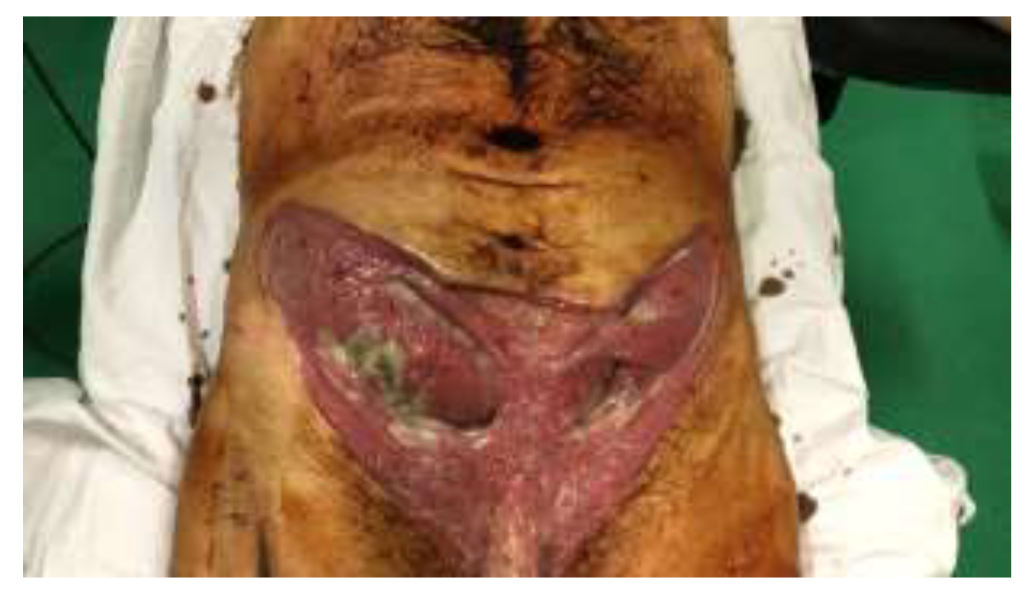
The patient was immediately taken to the operating theatre where he underwent emergency surgery by a mixed team of urologic and plastic surgeons, consisting of abscess drainage, wide necrectomy (from the perianal area up to the umbilicus), right orchiectomy, and suprapubic urinary drainage. During surgery, the point of origin was established as being the left ischiorectal fossa. A small portion of the peritoneum at the level of the right lower quadrant was borderline necrotic; the external oblique, internal oblique, and transversus abdominis muscles bilaterally, below the level of the umbilicus, were partially necrotic and had to be excised (
Figure 1). An incision was made superior to the umbilicus (for demarcation) and on both sides of the anus. Large, multi-perforated drain tubes were placed bilaterally running down from the supraumbilical incision, underneath the rectus abdominis to the paravesical spaces, which were completely necrotic, and then through the pelvis, through the pararectal spaces, and out through the perianal incisions. Extensive lavage with isotonic saline and hydrogen peroxide was performed at the end. The wounds were bandaged with large dressings soaked in isotonic saline.
Postoperatively, the patient received wide-spectrum antibiotics (Meropenem, Metronidazol, Gentamicin, and Linezolid), pain management medication, LMWH for DVT prophylaxis, and fluid replacement therapy. His dressings were changed twice daily in the OR and subsequent additional debridement was performed. On the 3rd day postop, the patient was taken out of the ICU and we started to apply Sulfadiazine 1% cream on the wounds. In the fifth week he was transferred to the plastic surgery ward for defect reconstruction.
The abdominal wall defect was reconstructed using two large pedicled tensor fascia lata (TFL) musculocutaneous flaps which were harvested with extra fascia to give support to the intraabdominal contents. The pubis and penis were covered using split thickness skin grafts and the perineal defects were left to heal by secondary intention (
Figure 2 and
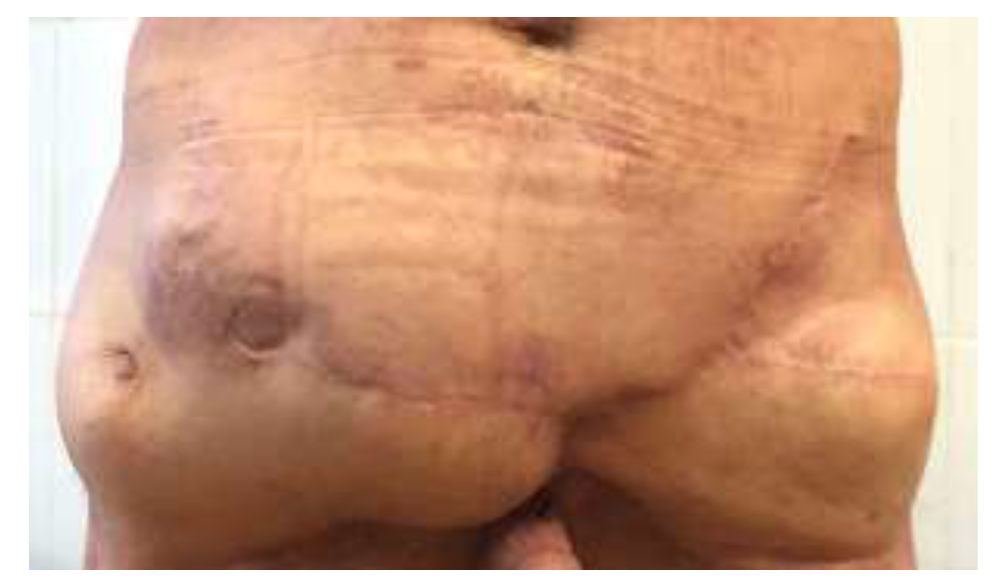
Figure 3). After 4 days, the wounds became infected with Pseudomonas aeruginosa which led to graft lysis and marginal wound breakdown. We treated the problem with daily wound dressings and the wounds eventually healed by granulation. The patient was discharged after 12 weeks. A year after, the patient developed an inferior right quadrant abdominal hernia with skin ulceration. He was once again operated with hernia reduction and the abdominal wall was reinforced with a large mesh.
Discussions
This was an extreme case of Fournier’s gangrene that we managed to treat successfully. The particularities of this case were that the patient did not have any comorbidities and was in good apparent health and that the infection progressed rapidly throughout the deeper planes involving the abdominal wall muscles besides the fascial layers. This may be explained by initial or rapid involvement of the superficial and deep spermatic fasciae alongside Buck’s fascia, thus permitting the infection to extend into the abdomen and the testicles.
Only one microorganism (Klebsiella spp.) was isolated from the purulent discharge that was obtained intraoperatively. This aspect differentiates this case form the majority of FG’s in which a polymicrobial infection consisting of aerobe and anaerobe bacteria is detected 54%-80% [
3,
8,
9]. Furthermore, this includes our case in the third category of necrotic fasciitis (NF) from the microbiological perspective [
10], with a prevalence of under 5% and presenting a rapid progression rate, leading to MSOF and mortality if not addressed quickly [
6,
7].
Another particularity of the case was that the patient did not present any of the comorbidities frequently associated with the FG, such as diabetes mellitus, obesity, immunosuppression, or chronic alcoholism [
2,
6,
7,
8,
9].
The cosmetic results were not very good, mostly because of the postoperative Pseudomonas infection that caused the failure of the skin grafts and the necessity to heal the wounds by secondary intention, thus leading to wound contracture and fibrosis in the pubic area and penis. However, the patient did not regard this as a complication and agreed to further corrections (
Figure 4).
The TFL flaps provided good coverage with good vascularity to the abdomen, but they did not provide the sufficient support to the abdominal contents, thus leading to an incisional hernia that needed surgical correction and abdominal wall reinforcement with surgical mesh. We also managed to keep the operation time to a minimum (total operative time – 3.5 hours) by using this flap, that needed not so tedious dissection as maybe an ALT, thus limiting the anaesthesia time for a patient that had already undergone 5 surgeries with general anaesthesia in the previous 5 weeks.
Conclusions
Fournier’s gangrene is a life-threatening condition that evolves rapidly and poses a challenge to the physician that must tackle it. Its high mortality and morbidity rates necessitate rapid diagnosis and decision making. Its management remains mainly surgical, despite the advances in antibiotic treatment. It needs a team approach consisting of different surgical specialties (urology, general surgery, plastic surgery) and ICU. If the patient manages to get through the initial debridement phase, then the plastic surgeon is often faced with a reconstructive challenge, to address all the body regions that may be involved. The reconstructive process must address not only the physical aspects of the surgery (covering the defect) but also the psychological aspects in a patient traumatized not only by an extremely aggressive disease but also by multiple surgeries, long bedrest and painful recovery.