Next Article in Journal
Unexpected Hope for a Multiple Myeloma Patient
Previous Article in Journal
Paroxysmal Nocturnal Hemoglobinuria: Pandora’s Box?
 
 
Journal of Mind and Medical Sciences is published by MDPI from Volume 12 Issue 1 (2025). Previous articles were published by another publisher in Open Access under a CC-BY (or CC-BY-NC-ND) licence, and they are hosted by MDPI on mdpi.com as a courtesy and upon agreement with Valparaiso University (ValpoScholar).
Case Report

Immunohistochemical Pattern—A Prognostic Factor for Synchronous Gastrointestinal Cancer

by
Catalin Alius
1,
Catalin Gabriel Cirstoveanu
2,3,
Cristinel Dumitru Badiu
2,4,*,
Valeriu Ardeleanu
5,6,7 and
Vasile Adrian Dumitru
8
1
IVth Department of Surgery, University Emergency Hospital Bucharest, Bucharest, Romania
2
Carol Davila University of Medicine and Pharmacy, Bucharest, Romania
3
Anesthesia and Intensive Neonatal Care Unit, Marie Curie Emergency Clinical Hospital, Bucharest, Romania
4
Bagdasar Arseni Emergency Clinical Hospital, 031298 Bucharest, Romania
5
Faculty of Medicine, Ovidius University, Constanta, Romania
6
Dunarea de Jos University, 47 Domneasca Street, Galati, Romania
7
General Hospital CFR, 4-6 Moruzzi Street, Galati, Romania
8
Department of Pathologic Anatomy, University Emergency Hospital Bucharest, Bucharest, Romania
*
Author to whom correspondence should be addressed.
J. Mind Med. Sci. 2020, 7(2), 250-256; https://doi.org/10.22543/7674.72.P250256
Submission received: 1 June 2020 / Revised: 25 August 2020 / Accepted: 4 September 2020 / Published: 29 September 2020

Abstract

:
Recent advancements in medical genetics and molecular biology are reflected in the modern understanding and approach to colorectal carcinoma (CRC). Understanding the cellular mechanisms and mutational patterns that promote carcinogenesis could enhance the predictive accuracy of the TNM classification. Furthermore, this will allow for a much more documented stratification and tailored oncological treatment. This paper presents an illustrative case of a relatively young patient (50 years old) with no family history of cancer who was diagnosed with four synchronous gastrointestinal (GI) adenocarcinomas displaying a wild type P53, negative BRAF testing, and mutated MLH1 and PMS2 proteins. This case report contributes to the relevant literature with a concise review of the role of micro-satellite instability (MSI), chromosomal instability (CIN), and CpG island methylator phenotype (CIMP) in carcinogenesis, hereditary and sporadic gastrointestinal cancers, a discussion over the importance of molecular sub-typing in predicting long term outcomes and choosing the most suitable adjuvant treatment regimen.

Introduction

Recent advancements in medical genetics and molecular biology are reflected in the modern understanding and approach to colorectal carcinoma (CRC). Although the intricacies of the subtle mechanisms of molecular carcinogenes are not yet fully clarified, emerging research suggests that similar to breast cancer, stratification of the patients based on molecular characteristics or subtypes of CRC could influence survival, response to treatment, and loco-regional recurrences.
Nowadays it is unanimously agreed that colonic carcinogenesis is multifactorial and multi-phased. Based on proven causal links between etiologic factors, genetic and epigenetic mutations, and prognostic factors, we believe that representative cases of multiple primary familial or hereditary CRC ought to be popularized in order to reiterate the importance of screening and genetic counseling in one of the most preventable malignancies of our times.
This paper presents an illustrative case of a relatively young patient (50 years old) with no family history of cancer who was diagnosed with four synchronous gastrointestinal (GI) adenocarcinomas displaying multiple mutational patterns. This case report contributes to the relevant literature with a concise review of the role of micro-satellite instability (MSI), chromosomal instability (CIN), and CpG island methylator phenotype (CIMP) in carcinogenesis, hereditary and sporadic gastrointestinal cancers, and a discussion of the importance of molecular sub-typing in predicting long term outcomes and choosing the most suitable adjuvant treatment regimen.

Case presentation

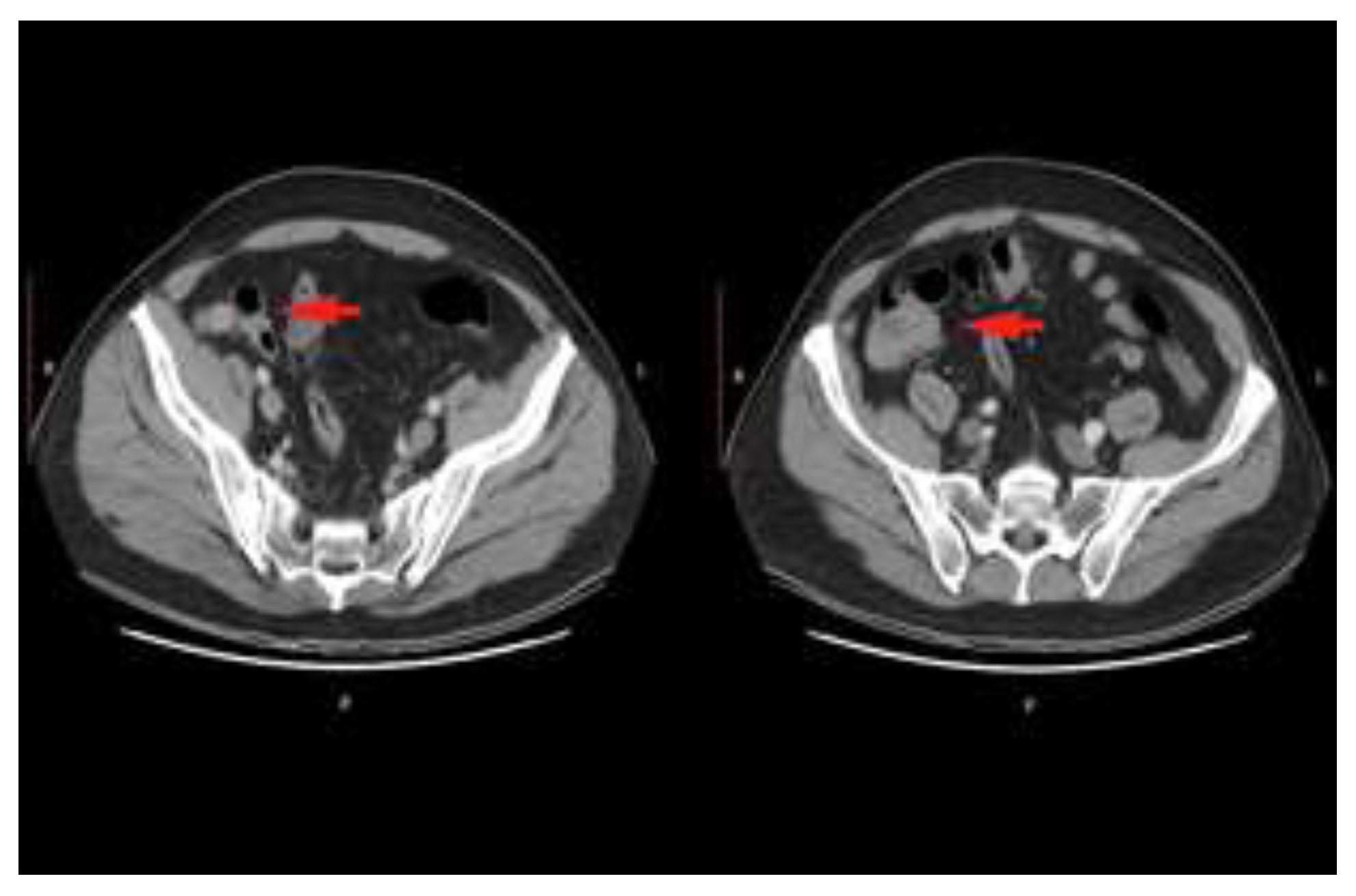
A 50 years old patient with no previous personal or family medical history and no associated comorbidities presented with recurrent left flank abdominal discomfort to his general practice doctor who, after an unremarkable clinical examination, recommended a renal ultrasound (USS), a complete blood count (CBC), and a urine sample. No abnormality was detected except for an incidental renal cyst of 25/14mm on the right side. Despite these, having a high index of suspicion of a kidney condition, the GP ordered a CT scan for further characterization of the renal tract. The CT exam unexpectedly revealed circumferential thickening of the splenic flexure extending over 9 centimeters in length, with intense iodophilia, adjacent fat stranding, and supracentrimetric perilesional lymph nodes (Figure 1). An additional endoluminal area in the caecum was described, but with minimal contrast enhancement and nonspecific features (Figure 2). No lesions suggestive of liver or bony metastases were reported.
The patient was admitted to our institution with the presumptive diagnosis of colorectal cancer—possible synchronous cecal and splenic flexure tumors—for further investigation and treatment. The clinical exam did not reveal significant pathological features, the main subjective sign was transient left hypocondrum and left flank pain of moderate intensity, and intestinal transit disturbances, with predominant constipation. The laboratory tests were normal, except mild anemia (Hb 12mg/dl). For completion of the preoperative evaluation, a thoracic CT scan (no pulmonary abnormality detected) and a colonoscopy were performed. The endoscopic examination revealed a small polypoid lesion in the sigmoid colon and a circumferential tumor at the splenic flexure which could not be negotiated. Biopsies showed moderately differentiated adenocarcinoma (G2) NOS type for both lesions.
After informed consent was signed, the patient underwent surgery with a planned exploratory laparotomy +/- proceed and repeat on table colonoscopy for a full assessment of the bowel. During the macroscopic intra-abdominal assessment, a supracentrimetric lesion was identified on the greater curvature of the stomach (antral area), with no connection to the splenic flexure mass.
After a second unsuccessful endoscopic attempt to pass above the splenic flexure lesion, a minimal colotomy was performed proximal to the tumor and a purse-string tie was placed to ensure air-tightness around the colonoscope. Completion of the colonoscopy revealed a caecal mass from which frozen sections biopsies confirmed adenocarcinoma. At this stage, we have identified three synchronous colonic adenocarcinomas and a suspicious gastric lesion for which endoscopic frozen sections confirmed moderately differentiated adenocarcinoma. Total colectomy with complete mesocolic excision (CME) and D1 gastrectomy were performed. The postoperative course was uneventful, with discharge after a week.
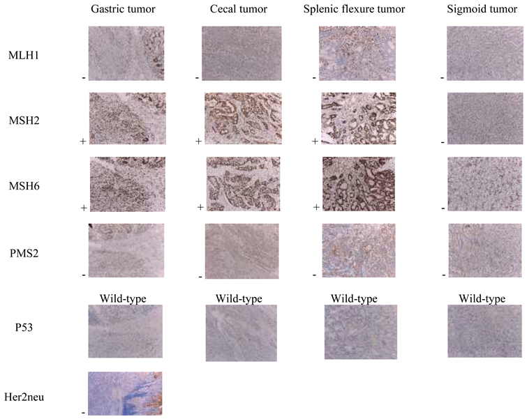
The ensuing commented images show the histopathological features of the tumors. An additional panel of surrogate gene assessment using IHC demonstrates a wild type of p53, the absence of MLH1 and PMS2 proteins, suggesting either a germ line mutation or inhibition by epigenetic modification (CIMP). Since CIMP mutations are associated with BRAF mutations, we believe that a negative BRAF testing is highly indicative of a possible Lynch syndrome. A positive diagnosis of non-polyposis hereditary colon cancer (HNPCC) means a 50% risk for the direct descendants to develop the disease since it is transmitted in an autosomal dominant manner.
All 4 tumors are solid-type proliferations, with weak-moderate differentiation, with ulcerated surface, and showing important areas of necrosis and intra-tumoral hemorrhage, as well as a rich inflammatory infiltrate formed by polymorphonuclear, lymphocytes and large histiocytes, which partially shield tumor proliferation at the level of the invasion front. The lymph nodes analyzed do not show tumor invasion, but show non-specific chronic inflammatory features.
Other histopathological findings were adenomatous colonic polyp with low grade intraepithelial dysplasia and chronic non-atrophic gastritis with moderate inflammatory activity and intestinal focal metaplasia.
Histopathological characteristics of the 4 synchronous tumors, in a young patient 50 years old, require the continuation of investigations by immunohistochemical testing to complete the diagnosis and establish a possible syndromic classification, such as Lynch Syndrome.
Figure 3. (A,B). Histopathological aspects of the 4 synchronous tumors (HE, x10). (C,D). Histopathological aspects of the 4 synchronous tumors (HE, x10). (A): moderately differentiated tubular gastric adenocarcinoma (G2), intestinal type (Lauren classification), stage IIB (pT4N0Mx), with cell proliferation with medium and large cells, with moderate cyto-nuclear atypia, which infiltrates the entire gastric wall passing the serosa. Tumor cells show atypical mitotic figures, some being multipolar, with severe dyskaryosis. (B): moderately differentiated colonic (cecal) adenocarcinoma (G2), stage IIA (pT3NoMx): large, atypical cells, with moderate cyto-nuclear pleomorphism that form numerous irregular glandular structures, of various sizes, with infiltrative and destructive character on adjacent structures, without invading the serosa. Bd1 tumor budding; absence of perineural invasion or angio-lymphatic tumor emboli. (C): poorly differentiated colonic adenocarcinoma (splenic flexure) (G3), stage IIA (pT3pN0Mx): numerous cellular atypia, cyto-nuclear pleomorphism, rare small glandular and pseudo-glandular structures with irregular appearance, typical and atypical mitotic figures, without serous infiltration, with Bd3 tumor budding and perineural invasion elements and lympho-vascular emboli. (D): colonic adenocarcinoma (sigmoid), moderately differentiated G2, stage IIB (pT3pN0Mx), with mucinous differentiation (in 15% of cases), with extracellular mucin lakes with basophilic tint containing glandular fragments and groups of tumor cells, Bd2 tumor budding and perineuronal invasion and vascular-lymphatic emboli.
Jmms 07 00039 g003
The immunohistochemical expression pattern performed for the 4 synchronous tumors completes and confirms the initial diagnosis and suggests high microsatellite instability in the MLH1 / PMS2 gene for all tumors and MSH2 / MSH6 for the sigmoid one. The wild-type expression of p53 is a favorable prognostic element, as is the absence of Her2neu expression in the gastric tumor (Table 1).
BRAF V600 mutation was further tested regarding the differentiation of sporadic colorectal cancer with high microsatellite instability (MSI-H) from hereditary non-polyposis colorectal carcinoma (HNPCC) or Lynch syndrome and the result was negative, suggesting a mutational pattern rather than inhibition by hypermethylation of MLH1.

Discussions

Colorectal cancer is a major cause of morbidity and mortality in the world and in Romania. Synchronous malignancies were first described by Billroth in 1889. The diagnostic criteria were developed in 1932 by Warren and Gates and set out the following conditions: i) the tumors must have histopathological confirmation of malignancy; ii) they must be anatomically distinct (be separated from each other by at least four cm of healthy tissue); iii) one tumor must not be the metastasis of the other; iv) they must be diagnosed simultaneously or at a maximum interval of one year from each other. [1,2,3,4] The incidence of synchronous tumors varies, in different studies, between 1.8 and 12.4%. [4] The frequency with which adenomatous polyps are associated with colorectal cancer is 15-50%. Known risk factors for synchronous tumors are male sex, family history, age, and the presence of synchronous adenomas. [2,3] Predisposing conditions are familial adenomatous polyposis, inflammatory bowel disease, and hereditary non-polyposis cancer (Lynch syndrome) [4,5,6,7].
Preoperative evaluation of the entire colon in patients with colorectal cancer is important, but not always possible. Because of this, proximal lesions synchronous with the initial tumor may be missed. This can change the intraoperative treatment plan or even cause a reoperation. The undetected lesions (polyps or cancers) continue their evolution and, at the time of subsequent diagnosis, they appear in an advanced stage or even surgically outdated. Synchronous colorectal cancers have rarely been studied in large cohorts; however, the analysis of synchronous tumors can provide special, unique data on the pathogenesis of colon and rectal cancer [4,7].

Molecular events associated with colon carcinogenesis

During malignancy, the colonic epithelium undergoes a series of genetic changes that are reflected in histological changes. Genetic mutations occur long before clinically obvious lesions. In many cancers, the cells acquire characteristics that favor malignant transformation, but do not produce visible morphological changes, creating only a predisposition for cancer. [8] The modified cells form areas of mucosa prone to malignancy; the process is called "field cancerization" and can lead to synchronous tumors. There are three main recognized carcinogenic mechanisms: chromosomal instability (CIN), micro-satellite instability (MI), involved with more than 95% of HNPCC’s, and aberrant DNA methylation (CIMP).
Most malignancies (70-75%) occur as a result of events that cause chromosomal instability (CIN) and progress to the suppressive pathway. The other colon cancers are caused by hypermethylation of the genes (the methylating phenotype of the CpG-CIMP islands) and evolve in the serum pathway; some of them have microsatellite instability (MSI), and the development of the latter takes place on the mutant path. One of the earliest changes in both oncogenesis pathways (MSI and CIN) is the loss of APC gene function, which predisposes people with germ-line mutations in APC to develop colon cancer [6,7,8,9].
Approximately 15% of colorectal adenocarcinomas are associated with MSI: 2.5% coming from a genetic inheritance and 12.5% being sporadic. [10]

Mismatch repair (MMR) genes and Microsatellite instability (MSI)

Microsatellite instability (MSI) is due to altered expression of MMR genes (mismatch repair), involved in the processes of recognition and repair of microsatellite errors during DNA synthesis to maintain genomic stability. There are 4 MMR genes and their encoded proteins, which form heterodimers 2 by 2: MLH1 / PMS2 and MSH2 / MSH6. Inactivation of MLH1 and MSH2 account for over 90% of deficient MMR cases. If any of the four major proteins (MSH2, MLH1, MSH6, or PMS2) is functionally inactive, mismatches are not repaired. A defective DNA MMR system increases the mutation rate and makes the cell vulnerable to mutations in genes controlling cell growth (including tumor suppressor genes and oncogenes), resulting in an elevated cancer risk. [11,12]
Immunohistochemical analysis revealed that when MLH1 or MSH2 is nonfunctional, their corresponding peers will also appear negative, as they disintegrate without their obligatory partners. In contrast, inactivation of MSH6 or PMS2 does not lead to absence of MSH2, respectively MLH1, as these proteins can dimerize also with other proteins, for example MLH3 [11].
Sporadic MSI is caused more frequently by epigenetic silencing of the MLH1 promoter by methylation, which is associated with a somatic BRAF V600E mutation. Hereditary MSI commonly happens due to autosomal dominant mutations in MMR. Lynch syndrome is associated in 90% of cases with MLH1 or MSH2 mutations [13].
In the presented case, the immunohistochemical pattern associated with the absence of the BRAF V600 mutation is suggestive of the MLH1 mutation in all 4 tumors, and, in addition, of MSH2 in the sigmoid tumor.
Testing the mismatch repair genes (MMR) profile by immunohistochemistry is particularly important for understanding the mechanism of carcinogenesis, planning cancer treatment, and evaluating the prognosis in synchronous digestive cancers. [14] The criteria for recommending testing for MSI are, according to the Dutch guideline for MSI testing (http://www.oncoline.nl):
  • CRC or endometrial carcinoma before the age of 50 years.
  • A second CRC before the age of 70 years.
  • CRC before the age of 70 years AND another synchronous or previous Lynch Syndrome associated tumor.

Clinical and prognostic features of MSI-H colorectal tumors

Cancers associated with MSI-H have special characteristics from a histopathological point of view; they are generally adenocarcinomas with medium-weak differentiation, with possible mucinous component and important associated inflammatory lymphocytic infiltrate. They are predominantly located on the right side of the colon [15,16].
Patients with MSI-H tumors have been associated with a more favorable prognosis compared to patients with MSI-L or MSS tumors, a significantly lower likelihood of metastasizing to regional lymph and distant organs [17,18]. Given our current level of understanding, this favorable prognosis could not be fully explained only by molecular mechanisms, but studies correlate better survival with the presence of lymphocytic infiltration seen in MSI-H tumors, an expression of an enhanced host immune response invoked by the presence of numerous mutated products [19,20,21,22].
Others have also suggested that the enormous mutational burden resulting from loss of MMR activity may be self-limiting in that essential cell functions may be hindered [16,21,22,23]. Regarding the response to 5-FU, it is considered that in stage II chemotherapy is not necessary for MSI-H tumors. The favorable prognosis and the evidence of lack of benefit from 5-FU based adjuvant chemotherapy in stage II CRC patients with deficient MMR indicate that these patients should not receive adjuvant chemotherapy. In stage III CRC, however, patients with deficient MMR cancers treated with adjuvant 5-FU had better outcomes. In stage III colon cancer patients, oxaliplatin combined with 5-FU is the current standard of care for adjuvant chemotherapy. In contrast to 5-FU, sensitivity to oxaliplatin was independent of the MMR system in CRC cell lines [24,25,26]. The absence of BRAF mutation expression is a favorable prognostic factor [27,28].
The particularity of the case resides in the absence of any family history of CRC and the lack of digestive symptoms. These led to the accidental detection of the colon tumors by CT examination, followed by subsequent exploration, but without paying preoperatory attention to upper digestive tract [29]. However, rare “de novo” cases of Lynch syndrome have been reported and prompted early familial screening potentially preventing CRC associated morbidity.
Incomplete possibilities of preoperative assessment, due to the stenotic behavior of splenic flexure tumor, led to intraoperative change of the surgical plan. Although bulky, tumors had histopathological and immunohistochemical features which were statistically associated with favorable prognosis. Regular follow-up in the surgical department is necessary for screening of recurrent tumors or metachronous cancers. [30,31,32].

Highlights

Careful pre- and intraoperative evaluation of patients with suspected synchronous cancers is particularly important in therapeutic planning.
MSI represents a promising disease marker for colorectal cancers because of the favorable prognosis associated with MSI-H and particularities of oncological management.

Conclusions

Careful pre- and intraoperative evaluation of patients with suspected synchronous cancers is of paramount importance in therapeutic planning. Additionally, employment of IHC testing for surrogate gene mutations identification represents a promising complementary tool for improvement of prognostic assessment and tailored oncological treatment.
Although more than 75% of colorectal cancers are sporadic, identification of familial and hereditary CRC could increase the efficacy of screening programs and reduce the economic pressure exerted on health systems worldwide. The perspective of developing new treatment options and optimized guidelines for CRC can become a reality only by incorporating gene assessment and ultra-stratification of patients.

Compliance with ethical standards

Any aspect of the work covered in this manuscript has been conducted with the ethical approval of all relevant bodies and that such approvals are acknowledged within the manuscript.

Conflict of Interest

There are no known conflicts of interest in the publication of this article. The manuscript was read and approved by all authors.

References

  1. Bittorf, B.; Kessler, H.; Merkel, S.; Brückl, W.; Wein, A.; Ballhausen, W.G.; Hohenberger, W.; Günther, K. Multiple primary malignancies: An epidemiological and pedigree analysis of 57 patients with at least three tumours. Eur J Surg Oncol 2001, 27, 302–313. [Google Scholar] [CrossRef] [PubMed]
  2. Suceveanu, A.I.; Mazilu, L.; Nitipir, C.; et al. Diabetes Mellitus raise the Risk for Interval Colorectal Cancer and Advanced Colorectal Adenomas. Revista Chimie 2019, 70, 1808–1811. [Google Scholar] [CrossRef]
  3. Nitipir, C.; Barbu, M.A.; Orlov, C.; et al. Type II Diabetes Mellitus—Associated Risk Factor in the Onset and Evolution of Digestive Tract Carcinoma. Romanian Biotechnological Letters 2019, 24, 140–146. [Google Scholar] [CrossRef]
  4. Şavlovschi, C.; Comandaşu, M.; Şerban, D. Specifics of diagnosis and treatment in synchronous colorectal cancers (SCC). Chirurgia 2013, 108, 43–45. [Google Scholar] [PubMed]
  5. Lam, A.K.; Chan, S.S.; Leung, M. Synchronous colorectal cancer: Clinical, pathological and molecular implications. World J Gastroenterol 2014, 20, 6815–6820. [Google Scholar] [CrossRef] [PubMed]
  6. Lee, B.C.; Yu, C.S.; Kim, J.; et al. Clinicopathological features and surgical options for synchronous colorectal cancer. Medicine 2017, 96, e6224. [Google Scholar] [CrossRef]
  7. Kato, T.; Alonso, S.; Muto, Y.; et al. Clinical characteristics of synchronous colorectal cancers in Japan. World J Surg Oncol 2016, 14, 272. [Google Scholar] [CrossRef]
  8. Mazilu, L.; Suceveanu, A.I.; Tomescu, D.; et al. Optimizing the indication for breast-conservative surgery (BCS) in patients with locally-advanced breast cancer. Chirurgia 2013, 108, 478–481. [Google Scholar] [PubMed]
  9. Savlovschi, C.; Serban, D.; Trotea, T.; Borcan, R.; Dumitrescu, D. Post-surgery morbidity and mortality in colorectal cancer in elderly subjects. Chirurgia 2013, 108, 177–179. [Google Scholar]
  10. Nguyen, H.T.; Duong, H.Q. The molecular characteristics of colorectal cancer: Implications for diagnosis and therapy. Oncol Lett. 2018, 16, 9–18. [Google Scholar] [CrossRef]
  11. van Lier, M.G.; Wagner, A.; van Leerdam, M.E.; et al. A review on the molecular diagnostics of Lynch syndrome: A central role for the pathology laboratory. J Cell Mol Med. 2010, 14, 181–197. [Google Scholar] [CrossRef] [PubMed]
  12. Lanza, G.; Gafà, R.; Maestri, I.; Santini, A.; Matteuzzi, M.; Cavazzini, L. Immunohistochemical pattern of MLH1/MSH2 expression is related to clinical and pathological features in colorectal adenocarcinomas with microsatellite instability. Mod Pathol 2002, 15, 741–749. [Google Scholar] [CrossRef] [PubMed]
  13. Dudley, J.C.; Lin, M.T.; Le, D.T.; Eshleman, J.R. Microsatellite Instability as a Biomarker for PD-1 Blockade. Clin Cancer Res. 2016, 22, 813–820. [Google Scholar] [CrossRef] [PubMed]
  14. Lynch, H.T.; Lynch, P.M.; Lanspa, S.J.; Snyder, C.L.; Lynch, J.F.; Boland, C.R. Review of the Lynch syndrome: History, molecular genetics, screening, differential diagnosis, and medicolegal ramifications. Clin Genet 2009, 76, 1–18. [Google Scholar] [CrossRef]
  15. Boland, C.R.; Goel, A. Microsatellite instability in colorectal cancer. Gastroenterology 2010, 138, 2073–2087.e3. [Google Scholar] [CrossRef]
  16. Lanza, G.; Gafà, R.; Maestri, I.; Santini, A.; Matteuzzi, M.; Cavazzini, L. Immunohistochemical pattern of MLH1/MSH2 expression is related to clinical and pathological features in colorectal adenocarcinomas with microsatellite instability. Mod Pathol 2002, 15, 741–749. [Google Scholar] [CrossRef]
  17. Jo, W.S.; Carethers, J.M. Chemotherapeutic implications in microsatellite unstable colorectal cancer. Cancer Biomark. 2006, 2, 51–60. [Google Scholar] [CrossRef]
  18. Stanciu, A.E.; Zamfir-Chiru-Anton, A.; Stanciu, M.M.; Pantea-Stoian, A.; Nitipir, C.; Gheorghe, D.C. Serum melatonin is inversely associated with matrix metalloproteinase-9 in oral squamous cell carcinoma. Oncol Lett 2020, 19, 3011–3020. [Google Scholar] [CrossRef]
  19. Smyrk, T.C.; Watson, P.; Kaul, K.; Lynch, H.T. Tumor-infiltrating lymphocytes are a marker for microsatellite instability in colorectal carcinoma. Cancer. 2001, 91, 2417–2422. [Google Scholar] [CrossRef]
  20. Savlovschi, C.; Serban, D.; Andreescu, C.; Dascalu, A.; Pantu, H. Economic analysis of medical management applied for left colostomy. Chirurgia 2013, 108, 666–669. [Google Scholar]
  21. Canna, K.; McArdle, P.A.; McMillan, D.C.; et al. The relationship between tumour T-lymphocyte infiltration, the systemic inflammatory response and survival in patients undergoing curative resection for colorectal cancer. Br J Cancer. 2005, 92, 651–654. [Google Scholar] [CrossRef] [PubMed]
  22. Savlovschi, C.; Brănescu, C.; Serban, D.; et al. Hernia Amyand—caz clinic [Amyand's hernia—a clinical case]. Chirurgia 2010, 105, 409–414. [Google Scholar] [PubMed]
  23. Kiss, L.; Kiss, R.; Porr, P.J.; et al. Pathological evidence in support of total mesorectal excision in the management of rectal cancer. Chirurgia 2011, 106, 347–352. [Google Scholar]
  24. Jo, W.S.; Carethers, J.M. Chemotherapeutic implications in microsatellite unstable colorectal cancer. Cancer Biomark. 2006, 2, 51–60. [Google Scholar] [CrossRef]
  25. Li, K.; Luo, H.; Huang, L.; Luo, H.; Zhu, X. Microsatellite instability: A review of what the oncologist should know. Cancer Cell Int. 2020, 20, 16. [Google Scholar] [CrossRef]
  26. Jover, R.; Zapater, P.; Castells, A.; et al. Mismatch repair status in the prediction of benefit from adjuvant fluorouracil chemotherapy in colorectal cancer. Gut 2006, 55, 848–855. [Google Scholar] [CrossRef]
  27. McGivern, A.; Wynter, C.V.; Whitehall, V.L.; et al. Promoter hypermethylation frequency and BRAF mutations distinguish hereditary non-polyposis colon cancer from sporadic MSI-H colon cancer. Fam Cancer 2004, 3, 101–107. [Google Scholar] [CrossRef]
  28. Tulin, A.; Slavu, I.; Tulin, R.; et al. Does sex of the patient play a role in survival for MSI colorectal cancer? J Mind Med Sci. 2018, 5, 278–283. [Google Scholar] [CrossRef]
  29. Serban, D.; Smarandache, A.M.; Cristian, D.; Tudor, C.; Duta, L.; Dascalu, A.M. Medical errors and patient safety culture-shifting the healthcare paradigm in Romanian hospitals. Rom J Leg Med 2020, 28, 195–201. [Google Scholar] [CrossRef]
  30. Mazilu, L.; Stanculeanu, D.L.; Gheorghe, A.D.; Voinea, F.; Suceveanu, A.P.; Pituru, S.; Diaconu, C.C.; Parepa, I.R.; Pantea Stoian, A.; Pop, C.S.; et al. Incidenced of Chemotherapy Induced Peripheral Neuropathy in Cancer Patients in clinical Practice. Farmacia. 2018, 66, 904–908. [Google Scholar]
  31. Dumitrescu, D.; Savlovschi, C.; Borcan, R.; et al. Caz clinic—hernie diafragmatică voluminoasă—abdomen acut chirurgical: Dificultăţi diagnostice şi terapeutice [Clinical case—voluminous diaphragmatic hernia—Surgically acute abdomen: Diagnostic and therapeutical challenges]. Chirurgia 2011, 106, 657–660. [Google Scholar] [PubMed]
  32. Ciuhu, A.N.; Pantea-Stoian, A.M.; Nitipir, C.; et al. Assessment of cachexia in cancer patients with advanced disease. In Proceedings of the 3rd International Conference on Interdisciplinary Management of Diabetes Mellitus and its Complications (INTERDIAB), Sponsor(s): Assoc Renal Metab & Nutrit Studies; AstraZeneca Diabetes; MSD Diabetes; novo nordisk; Sanofi Interdiab 2017: Diabetes Mellitus in Internal Medicine Book Series: International Conference on Interdisciplinary Management of Diabetes Mellitus and its Complications. Bucharest, Romania, 2–4 March 2017; 2017; pp. 139–147. [Google Scholar]
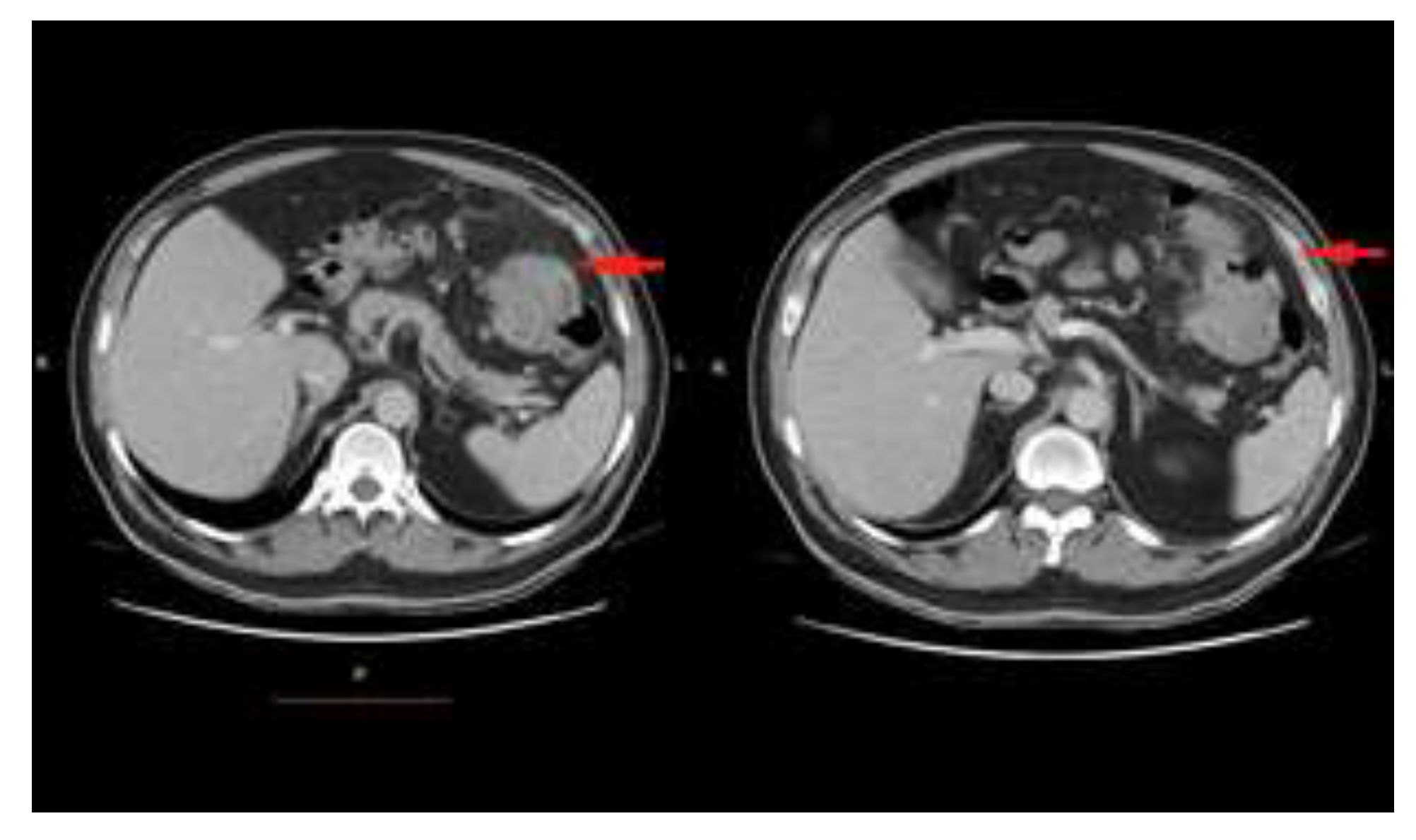
Figure 1. CT scan: splenic flexure tumoral mass, with severe narrowing of the lumen.
Jmms 07 00039 g001
Figure 2. CT scan: caecal mass and the ileocaceal-valve.
Jmms 07 00039 g002
Table 1. Immunohistochemical MMR pattern in the 4 synchronous tumors.
Jmms 07 00039 i001