Next Article in Journal
Hepatitis C Virus: Host, Environmental and Viral Factors Promoting Spontaneous Clearance
Previous Article in Journal
Oral Pathology Induced by Excess or Deficiency of Glucocorticoids in Adults
 
 
Journal of Mind and Medical Sciences is published by MDPI from Volume 12 Issue 1 (2025). Previous articles were published by another publisher in Open Access under a CC-BY (or CC-BY-NC-ND) licence, and they are hosted by MDPI on mdpi.com as a courtesy and upon agreement with Valparaiso University (ValpoScholar).
Review

Diagnosis and Management of Pericardial Effusion

by
Maria Manea
1,
Ovidiu Gabriel Bratu
2,
Nicolae Bacalbasa
3 and
Camelia Cristina Diaconu
4,*
1
University Emergency Central Military Hospital, Cardiology Clinic, Bucharest, Romania
2
Carol Davila University of Medicine and Pharmacy, Department of Urology, University Emergency Central Military Hospital, Academy of Romanian Scientists, Bucharest, Romania
3
Carol Davila University of Medicine and Pharmacy, Department of Visceral Surgery, Fundeni Clinical Institute, Bucharest, Romania
4
Carol Davila University of Medicine and Pharmacy, Department of Internal Medicine, Clinical Emergency Hospital of Bucharest, Calea Floreasca no. 8, Bucharest, Romania
*
Author to whom correspondence should be addressed.
J. Mind Med. Sci. 2020, 7(2), 148-155; https://doi.org/10.22543/7674.72.P148155
Submission received: 26 March 2020 / Revised: 3 June 2020 / Accepted: 3 June 2020 / Published: 29 September 2020

Abstract

:
Pericardial effusion is a common pathology in clinical practice. The etiology of pericardial effusion is diverse, from infectious diseases, malignancies, autoimmune diseases, chronic renal failure, to drug-related or after an invasive cardiac intervention. Clinical presentation of pericardial effusion is variable, depending on the volume of the fluid and the rate of accumulation. Clinical manifestations are dramatic if the pericardial fluid accumulates rapidly. Classical symptoms in pericardial effusion include pleuritic chest pain associated with dyspnea. In case of cardiac tamponade, orthopnea, jugular venous distension, pulsus paradoxus, hypotension, and shock appear. Transthoracic echocardiography is the gold standard investigation for the diagnosis of pericardial effusions. The echocardiographic signs of cardiac tamponade are represented by the collapse of the right atrium and right ventricle, respiratory alteration of mitral and tricuspid flow, and changes of the inferior vena cava. Management of pericardial effusion is guided by several factors, including hemodynamic impact and etiology. Pericardiocentesis guided by echocardiography is a life-saving procedure in cases of large pericardial effusions and cardiac tamponade. This is a review of the diagnosis and treatment of pericardial effusion.

Introduction

The pericardium is a double-layered membrane that covers the heart. It consists of a fibrous and a serous layer and encloses the pericardial cavity, which contains, physiologically, 15-50 mL of pericardial fluid. The pericardium provides mechanical protection of the heart and against infections.
Pericarditis is a common pathology, caused by inflammation of the pericardium. Usually, pericarditis is associated with pericardial effusion, by accumulation of pericardial fluid that can be serous, hemorrhagic, or purulent, depending on the etiology. Snyder et al observed that acute pericarditis is reported in 5% of patients presented to the emergency room for chest pain [1]. Cardiac tamponade is a critical condition, that occurs after rapid or excessive accumulation of pericardial fluid, associated with hemodynamic instability.
The prevalence of pericarditis can be difficult to estimate because in some cases the accumulation of pericardial fluid can be clinically insignificant. The incidence of pericardial diseases is estimated to be 2 cases per 10 000 persons [2].
Pericardial effusion is classified by its onset as acute, subacute, or chronic and by its distribution in circumferential or loculated. Pericardial effusion is classified also based on hemodynamic impact (with or without cardiac tamponade) or by its composition (exudate, transudate, hemopericardium, pyo-pericardium). Depending on the effusion volume, pericardial effusion can be mild, moderate, or large [1].

Discussions

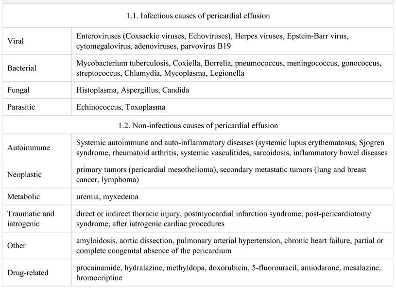
Etiology of pericardial effusion

The etiology of pericardial effusion is varied (Table 1). The 2015 European Society of Cardiology Guideline for the management of pericardial effusion classified the etiology into infectious and non-infectious [3]. Among infectious causes, the most common agents are viruses, such as Coxsackie A and B, echovirus, adenovirus, influenza, and human herpes virus. Bacterial infections that can lead to pericardial effusion are represented by Coxiella burnetii, meningococcus, pneumococcus, staphylococcus and streptococcus [4].
A review by Mayosi et al in 2005 reported that tuberculosis is the most common cause of pericarditis in developing countries [5]. Another review reported that the incidence of tuberculous pericarditis is increasing in Africa, as a result of the HIV epidemic [6].
Table 1. The etiology of pericardial effusion [3,7].
Jmms 07 00023 i001
Pericardial effusion can be secondary to noninfectious diseases, such as autoimmune diseases (systemic lupus erythematosus, rheumatoid arthritis, Sjogren syndrome, sarcoidosis), primary tumors (pericardial mesothelioma) or secondary tumors (most common lung cancer and breast cancer, lymphomas and melanoma), acute myocardial infarction (Dressler syndrome), chronic renal failure [8], hypothyroidism (myxedema), mediastinal radiation, direct injury (penetrating thoracic injury), drugs (especially procainamide, hydralazine), and after an invasive cardiac procedure (post-pericardiotomy syndrome), or it may have hemodynamic causes (heart failure, pulmonary hypertension) [3]. In a study that enrolled 114 patients with neoplasia, pericardial diseases were diagnosed in 18% of cases throughout the monitoring period [9].
In many cases, the etiology of pericarditis is difficult to establish. Multiple studies on this issue have revealed that the cause of pericarditis is idiopathic in 90% of cases [4]. Cardiac tamponade can be caused by chest trauma complicated with pericardial hemorrhage (hemopericardium), ventricular wall rupture after acute myocardial infarction, or after invasive cardiac procedures [3].

Pathophysiology of pericardial effusion

The pericardium has the role to provide mechanical protection to the heart, to act as a barrier against infections, and to ensure dynamic interactions of the heart chambers. The normal pericardium contains 10-50 mL of pericardial fluid, which is an ultrafiltrate of plasma. The interactions of the pericardium with pleural lymphatics and the lymphatic drainage of the pericardial fluid to the mediastinal and tracheobronchial lymph nodes explain the pathological involvement of the pericardium in pulmonary diseases [9].
Depending on the mechanism of appearance, the pericardial effusion can be classified into exudate and transudate. Pericardial fluid is an exudate when it is the result of an inflammatory pericardial process, which determines an increased production of pericardial fluid. The transudate appears when the reabsorption of the pericardial fluid decreases due to the increased systemic venous pressure; thus, the fluid accumulates in the pericardium, as in heart failure or pulmonary hypertension [4].
The normal pericardium has a J-shaped pressurevolume curve. A slow accumulation of 1-2 L of pericardial fluid, over several weeks or months, will lead to a modest increase in pericardial pressure, without cardiac tamponade until advanced stages [10]. Usually, pericardial effusions caused by infections, autoimmune diseases, or malignancies are associated with a slow accumulation of the fluid. In these cases, the compliance of the pericardium increases, and the pericardial sac dilates without compression of the heart [4].
A sudden increase in the pericardial fluid is associated with increased intrapericardial pressure. Intrapericardial pressure increases rapidly to 20-30 mmHg or more at a sudden increase of 100-200 mL of pericardial fluid (in cases of hemopericardium) and this can lead to cardiac tamponade [11]. Cardiac tamponade results from the rapid accumulation of the pericardial fluid. In the development of cardiac tamponade, some factors influence the hemodynamic consequences. These factors are represented by the rapidity of fluid accumulation, subjacent cardiac disease and the pericardial pressure-volume relationship. The ongoing accumulation of pericardial fluid increases the intrapericardial pressure. The compensatory mechanisms are the increased central systemic pressure and pulmonary venous pressure, for equalizing the pericardial pressure and preventing the collapse of the heart. Stroke volume is reduced, but in the first stages, the cardiac output is maintained. Also, the increased ventricular pressure leads to pulmonary hypertension to maintain pulmonary circulation. In these cases, pulmonary arterial pressure increases to 40 mmHg [12,13].
As the pericardial fluid continues to accumulate, the pericardium becomes inextensible. A discordance in filling and ejection between the left and the right ventricle will appear. This is called ventricular interdependence, meaning that any changes that appear in the volume of one side of the heart determine the opposite changes in the volume of the other side. The clinical manifestation that appears in this context is represented by pulsus paradoxus. The dilatation of the right ventricle leads to the bowing of the interventricular septum into the left ventricle, with a reduced left ventricle preload. When the pericardial pressure is above 25 mmHg, the cardiac output is decreased and the blood pressure is reduced, resulting in cardiogenic shock and circulatory collapse [14]. The pericardial drainage and removal of 100 mL of fluid may reduce the pericardial pressure, which is lifesaving in cardiac tamponade [15].

Clinical presentation and paraclinical tests

Clinical presentation of pericardial effusion is variable, depending on the volume of fluid and rate of accumulation. Clinical manifestations are dramatic if the pericardial fluid accumulates rapidly. Classical symptoms in pericardial effusion include pleuritic chest pain, relieved when the patient leans forward, associated with dyspnea at exertion that can progress to orthopnea. Other symptoms may appear due to local compression, such as nausea and dysphagia. Depending on the etiology of pericardial fluid, nonspecific symptoms may appear, such as cough, weakness, palpitations, or fatigue [3].
Cardiac auscultation reveals diminished heart sounds. Pulmonary auscultation identifies sometimes a left parasternal pericardial friction rub. This is a sound corresponding to friction between the pericardium layers. In the literature, the friction rub is reported in 35-85% of the cases of pericarditis [16]. Physical examination in cardiac tamponade reveals jugular venous distension, tachypnea, tachycardia, pulsus paradoxus, hypotension, and cold extremities. Pulsus paradoxus means a decrease in systolic blood pressure by more than 10 mmHg with inspiration [6]. Pulsus paradoxus appears in cardiac tamponade due to ventricular interdependence, when the volume of the cardiac chambers is fixed. The classical finding at the physical examination in patients with cardiac tamponade is Beck’s triad, which includes arterial hypotension, jugular venous distension, and muffled heart sounds [17]. Data from the literature reveal that signs of Beck’s triad are identified especially in cases of cardiac tamponade due to hemopericardium, as a result of thoracic injury or acute rupture of the ventricular wall [4].
Laboratory tests may show leukocytosis and high levels of inflammatory markers in pericardial effusion caused by infectious and inflammatory diseases. Also, in these cases, liver and renal function must be evaluated, together with thyroid stimulating hormone, and different markers in case of suspected viral infection. In cases of high suspicion of tuberculosis, tests such as PPD or quantiferon TB will be performed [10].
The electrocardiogram (ECG) performed in patients with pericarditis shows changes that evolve through stages [11]. In stage I, the ECG presents a diffuse elevation of the ST-segment. In stage II, the ST segment normalizes. Stage III and stage IV are characterized by T wave inversions, followed by T wave normalization. ECG changes appear in more than half of the patients [18]. In cardiac tamponade, the ECG reveals sinus tachycardia and low-voltage complexes. Electrical alternation on ECG is characteristic in cases of large pericardial effusion.
The European Society of Cardiology guideline for pericardial diseases requires two out of the following four criteria for the diagnosis of acute pericarditis: pericardial chest pain, pericardial rub, ST-segment elevation on ECG and pericardial effusion [3]. In cases of tuberculous pericardial effusion, the diagnosis is made when there are a lymphocytic pericardial exudate and elevated ADA levels [6].
Transthoracic echocardiography is the gold standard method for the diagnosis of pericardial effusions. M-mode and two-dimensional Doppler ecocardiography should be performed to evaluate the pericardial fluid, its distribution, size, and hemodynamic effects [16]. On two-dimensional echocardiography, pericardial effusion appears as an echofree space. In this case, the clinician should assess the location of the fluid and the hemodynamic impact. Small pericardial effusions are localized over the postero-basal left ventricle. Then, as the pericardial fluid increases, it will be visible anterior or lateral. Any pericardial effusion from 5 to 10 mm in diastole is considered small, 10 to 20 mm is moderate, and greater than 20 mm is considered large. Swinging heart is a sign of large effusion at echocardiography [10].
In cardiac tamponade, echocardiography will show the diastolic collapse of the right heart cavities, the most characteristic ultrasonographic sign [19]. Usually, the compression of the right atrium for more than one-third of the cardiac cycle is specific for cardiac tamponade [20]. The collapse of the left atrium appears in 25% of the cases of tamponade [21]. Also, Doppler echocardiography is used to estimate respiratory variations in the mitral and tricuspid flow –a decrease in the peak mitral inflow velocity with >30% and an inspiratory increase of the peak of tricuspid inflow velocity with >50% suggest an increased ventricular interdependence [22]. The inferior vena cava is dilated in these cases and shows a less than 50% reduction of its diameter during inspiration, as a sign of an increased central venous pressure [23]. The echocardiographic changes of the inferior vena cava associated with pulsus paradoxus at the physical examination are present in 92% of cases of cardiac tamponade [24]. Also, echocardiography will show abnormal septal motion in the case of cardiac tamponade.
The clinician should correlate the echocardiographic signs of cardiac tamponade, such as collapse of the right atrium and right ventricle, respiratory alteration of mitral and tricuspid flow, and changes of the inferior vena cava, with the clinical signs of tamponade – dyspnea, tachycardia, jugular venous distension, pulsus paradoxus, hypotension, and shock [25].
Other imaging tests, such as computed tomography (CT) scan or magnetic resonance imaging (MRI) can describe the characteristics of the pericardial effusion and cardiac tamponade. The cardiac CT scan may reveal pericardial fluid accumulation, but this method cannot be used to assess hemodynamic parameters. The cardiac MRI evaluates the pericardium morphology but requires more time and has higher costs [26].
The biochemical analysis of the pericardial fluid is recommended to assess the etiology of the pericardial effusion [3]. These tests can determine whether the etiology of the pericardial fluid is infectious or neoplastic. The pericardial fluid required for these tests will be obtained by pericardial drainage, and cultures for aerobes and anaerobes will be performed, together with other tests, depending on the suspected etiology. Also, the biochemical analysis of the pericardial fluid can distinguish the exudate from transudate [27].

Differential diagnosis of pericardial effusion

There are several diseases with similar symptoms that should be considered in the differential diagnosis of pericardial effusion, such as pulmonary embolism, congestive heart failure, acute coronary syndrome, and cardiogenic pulmonary edema. In these cases, the differential diagnosis is based on the chest pain characteristics, laboratory tests (cardiac enzymes, Ddimers, NT-proBNP), and imaging investigations (echocardiography, CT scan). Other causes of chest pain that should be considered in these patients are represented by gastritis, esophagitis, or peptic ulcer disease [11]. Also, the differential diagnosis of cardiac tamponade includes other causes of cardiogenic shock, such as pulmonary embolism or type A aortic dissection with extension into the pericardium [2].

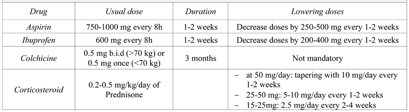
Treatment of pericardial effusion

Medical treatment

Medical treatment in patients diagnosed with pericardial effusion whose tests show elevated inflammatory markers and develop inflammatory signs and symptoms, such as fever, is represented by high dose of non-steroidal anti-inflammatory drugs (NSAIDs) or aspirin, associated with colchicine [3] (Table 2). Several studies showed good results when colchicine is associated with aspirin or NSAIDs in the treatment of pericarditis [28]. Idiopathic and viral pericarditis associated with pericardial effusion is often responsive to this combination of drugs [29,30]. The treatment will be targeted for the causative agent in cases where the specific etiology is known (viral or bacterial infections). Low or moderate doses of corticosteroids are not recommended as first-line treatment, because this class is associated with the risk of recurrent pericarditis. Corticosteroids are administered usually when there are specific indications or in cases of intolerance to aspirin or NSAIDs [28]. The therapeutic response is evaluated based on the relief of the symptoms, associated with an improvement of the biological parameters.
When a complete response is obtained, lowering doses should be considered, initially with a single class of drugs at a time. Each tapering is done if the patient is asymptomatic and the values of inflammatory markers are normal [3].
Pericardial effusion developed after acute myocardial infarction complicated with rupture of the ventricle wall may result in hemopericardium. Studies show that up to two-thirds of these patients may present cardiac tamponade in the context of ventricular free wall rupture. In these cases, urgent treatment is life-saving [31].
Purulent pericardial effusion is associated with high mortality, so these patients should be treated aggressively. In the context of a tuberculous etiology of pericardial effusion, the treatment includes anti-tuberculosis drugs administered for at least 6 months. In these cases, the clinician should consider pericardiectomy if there is a failure of clinical improvement with medical therapy [32]. If the medical treatment is not effective in case of symptomatic pericardial effusion with no inflammatory signs, the clinician may consider pericardial drainage.

Pericardial drainage

Pericardial drainage can be performed by pericardiocentesis or by an open surgical method. Percutaneous needle pericardiocentesis is a procedure used for diagnosis or therapeutic purposes. In cases of symptomatic large pericardial effusion or cardiac tamponade, pericardiocentesis is the most useful therapeutic procedure. This is a life-saving method, indicated when echocardiography shows diastolic pericardial effusion >20 mm associated with hemodynamic impact [18].
Also, pericardiocentesis is performed in patients with chronic idiopathic pericardial effusion who remain asymptomatic, but who can develop unexpected tamponade. In a study performed by Sagrista et al, who followed patients diagnosed with chronic idiopathic pericardial effusion, cardiac tamponade appeared in 29% of cases during the course of the disease [7]. Numerous studies on this issue have confirmed that complications are reduced with 20% when percutaneous pericardiocentesis is guided by transthoracic echocardiography [33]. The feasibility of pericardiocentesis is 90% in cases when the pericardial fluid is localized anterior to the ventricle and <60% in cases of posterior localization [34]. Pericardiocentesis involves the removal of pericardial fluid by percutaneous catheterization of the pericardium, being an urgent procedure in cases of acute tamponade [18]. There are three approaches for pericardiocentesis – left parasternal, sub-xyphoid and left apical. Usually, the subxyphoid approach is used to avoid injury of the major arteries [33].
Halpern et al suggested a pericardial effusion scoring index, which consists of 3 components: the etiology of pericardial effusion, the size of the effusion, and echocardiographic signs. According to this index, pericardiocentesis could be performed when the score is 4 or higher [35]. Hyun et al conducted a study in 2007 on 116 patients diagnosed with symptomatic pericardial effusions, who were treated by pericardiocentesis guided by echocardiography. Common causes of pericardial effusion in these patients were lung cancer in 27% of cases, tuberculosis in 13.8%, and uremia in 6.9% of patients. Pericardiocentesis performed in these cases had a rate of success of 99.1% [18].
Malignant pericardial effusion can be a complication of lung or breast cancer or lymphomas. In these cases, pericardial drainage can be performed by pericardiocentesis or by surgical drainage, depending on the patient’s prognosis and risks. Many patients with a malignant pericardial effusion have a poor prognosis; in this context, the purpose of the treatment is to improve the quality of life [20].
Tsang et al confirmed that echocardiography can rapidly confirm the presence of pericardial effusion after cardiothoracic surgery. In these cases, pericardiocentesis performed under echocardiographic guidance is safe and efficient. Also, it was confirmed that the use of a pericardial catheter for drainage is associated with lower recurrence rates [36]. Lindenberger et al conducted a retrospective study, which included 252 patients with pericardial effusion. This study confirmed that pericardiocentesis guided by echocardiography is a safe and efficient method for patients with malignant etiology of the pericardial effusion [37].
Retrospective studies and case reports indicate pericardiocentesis with prolonged drainage (<30 mL/24h) to prevent re-accumulation of the fluid [35]. In cases when pericardiocentesis fails or is not feasible, the clinician should consider performing a pericardial window [24]. This technique uses sub-xyphoid approach under echocardiographic guidance and involves the insertion of a catheter into the pericardial space. This technique is efficient in preventing recurrence in >80% of cases [38].
The most important complications of pericardiocentesis are represented by perforation of the myocardium, injury of the coronary arteries, pneumothorax, air embolism, or puncture of the peritoneal cavity [34]. These complications are reduced by using the echocardiographic guidance. In a large review on this issue, the incidence of complications in echocardiographyguided pericardiocentesis was 1-1.6% [38]. Pericardiocentesis is contraindicated in aortic dissection.
Surgical drainage is a method used when pericardial drainage performed by pericardiocentesis is not feasible or has failed. This method is used in cases of hemopericardium, purulent pericarditis, recurrent malignant effusion, or when the pericardial effusion is localized posterior [39]. Surgical drainage can be performed by different techniques: complete or partial pericardectomy, subxyphoid pericardiotomy, anterior transthoracic window, all methods having the same efficiency [40].
In cases of idiopathic moderate pericardial effusions, an echocardiographic follow-up will be necessary every 6 months. Patients with severe pericardial effusion require an echocardiographic reassessment every 3-6 months after discharge [41].

Evolution and prognosis

The prognosis for patients with pericardial effusion depends on etiology. Patients with a neoplastic pericardial effusion have a poor prognosis. The evolution is favorable in patients with viral or idiopathic pericardial effusion. Also, the size of the effusion is correlated with prognosis. A mild idiopathic effusion is associated with a good prognosis and does not need special monitoring, the patients being mostly asymptomatic. Large pericardial effusions are associated with a high risk of progression to cardiac tamponade. Moderate to large pericardial effusions may evolve to cardiac tamponade in up to one-third of cases [3].
Recurrent pericardial effusion can occur in 30% of cases when patients are not treated with colchicine [3]. Another complication is constrictive pericarditis, which occurs in <1% of cases [5]. The most feared complication in patients with pericardial effusion is represented by cardiac tamponade. This complication rarely occurs in cases of idiopathic pericardial effusion but is more common in neoplastic forms [4].

Highlights

The etiology of pericardial effusion is diverse, from infectious diseases, malignancies, autoimmune diseases, chronic renal failure, to drug-related or after an invasive cardiac intervention.
Classical symptoms in pericardial effusion include pleuritic chest pain, associated with dyspnea.
Transthoracic echocardiography is the gold standard investigation for the diagnosis of pericardial effusions. The echocardiographic signs of cardiac tamponade are represented by the collapse of the right atrium and right ventricle, respiratory alteration of mitral and tricuspid flow, and changes of the inferior vena cava.
Pericardiocentesis guided by echocardiography is a life-saving procedure in cases of large pericardial effusions and cardiac tamponade.

Conclusions

Pericardial effusion may have different etiologies, such as infectious diseases, malignancies, chronic renal failure, thyroid disease, or autoimmune diseases, after cardiac interventions, or it can be idiopathic. Transthoracic echocardiography is the most useful investigation for the diagnosis of pericardial effusion. Large pericardial effusions require pericardial drainage performed by percutaneous pericardiocentesis or by surgical procedure. Pericardiocentesis guided by echocardiography is a lifesaving procedure in cases of large pericardial effusions and cardiac tamponade.

Conflicts of Interest disclosure

There are no known conflicts of interest in the publication of this article. The manuscript was read and approved by all authors.

Compliance with ethical standards

Any aspect of the work covered in this manuscript has been conducted with the ethical approval of all relevant bodies and that such approvals are acknowledged within the manuscript.

References

  1. Snyder, M.J.; Bepko, J.; White, M. Acute pericarditis: Diagnosis and management. Am Fam Physician. 2014, 89, 553–560. [Google Scholar] [PubMed]
  2. Sharma, N.K.; Waymack, J.R. Acute cardiac tamponade. [Updated 2019 Dec 17]. In StatPearls [Internet]; StatPearls Publishing: Treasure Island, FL, USA, 2020. Available online: https://www.ncbi.nlm.nih.gov/books/NBK534806/.
  3. Adler, Y.; Charron, P.; Imazio, M.; et al. 2015 ESC Guidelines for the diagnosis and management of pericardial diseases: The Task Force for the diagnosis and management of pericardial diseases of the European Society of Cardiology (ESC) endorsed by: The European Association for Cardio-Thoracic Surgery (EACTS). Eur Heart J. 2015, 36, 2921–2964. [Google Scholar]
  4. Dababneh, E.; Siddique, M.S. Pericarditis. In StatPearlsPublishing [Internet]; StatPearls Publishing: Treasure Island, FL, USA, 2019. Available online: https://www.ncbi.nlm.nih.gov/pubmed/28613734.
  5. Mayosi, B.M.; Burgess, L.J.; Doubell, A.F. Tuberculous pericarditis. Circulation. 2005, 112, 3608–3616. [Google Scholar] [CrossRef] [PubMed]
  6. Ntsekhe, M.; Mayosi, B.M. Tuberculous pericarditis with and without HIV. Heart Fail Rev. 2013, 18, 367–373. [Google Scholar] [CrossRef] [PubMed]
  7. Zaha, D.C.; Bungau, S.; Aleya, S.; et al. What antibiotics for what pathogens? The sensitivity spectrum of isolated strains in an intensive care unit. Sci Total Environ. 2019, 687, 118–127. [Google Scholar] [CrossRef]
  8. Moisi, M.I.; Rus, M.; Bungau, S.; et al. Acute coronary syndromes in chronic kidney disease: Clinical and therapeutic characteristics. Medicina 2020, 56, 118. [Google Scholar] [CrossRef]
  9. Sagrista-Salueda, J.; Merce, A.S.; Soler-Soler, J. Diagnosis and management of pericardial effusion. World J Cardiol. 2011, 3, 135–143. [Google Scholar] [CrossRef]
  10. Imazio, M.; Adler, Y. Management of pericardial effusion. Eur Heart Journal. 2013, 34, 1186–1197. [Google Scholar] [CrossRef]
  11. Saito, Y.; Donohue, A.; Attai, S.; et al. The syndrome of cardiac tamponade with ‘small’ pericardial effusion. Echocardiography. 2008, 25, 321–327. [Google Scholar] [CrossRef]
  12. Shabetai, R. Pericardial effusion: Haemodynamic spectrum. Heart. 2004, 90, 255–256. [Google Scholar] [CrossRef]
  13. Stoicescu, M.; Csepento, C.; Mutiu, G.; et al. The role of increased level of plasma renin in etiopathogenic arterial hypertension in the young. Rom J Morphol Embryol 2011, 52 (Suppl. S1), 419–423. [Google Scholar] [PubMed]
  14. Stashko, E.; Meer, J.M. Cardiac Tamponade. [Updated 2019 Dec 5]. In StatPearls [Internet]; StatPearls Publishing: Treasure Island, FL, USA, 2020. Available online: https://www.ncbi.nlm.nih.gov/books/NBK431090/.
  15. Maisch, B. Management of pericarditis and pericardial effusion, constrictive and effusive-constrictive pericarditis. Herz. 2018, 43, 663–678. [Google Scholar] [CrossRef]
  16. Imazio, M.; Gaita, F. Diagnosis and treatment of pericarditis. Heart. 2015, 101, 1159–1168. [Google Scholar] [CrossRef]
  17. Bodson, L.; Bouferrache, K.; Vieillard-Baron, A. Cardiac tamponade. Curr Opin Crit Care. 2011, 17, 416–424. [Google Scholar] [CrossRef] [PubMed]
  18. Rosselo, X.; Wiegerinck, R.F.; Alguersuari, J.; et al. New electrocardiographic criteria to differentiate acute pericarditis and myocardial infarction. Am J Med. 2014, 127, 233–239. [Google Scholar] [CrossRef] [PubMed]
  19. Hae-Ok, J. Pericardial effusion and pericardiocentesis: Role of echocardiography. Korean Circ J. 2012, 42, 725–734. [Google Scholar]
  20. Little, W.C.; Freeman, G.L. Pericardial disease. Circulation. 2006, 113, 1622–1632. [Google Scholar] [CrossRef]
  21. Goodman, A.; Perera, P.; Mailhot, T.; Mandavia, D. The role of bedside ultrasound in the diagnosis of pericardial effusion and cardiac tamponade. J Emerg Trauma Shock. 2012, 5, 72–75. [Google Scholar]
  22. Imazio, M.; Spodick, D.H.; Brucato, A.; et al. Controversial issues in the management of pericardial diseases. Circulation. 2010, 121, 916–928. [Google Scholar] [CrossRef]
  23. Cosyns, B.; Plein, S.; Nihoyanopoulos, P.; et al. Multimodality imaging in pericardial disease. Eur J Heart Cardiovasc Imaging. 2015, 16, 12–31. [Google Scholar] [CrossRef]
  24. Pepi, M.; Muratori, M. Echocardiography in the diagnosis and management of pericardial disease. J Cardiovasc Med. 2006, 7, 533–544. [Google Scholar] [CrossRef]
  25. Abdel-Daim, M.M.; El-Tawil, O.S.; Bungau, S.G.; Atanasov, A.G. Applications of antioxidants in metabolic disorders and degenerative diseases: Mechanistic approach. Oxid Med Cell Longev. 2019, 2019, 1–3. [Google Scholar] [CrossRef] [PubMed]
  26. Bogaert, J.; Francone, M. Cardiovascular magnetic resonance in pericardial disease. J Cardiovasc Magn Reson. 2009, 11, 14. [Google Scholar] [CrossRef] [PubMed]
  27. Ben-Horin, S.; Bank, I.; Shinfeld, A. Diagnostic value of the biochemical composition of pericardial effusions in patients undergoing pericardiocentesis. Am J Cardiol. 2007, 99, 1294–1297. [Google Scholar] [CrossRef] [PubMed]
  28. Imazio, M.; Brucato, A.; Trinchero, R. Colchicine for pericarditis: Hype or hope? Eur Heart J. 2009, 30, 532–539. [Google Scholar] [CrossRef]
  29. Imazio, M.; Brucato, A.; Trinchero, R.; et al. Individualized therapy for pericarditis. Expert Rev Cardiovasc Ther. 2009, 7, 965–975. [Google Scholar] [CrossRef]
  30. Suceveanu, A.I.; Pantea Stoian, A.; Parepa I et, a.l. Gut microbiota patterns in obese and type 2 Diabetes (T2D) patients from Romanian Black Sea coast region. Rev Chim. 2018, 69, 2260–2267. [Google Scholar] [CrossRef]
  31. Seferovic, P.M.; Ristic, A.D.; Imazio, M.; et al. Management strategies in pericardial emergencies. Herz. 2006, 31, 891–900. [Google Scholar] [CrossRef]
  32. Mayosi, B.M.; Ntsekhe, M.; Volmink, J.A.; et al. Interventions for treating tuberculous pericarditis. Cochrane Database Syst Rev 2002, CD000526. [Google Scholar]
  33. Kil, U.H.; Jung, H.O.; Koh, Y.S.; et al. Prognosis of large, symptomatic pericardial effusion treated by echo-guided percutaneous pericardiocentesis. Clin Cardiol. 2008, 31, 531–537. [Google Scholar] [CrossRef]
  34. Kim, D.Y.; Park, J.H.; Shin, J.D.; et al. Long-term follow-up results and clinical manifestations of patients with a moderate to large amount of pericardial effusion. Korean J Med. 2008, 74, 154–161. [Google Scholar]
  35. Halpern, D.G.; Argulian, E.; Briasoulis, A.; et al. A novel pericardial effusion scoring index to guide decision for drainage. Crit Pathw Cardiol. 2012, 11, 85–88. [Google Scholar] [CrossRef] [PubMed]
  36. Tsang, T.S.; Enriquez-Sarano, M.; Freeman, W.K.; et al. Consecutive 1127 therapeutic echocardiographically guided pericardiocenteses: Clinical profile, practice patterns and outcomes spanning 21 years. Mayo Clin Proc. 2002, 77, 429–436. [Google Scholar] [CrossRef] [PubMed]
  37. Lindenberger, M.; Kjellberg, M.; Karlsson, E.; Wranne, B. Pericardiocentesis guided by 2-D echocardiography: The method of choice for treatment of pericardial effusion. J Intern Med. 2003, 253, 411–417. [Google Scholar] [CrossRef]
  38. Loukas, M.; Walters, A.; Boon, J.M.; et al. Pericardiocentesis: A clinical anatomy review. Clin Anat. 2012, 25, 872–881. [Google Scholar] [CrossRef]
  39. Manea, M.; Marcu, D.; Pantea Stoian, A.; et al. Heart failure with preserved ejection fraction and atrial fibrillation: A review. Rev Chim. 2018, 69, 41804184. [Google Scholar] [CrossRef]
  40. Khandaker, M.H.; Schaff, H.V.; Greason, K.L.; et al. Pericardiectomy vs medical management in patients with relapsing pericarditis. Mayo Clin Proc. 2012, 87, 1062–1070. [Google Scholar] [CrossRef]
  41. Hota, S.S.; Chow, C.M.; Bonneau, D. Surgical treatment for incessant pericarditis. Can J Cardiol. 2009, 25, 161162. [Google Scholar] [CrossRef]
Table 2. Drug treatment options for pericardial effusion [3].
Jmms 07 00023 i002

