Next Article in Journal
Conservative Surgical Treatment in Cervical Dysplastic Lesions Associated with Cystorectocele
Previous Article in Journal
Pigmented Melanoma Cell Migration Study on Murine Syngeneic B16F10 Melanoma Cells or Tissue Transplantation Models
 
 
Journal of Mind and Medical Sciences is published by MDPI from Volume 12 Issue 1 (2025). Previous articles were published by another publisher in Open Access under a CC-BY (or CC-BY-NC-ND) licence, and they are hosted by MDPI on mdpi.com as a courtesy and upon agreement with Valparaiso University (ValpoScholar).
Article

Laparoscopic Resection of Gastric GISTs. Where Do We Stand Now? A Single-Centered Experience

by
Iulian Slavu
1,
Lucian Alecu
2,
Adrian Tulin
2,*,
Dana L. Stanculeanu
3 and
Cornelia Nitipir
4
1
Department of General Surgery, Clinical Emergency Hospital Bucharest, Bucharest, Romania
2
Department of General Surgery, Agrippa Ionescu Clinical Emergency Hospital, Bucharest, Romania
3
The Oncology Clinic, Al. Trestioreanu Institute of Oncology, Bucharest, Romania
4
The Oncology Clinic, Elias University Emergency Hospital, Bucharest, Romania
*
Author to whom correspondence should be addressed.
J. Mind Med. Sci. 2019, 6(2), 334-339; https://doi.org/10.22543/7674.62.P334339
Submission received: 5 February 2019 / Revised: 7 May 2019 / Accepted: 7 May 2019 / Published: 9 October 2019

Highlights

  • Complete surgical resection independent of tumor size represents the current optimal treatment of GISTs.
  • Laparoscopic resection is possible, although the surgeon’s experience along with careful patient selection plays a critical role.

Abstract

:

Highlights

  • Complete surgical resection independent of tumor size represents the current optimal treatment of GISTs.
  • Laparoscopic resection is possible, although the surgeon’s experience along with careful patient selection plays a critical role.

Abstract

Introduction. Gastrointestinal stromal tumors (GISTs) represent a rare type of gastrointestinal neoplasms. Resection with negative margins has been established as a mainstay treatment, but laparoscopic resections are still open to debate. Material and method. This retrospective study was conducted at a single institution, with data collected over 2 years (01.01.2017–01.01.2019). The variables examined were age, tumor location with regard to the gastric wall, the results of the intraoperative endoscopy, intraoperative and postoperative complications, the surgical technique, and histopathological reports. Results. We identified 12 relevant cases, of which 8 were females and 4 males. The average tumor diameter was 2.3 cm. The majority of the lesions were located on the anterior gastric wall (8 cases), on the small curvature (2 cases), and in the pyloric region (2 cases). Intraoperative endoscopy was performed successfully in 10 cases in order to identify the lesions and guide the resection. The average operative time was 120 min and the average hospital stay was 5 days. The gastric wall with the lesion was resected using an Ultrasonic device, a 2-cm oncological safety margin was preserved. Conclusion. Complete surgical resection independent from the tumor size represents the current optimal treatment. From a surgical point of view, these tumors must be considered malignant and the surgeon must respect principles of oncological surgery. Maintaining tumor integrity at dissection is critical for the patient’s long-term prognosis. Laparoscopic resection independent of the tumor size is feasible.

Introduction

Mesenchymal gastrointestinal tumors or GISTs are a particular subtype of gastrointestinal tumors [1]. Although these tumors have historically been considered benign or borderline malignant, with the recent evolution of immunohistochemical and histopathological diagnostic methods, GISTs have been redefined as malignant tumors, meaning that the oncological principles of resection must be respected [2].
Regarding their location, these tumors can develop anywhere in the gastrointestinal tract but are most commonly identified at the gastric level in up to 60% of the cases. The stomach is more accessible for endoscopy than the small intestine which this may account for its increased incidence [3]. Another cause may be the embryological origin of these tumors that evolve from the Cajal cells – which represent the pacemaker of the digestive tract located at the level of the gastric fornix [3].
The degree of malignancy of these tumors is given by the tumor macroscopic characteristics (size), but also by the cells themselves, i.e. the number and frequency of mitoses, or the degree of differentiation [4].
Taking these factors into consideration, the surgeon must avoid rupturing the tumor capsule during surgery and maintain negative resection margins of 1-2 cm. Lymphadenectomy is not required when a GIST tumor is identified, since tumor cells rarely disseminate through the lymphatic system [5]. Given these recommendations, if a gastric GIST is confirmed, a wedge resection can be performed – which is a straight-forward intervention. Since laparoscopy has evolved and increasingly complex interventions are now being performed, the question regarding the application of this technique to gastric GISTs has arisen. [6]. Several authors such as Joensuu et al. and Nguyen et al. have confirmed the feasibility of this intervention, which implies a full-thickness resection of the gastric wall through a minimally invasive approach [7,8]. Also, a large-scale meta-analysis has been published, which confirms the aforementioned results [9,10].
Regarding the size of the tumors, laparoscopy has been recommended only for tumors under 2 cm due to the high risk of rupture and contamination at dissection, but this approach has not been supported by all authors. De Vogelaere et al. demonstrated, on a group of 31 patients, that the size of the tumor does not impact the relapse rate for laparoscopic operations [11,12].
Our study aimed to reevaluate the oncological outcome and technique when resecting gastric GISTs, based on experience from our clinic. Due to widespread endoscopic evaluations, gastric tumors are identified in the early stages limited to the submucosa, with no expression at the level of the serosa. Thus, they are difficult to identify and resect using oncological safety margins. Through this study, we aimed to evaluate the use of intraoperative endoscopy in order to identify these tumors and guide the resection process.

Materials and Methods

The study is a retrospective analysis conducted in a single institution, with data obtained over a 2-year period (01.01.2017-01.01.2019). Variables examined were age, tumor location with regard to the gastric wall, results of the intraoperative endoscopy, intraoperative and postoperative complications, surgical technique, and histopathological reports. In addition, the efficacy of the intraoperative endoscopical procedure in identifying the tumors and guiding the resection was evaluated. The results of the 1- year evaluation, which included a CT with contrast media and upper endoscopy, were also examined. All patients were operated on laparoscopically. Tumor size did not represent a contraindication.

Results

We identified 12 cases, of which 8 were females and 4 males. Age at diagnosis ranged between 32 and 64 years. Common symptoms were gastric bleeding, anemia, and weight loss.
The average tumor diameter was 2.3 cm, with a maximum of 15 cm and a minimum of 1.7 cm (Figure no. 4). Gastrostomy has always been sutured with a double layer Vicryl wire. The integrity of the suture line was verified intraoperatively with the use of methylene blue, which was administered through the nasogastric tube.
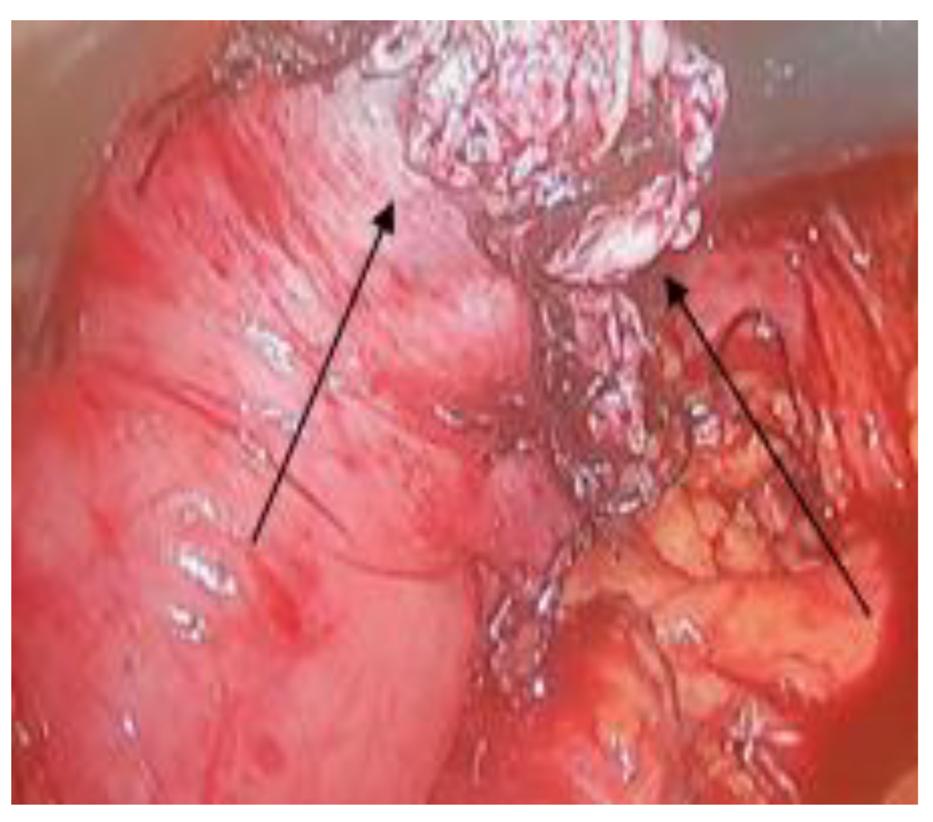
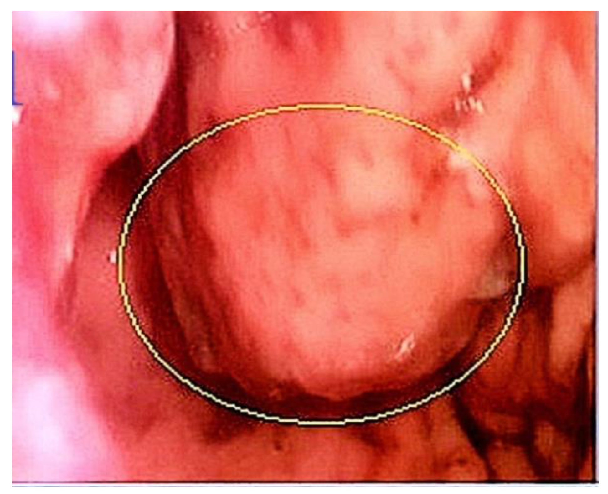
The intraoperative laparoscopic exploration did not reveal peritoneal metastasis, which correlated with the preoperative investigations. Regarding the location of the tumor, the majority of the lesions were located on the anterior gastric wall (8 cases), on the small curvature (2 cases), and in the pyloric region (2 cases). Some of the tumors were large and had an important expression on the level of the gastric serosa which made them easily identifiable, as seen in Figure 1. In other cases, the tumors were limited to the submucosa, thus being nearly impossible to identify laparoscopically. Frequently, tumors are located between the gastric mucosal folds. In such cases, intraoperative endoscopy is very helpful.
In our series, intraoperative endoscopy was performed successfully in 10 cases in order to identify the lesions and guide the resection (Figure no. 1). In 4 cases, preoperative endoscopic staining was used to identify the lesion (Figure no 2). Endoscopy proved helpful in identifying the small lesions located in the submucosa, which had no expression at the level of the serosa.
Great care was taken at dissection to avoid intraoperative tumor ruptures. The average operative time was 120 minutes, but depended on whether gastric suture was done with a stapler or manually. All patients had a contrast barium Rx on the third day to identify any potential leakage. There was none.
All patients had a drainage tube placed in the Morisson pouch and which was removed on the second postoperative day, with no complications. The average hospital stay was 5 days. All patients had a nasogastric tube placed intraoperatively which was removed on the third day. The gastric wall with the lesion was resected using an Ultrasonic device, with a 2-cm oncological safety margin. All resections were R0. A one-year reevaluation through endoscopy and CT did not reveal any sign of relapse. Regarding the postoperative complications, we encountered one case of intraluminal bleeding, which was terminated with clips applied endoscopically on the third postoperative day.
Figure 1. Large GIST (8cm) located on the anterior gastric wall (arrow).
Jmms 06 00046 g001
Figure 2. Small gastric GIST (circle) located between the mucosal folds of the stomach, identified through intraoperative endoscopy.
Jmms 06 00046 g002
Figure 3. An aspect of gastrotomy after the resection of a GIST identified through the preoperative endoscopic instillation of methylene blue.
Jmms 06 00046 g003
Figure 4. The aspect of a large gastric GIST after resection with safety margins of 2 cm. The surface is round, smooth with no macroscopic signs of invasion.
Jmms 06 00046 g004

Discussions

The first description of a gastrointestinal stromal tumor was published in 1983 by Clar and Mazur who defined it as a new type of sarcoma [13]. Although GISTs were initially considered benign, they are now considered malignant, although they rarely have a diffuse infiltrating evolution in the organs in which they originate. These tumors rarely lead to lymphatic metastasis [14,15].
Due to these characteristics, local excision is considered sufficient as a final treatment. Some authors even recommend enucleation for small tumors. We, however, do not recommend this technique in laparoscopy due to the rigidity of the instruments which may perforate and disseminate the tumor cells in the peritoneal cavity. Rupturing the capsule of the tumor can have catastrophic results for the patient, so one should avoid manipulating the tumor directly during surgery and should grasp only the tissue around the tumor. Also, an endo-bag is mandatory when extracting the specimen so as not to contaminate the abdominal wall [16].
As demonstrated in our series of patients, a laparoscopic wedge resection is technically feasible, ensuring good oncological results without affecting the functionality of the gastric tube [17]. Due to the constant advancements in stapler technology and laparoscopic gastric surgery, this technique has also been recommended by other authors [18,19]. However, depending on the tumor location, this technique is not always feasible; small tumors located on the anterior gastric wall are usually ideal for this resection. In our series, when a wedge resection could not be performed, a gastric wall resection using an ultrasonic device with subsequent suturing of the defect using a double layer, resorbable running suture gave the best results with no postoperative complications. It did, however, extend operative time considerably.
Regarding the size of the tumors, past practice avoided laparoscopy when tumor size exceeded 2 cm [20]. From our standpoint, if the laparoscopic instruments are available and the surgeon has the necessary experience, the size of the tumor should not represent a contraindication for laparoscopy. As demonstrated in our study, tumors up to 8-10 cm were resected laparoscopically with good oncological and functional outcomes. Multiple studies in the literature have confirmed its feasibility, although the NCCN guidelines and the European ESMO guidelines do not recommend laparoscopy for resection of large GIST tumors [21]. Consistent with our perspective, Ye et al. also note that the surgeon’s experience is paramount when selecting to laparoscopically operate on a GIST in order to avoid capsule tears and peritoneal spillage of the tumor cells [22]. Perhaps in the near future, the guidelines will adapt to the experience gained by surgeons in the field of laparoscopy [23,24,25].
Some studies have shown good results with hand-assisted laparoscopic resections, to gently manipulate the tumor and dissect it from the surrounding structures. This technique was not used on the patients in our series since the tumors were dissected safely only with the laparoscopic instruments. Regarding the oncological safety margins, our resections involve a 1-2cm distance from the tumor, although research has demonstrated that the rate of recurrence does not seem to be influenced by the resection distance and varies rather according to the size of the tumor. Keeping these aspects in mind, the surgeon must obtain a clear macroscopic resection without lymphadenectomy [26]. Although our sample size was small, our group had a postoperative survival rate of 100 % at 1 year.
When smaller tumors are encountered, intraoperative endoscopy is helpful in guiding the surgeon to the location of the lesion. For example, we encountered gastric submucosal tumors of 1-2cm, with no clear expression at the level of the serosa. Endoscopy is paramount in these situations. If one does not identify the lesion, the use of staples should be avoided and a circumferential resection under the direct guidance of the endoscopist is recommended to avoid the rupture of the tumor with subsequent spillage. In four cases, we used the preoperative marking of the lesion with methylene blue, but the endoscopist had to pay close attention to the depth of the gastric layers and the quantity when administering the substance, since it can always dissipate in a large area of the gastric wall [27,28]. This makes it difficult for the surgeon to correctly resect the area where the tumor is located.
Protocols for postoperative monitoring are open to discussion, since the frequency and the instruments used vary according to several criteria. Recent data tend to ignore absolute definitions of benign or malignant. Moreover, we noticed that this field is continuously changing [29]: the tumors were initially defined as benign, then malignant, and now benign or malignant, with differentiating factors being their size (above 10 cm) and rate of division [30]. A high index of replication increases the mortality rate of up to 50% when compared to low mitotic tumors [30].
A high mitotic index is defined as 10 mitoses per 50 HPF (high power fields), and this index predicts the recurrence rate. Thus, due to the ongoing redefinition of these tumors and reclassifications, no clear follow-up protocol has been standardized. Therefore, since these tumors have an unpredictable evolution, long-term monitoring is crucial, from our point of view. After surgery, we evaluated the patients at 6 months when endoscopy was done, with an annual CT of the thorax, abdomen, and pelvis in the first 2 years. Nowain et al. noted that most recurrences usually evolved within this time period [31].
The global overall survival at 5 years after laparoscopic resection reaches 92%-93%, while the 3-year survival rate is 96% after the laparoscopic resection [32].

Conclusions

Knowledge regarding GISTs is continuously evolving. Complete surgical resection independent of tumor size represents the current optimal treatment. These tumors must be considered malignant from a surgical standpoint and the surgeon must respect the oncological surgery principles.
Maintaining tumor integrity at dissection is critical for the patient’s long-term prognosis. Laparoscopic resection independent of the tumor size is possible, although the surgeon’s experience along with careful patient selection plays a critical role. Currently, no clear post-operative follow-up protocol exists, but long-term survival is good. The rate of tumor relapse varies according to the tumor size and mitotic index.

Conflicts of Interest Disclosure

There are no known conflicts of interest in the publication of this article. The manuscript was read and approved by all authors.

Compliance with Ethical Standards

Any aspect of the work covered in this manuscript has been conducted with the ethical approval of all relevant bodies and that such approvals are acknowledged within the manuscript.

References

  1. Corless, C.L.; Fletcher, J.A.; Heinrich, M.C. Biology of gastrointestinal stromal tumors. J Clin Oncol. 2004, 22, 3813–3825. [Google Scholar] [CrossRef] [PubMed]
  2. Nowain, A.; Bhakta, H.; Pais, S.; et al. Gastrointestinal stromal tumors: clinical profile, pathogenesis, treatment strategies and prognosis. J Gastroenterol Hepatol. 2005, 20, 818–824. [Google Scholar] [CrossRef]
  3. Sircar, K.; Hewlett, B.R.; Huizinga, J.D.; Chorneyko, K.; Berezin, I.; Riddell, R.H. Interstitial cells of Cajal as precursors of gastrointestinal stromal tumors. Am J Surg Pathol. 1999, 23, 377–389. [Google Scholar] [CrossRef] [PubMed]
  4. Lai, I.R.; Lee, W.J. SCY Minimally invasive surgery for gastric stromal cell tumors: intermediate follow-up results. J Gastrointest Surg. 2006, 10, 563–566. [Google Scholar] [CrossRef]
  5. Yang, J.; Feng, F.; Li, M.; Sun, L.; Hong, L.; Cai, L.; Wang, W.; Xu, G.; Zhang, H. Surgical resection should be taken into consideration for the treatment of small gastric gastrointestinal stromal tumors. Word J Surg Oncol. 2013, 11, 273. [Google Scholar] [CrossRef] [PubMed]
  6. Lin, J.; Huang, C.; Zheng, C.; Li, P.; Xie, J.; Wang, J.; Lu, J. Laparoscopic versus open gastric resection for larger than 5 cm primary gastric gastrointestinal stromal tumors (GIST): a size-matched comparison. Surg Endosc. 2014, 28, 2577–2583. [Google Scholar] [CrossRef]
  7. Joensuu, H.; Fletcher, C.; Dimitrijevic, S.; Silberman, S.; Roberts, P.; Demetri, G. Management of malignant gastrointestinal stromal tumours. Lancet Oncol. 2002, 3, 655–664. [Google Scholar] [CrossRef]
  8. Nguyen, N.T.; Jim, J.; Nguyen, A.; Lee, J.; Chang, K. Laparoscopic resection of gastric stromal tumor: a tailored approach. Am Surg. 2003, 69, 946–950. [Google Scholar] [CrossRef]
  9. Koh, Y.X.; Chok, A.Y.; Zheng, H.L.; Tan, C.S.; Chow, P.K.; Wong, W.K.; Goh, B.K. A systematic review and meta- analysis comparing laparoscopic versus open gastric resections for gastrointestinal stromal tumors of the stomach. Ann Surg Oncol. 2013, 20, 3549–3560. [Google Scholar] [CrossRef]
  10. Liang, J.W.; Zheng, Z.C.; Zhang, J.J.; Zhang, T.; Zhao, Y.; Yang, W.; Liu, Y.Q. Laparoscopic versus open gastric resections for gastric gastrointestinal stromal tumors: a meta-analysis. Surg Laparosc Endosc Percutan Tech. 2013, 23, 378–387. [Google Scholar] [CrossRef]
  11. De Vogelaere, K. 1.; Van Loo, I.; Peters, O.; Hoorens, A.; Haentjens, P.; Delvaux, G. Laparoscopic resection of gastric gastrointestinal stromal tumors (GIST) is safe and effective, irrespective of tumor size. Surg Endosc. 2012, 26, 2339–2345. [Google Scholar] [CrossRef] [PubMed]
  12. Matthews, B.D.; Walsh, R.M.; Kercher, K.W.; et al. Laparoscopic vs. open resection of gastric stromal tumors. Surg Endosc. 2002, 16, 803–807. [Google Scholar] [CrossRef]
  13. Mazur, M.T.; Clark, H.B. Gastric stromal tumors: reappraisal of histogenesis. Am J Surg Pathol. 1983, 7, 507–519. [Google Scholar] [CrossRef] [PubMed]
  14. Bolocan, A.; Paduraru, D.N.; Nitipir, C.; et al. Mixed adenoneuroendocrine carcinoma of the gastrointestinal tract-features, diagnosis, management and prognostics. Romanian Biotechnological Letters. 2018, 23, 14193–14202. [Google Scholar]
  15. Ng, E.H.; Pollock, R.E.; Munsell, M.F.; et al. Prognostic factors influencing survival in gastrointestinal leiomyosarcomas: implications for surgical management and staging. Ann Surg. 1992, 215, 68–77. [Google Scholar] [CrossRef] [PubMed]
  16. Kong, S.H.; Yang, H.K. Surgical Treatment of Gastric Gastrointestinal Stromal Tumor. J Gastric Cancer. 2013, 13, 3–18. [Google Scholar] [CrossRef]
  17. Heinrich, M.C.; Corless, C.L. Gastric GI stromal tumors (GISTs): the role of surgery in the era of targeted therapy. J Surg Oncol. 2005, 90, 195–207, discussion 207. [Google Scholar] [CrossRef]
  18. Yoshida, M.; Otani, Y.; Ohgami, M.; et al. Surgical management of gastric leiomyosarcoma: evaluation of the propriety of laparoscopic wedge resection. World J Surg. 1997, 21, 440–443. [Google Scholar] [CrossRef]
  19. Heinrich, M.C.; Corless, C.L. Gastric GI stromal tumors (GISTs): the role of surgery in the era of targeted therapy. J Surg Oncol. 2005, 90, 195–207. [Google Scholar] [CrossRef]
  20. Gastrointestinal stromal tumours. Gastrointestinal stromal tumours. ESMO clinical practice guidelines for diagnosis, treatment and follow- up. Ann Oncol 2014, 25 (Suppl. S3), iii21–iii26. [Google Scholar] [CrossRef]
  21. Ye, L.; Wu, X.; Wu, T.; Wu, Q.; Liu, Z.; Liu, C.; Li, S.; Chen, T. Meta-analysis of laparoscopic vs. open resection of gastric gastrointestinal stromal tumors. PLoS One. 2017, 12, e177193. [Google Scholar] [CrossRef]
  22. Otani, Y.; Furukawa, T.; Yoshida, M.; Saikawa, Y.; Wada, N.; Ueda, M.; Kubota, T.; Mukai, M.; Kameyama, K.; Sugino, Y.; Kumai, K.; Kitajima, M. Operative indications for relatively small (2–5 cm) gastrointestinal stromal tumor of the stomach based on analysis of 60 operated cases. Surgery. 2006, 139, 484–492. [Google Scholar] [CrossRef]
  23. Catena, F.; Di Battista, M.; Fusaroli, P.; Ansaloni, L.; Di Scioscio, V.; Santini, D.; Pantaleo, M.; Biasco, G.; Caletti, G.; Pinna, A. Laparoscopic treatment of gastric GIST: report of 21 cases and literature’s review. J Gastrointest Surg. 2008, 12, 561–568. [Google Scholar] [CrossRef]
  24. Novitsky, Y.W.; Kercher, K.W.; Sing, R.F.D.O.; Todd Heniford, B. Long-term outcomes of laparoscopic resection of gastric gastrointestinal stromal tumors. Ann Surg 2006, 243, 738–745. [Google Scholar] [CrossRef] [PubMed]
  25. DeMatteo, R.P.; Lewis, J.J.; Leung, D.; et al. Two hundred gastrointestinal stromal tumors: recurrence patterns and prognostic factors for survival. Ann Surg. 2000, 231, 51–58. [Google Scholar] [CrossRef] [PubMed]
  26. Stefanescu, D.C.; Ciucu, A.A.; Rabinca, A.A.; et al. An integrative medical perspective on novel dopamine detection. Rev Chim. (Bucharest) 2018, 69, 277–281. [Google Scholar] [CrossRef]
  27. Hainarosie, R.; Zainea, V.; Hainarosie, M.; et al. Methylene Blue Test in Assessing Disease Free Margins in Lingual Carcinoma Resection. Rev Chim. (Bucharest). 2017, 68, 2879–2880. [Google Scholar] [CrossRef]
  28. Ciocirlan, M.; Draghia, L.; Manuc, D.; et al. Nutritional status of patients with digestive cancers. Conference: 3rd International Conference on Interdisciplinary Management of Diabetes Mellitus and its Complications (INTERDIAB) Location: Bucharest, ROMANIA Date: MAR 02-04, 2017Sponsor(s): Assoc Renal Metab & Nutrit Studies; AstraZeneca Diabetes; MSD Diabetes; novo nordisk; SANOFI INTERDIAB 2017: DIABETES MELLITUS IN INTERNAL MEDICINE Book Series: International Conference on Interdisciplinary Management of Diabetes Mellitus and its Complications. 2017, 132–138.
  29. Miettinen, M.; Sobin, L.H.; Lasota, J. Gastrointestinal stromal tumors of the stomach: a clinicopathologic, immunohistochemical, and molecular genetic study of 1765 cases with long-term follow-up. Am J Surg Pathol. 2005, 29, 52–68. [Google Scholar] [CrossRef]
  30. Poesina, N.D.; Bălălău, C.; Bârcă, M.; Ion, I.; Baconi, D.; Baston, C.; Băran Poesina, V. Testicular histopathological changes following sodium fluoride administration in mice. Rom J Morphol Embryol. 2013, 54, 1019–1024. [Google Scholar]
  31. Novitsky, Y.W.; Kercher, K.W.; Sing, R.F.; Heniford, B.T. Long-term Outcomes of Laparoscopic Resection of Gastric Gastrointestinal Stromal Tumors. Ann Surg. 2006, 243, 738–747. [Google Scholar] [CrossRef]
  32. Fujimoto, Y.; Nakanishi, Y.; Yoshimura, K.; et al. Clinicopathologic study of primary malignant gastrointestinal stromal tumor of the stomach, with special reference to prognostic factors: analysis of results in 140 surgically resected patients. Gastric Cancer. 2003, 6, 39–48. [Google Scholar] [CrossRef] [PubMed]