Next Article in Journal
Laparoscopic Resection of Gastric GISTs. Where Do We Stand Now? A Single-Centered Experience
Previous Article in Journal
Exploring the Factors Affecting the Motivation for Learning from the Perspective of Public Health Students: A Qualitative Study
 
 
Journal of Mind and Medical Sciences is published by MDPI from Volume 12 Issue 1 (2025). Previous articles were published by another publisher in Open Access under a CC-BY (or CC-BY-NC-ND) licence, and they are hosted by MDPI on mdpi.com as a courtesy and upon agreement with Valparaiso University (ValpoScholar).
Article

Pigmented Melanoma Cell Migration Study on Murine Syngeneic B16F10 Melanoma Cells or Tissue Transplantation Models

by
Maria-Cristina Predoi
1,
Ion Mîndrilă
1,*,
Sandra Alice Buteică
2,
Ovidiu Marcel Mărginean
3,
Bogdan Mîndrilă
3 and
Mihaela Niculescu
3
1
Department of Morphological Sciences, Craiova University of Medicine and Pharmacy, Petru Rares 2, 200349 Craiova, Romania
2
Faculty of Pharmacy, Craiova University of Medicine and Pharmacy, 200349 Craiova, Romania
3
Craiova University of Medicine and Pharmacy, 200349 Craiova, Romania
*
Author to whom correspondence should be addressed.
J. Mind Med. Sci. 2019, 6(2), 327-333; https://doi.org/10.22543/7674.62.P327333
Submission received: 11 March 2019 / Revised: 29 August 2019 / Accepted: 29 August 2019 / Published: 9 October 2019

Highlights

  • The syngeneic B16F10 melanoma tissue transplantation in C57BL/6 mice was a viable technique for the study of melanoma cell behavior.
  • The melanic pigment loading level and the ability to remove the pigment seem to be defining elements of the migratory behavior and aggressiveness of the B16F10 melanoma cells.

Abstract

:

Highlights

  • The syngeneic B16F10 melanoma tissue transplantation in C57BL/6 mice was a viable technique for the study of melanoma cell behavior.
  • The melanic pigment loading level and the ability to remove the pigment seem to be defining elements of the migratory behavior and aggressiveness of the B16F10 melanoma cells.

Abstract

Melanoma is a lethal form of skin cancer with poor prognosis, especially due to the early metastatic feature. Recent studies have shown that the melanin pigment influences the nanomechanical properties and, therefore, the metastatic behavior of the melanoma cells. We aimed to study the growth of subcutaneously transplanted syngeneic melanoma tissue in female C57BL/6 mice harvested from a mouse with a four-week B16F10 melanoma. Also, we studied the effect of the melanin pigment loading on the peritumoral migratory abilities of melanoma cells. Even when the syngeneic transplant was different (cultured cells vs. tumor tissue), the morphological features and the tumor growth were similar in both groups of mice. Heavily pigmented melanoma cells had low migration abilities. Angiogenesis, the depigmentation phenomenon, and the cell shape changes were related to pigmented melanoma cell migration along the matrix collagen fibers of peritumoral structures: the abluminal face of the vessels (angiotropism), the endomysium, and the nerves (neurotropism). The replacement of the histopathological growth pattern, the absence of angiogenesis, and rapidly tumor-bearing emboli were correlated with amelanotic and low pigmented melanoma cells. This study demonstrated that syngeneic melanoma tissue transplantation was a viable technique, and that the melanin pigment loading level can affect the melanoma cell migration profile.

Introduction

Melanoma is an aggressive and lethal form of skin cancer with a poor prognosis, especially due to its susceptibility to early distant metastasis [1], even from primary non-identifiable tumors [2]. Several murine melanoma models, including syngeneic or xenograft transplantation, genetically modified models, ultraviolet radiation, or chemically induced melanoma, have been developed in recent decades to study the tumor line selection and cancer immunology [3,4], to elucidate the mechanisms of melanoma progression and metastasis [5,6,7,8], and to design and evaluate the novel anticancer therapeutic agents [9,10,11].
The syngeneic B16 melanoma induced in C57BL/6 mice through intradermic, subcutaneous, intraperitoneal, or intravascular injection of cancer cells is the most commonly used model for tumor proliferation, invasion, and metastatic analysis [12,13,14].
Murine models transplanted with B16F1 or B16F10 melanoma cell clones provide a valuable tool for the study of primary tumor growth and metastatic potential to distant visceral organs [15,16]. Widespread metastasis is the main cause of death from melanoma [17], and it is associated with resistance to therapeutic agents and increased disease recurrence [18]. The study of the local invasion of melanoma cells, the first stage of the metastatic process that occurs in the primary tumor [18], can be a way to better understand why the malignant melanoma is such an aggressive disease and can help design new therapeutic strategies, including nanotechnology [19,20].
Recently, several studies have shown that the nanomechanical phenotype of the cell can be an important indicator of metastatic abilities, and that cell elasticity may play a key role in determining the invasive potential of cancer cells [21,22,23]. Thus, the presence of the melanin pigment altered the elastic properties of the cells and reduced the cell migration capabilities of the melanoma cells [24].
In this work, we aimed to study the development of the melanoma tumor obtained through the subcutaneous transplantation of syngeneic melanoma tissue, and also the influence of melanin pigment loading on the metastatic migratory abilities of the melanoma cell. We used the syngeneic model of melanoma obtained in two groups of C57BL/6 mice using two different transplantation techniques: one group was subcutaneously injected with B16F10 cultured melanoma cells, and the other group was subcutaneously injected with melanoma tumor tissues harvested from a randomly selected 4-week tumor developed in the first group of mice.

Materials and Methods

Cell culture
For this study, we used the B16F10 mouse melanoma cell line obtained from The Institute of Biology of the Romanian Academy. Melanoma cells were grown in Dulbecco’s Modified Eagle Medium® supplemented with 10% fetal bovine serum and antibiotics (1ooU/ml penicillin and streptomycin) and maintained at 37°C in a humidified atmosphere with 5% CO2. The B16F10 cells were harvested from culture during the logarithmic growth phase (flasks should be ≤50% confluent) by trypsinization, washed and re-suspended in cold saline. The viable cells were counted by using trypan blue exclusion and finally the cell concentration was adjusted to 1x10⁶ cells/mL.
Murine model
Experiments on animals were carried out after the approval of the Committee of Ethics and Scientific Deontology of the University of Medicine and Pharmacy of Craiova. Female C57BL/6 mice (n=19) obtained from the Animal Facility of the University (aged 8 weeks, weighing between 17g and 22 g) were used. The mice were housed in stainless steel cages at 21°C under a 12:12 light-dark cycle and had free access to standard laboratory chow and water.
Under Ketamine (100 mg/Kg) and Xylazine (10 mg/Kg) anesthesia, a first group of 9 mice were injected subcutaneously into previously shaved left lateral flank with 0.2 ml of saline melanoma B16F10 cell suspension (2 x 10⁵ cells) using a 22G needle. The mice were monitored twice a week for 6 weeks to assess the local tumor growth by ultrasonography and measurement of the tumor diameter using a caliper. The tumor volume was calculated as 0.52 x (long diameter) x (short diameter) 2 [25]. Three randomly selected mice were sacrificed through intraperitoneal administration of pentobarbital at a dose of 120 mg/Kg at an interval of 2, 4, and 6 weeks after the initial inoculation with B16F10 melanoma cells.
Before the sacrifice, one of the randomly selected mice with a 4-week old tumor was anesthetized with Ketamine/Xylazine and used for harvesting malignant melanoma tissue with an 18G needle assembled onto a 10 mL syringe. With the tip of the needle inserted into the tumor and the full retraction of the plunger of the syringe to generate vacuum, 1mL tumor tissue was harvested by maneuvering the needle back and forth in various directions into the tumor mass without letting the needle come out of the skin. After suction, 1.5 mL of saline solution was added and the syringe content was firmly agitated.
A second group of female C57BL/6 mice (n=10) were each injected with a 0.2 mL melanoma tissue suspension using the same protocol described above. This group was monitored for 6 weeks after which the mice were sacrificed through the intraperitoneal administration of pentobarbital. For each sacrificed mouse, autopsy was performed to assess primary tumor extension and distant metastasis. Primary tumor and surrounding tissue were harvested for histological studies.
Histopathology
Formalin fixed samples were processed for paraffin embedding through standard methods, cut into 6 µm thick sections, and stained with Hematoxylin & Eosin and azan trichrome for the light microscopic examination.

Results

All mice inoculated with B16F10 cells developed tumors at the site of the implant. One mouse failed to develop a tumor at the implant site (Figure 1b), and one tumor stopped growing after a week (Figure 1c) in the group inoculated with tumor tissue. In the first group of mice, tumors showed exponential growth, becoming palpable after 6 days and then doubling in bulk weekly to reach an average volume of 15.28 ± 3.68 cm3 at 6 weeks.
Tumors of the second group of mice also had an exponential evolution, although the mean tumor volume at 6 weeks was smaller than the first group, subsequently reaching the value of 10.37 ± 0.85 cm3 (Figure 1a). The necrotic lesions of the skin overlying the implant site were observed in mice with tumors older than 5 weeks. Similar macroscopic aspects of tumors in both studied groups were revealed at autopsy (Figure 1d,e).
The migration of pigmented melanoma cells through the connective tissue
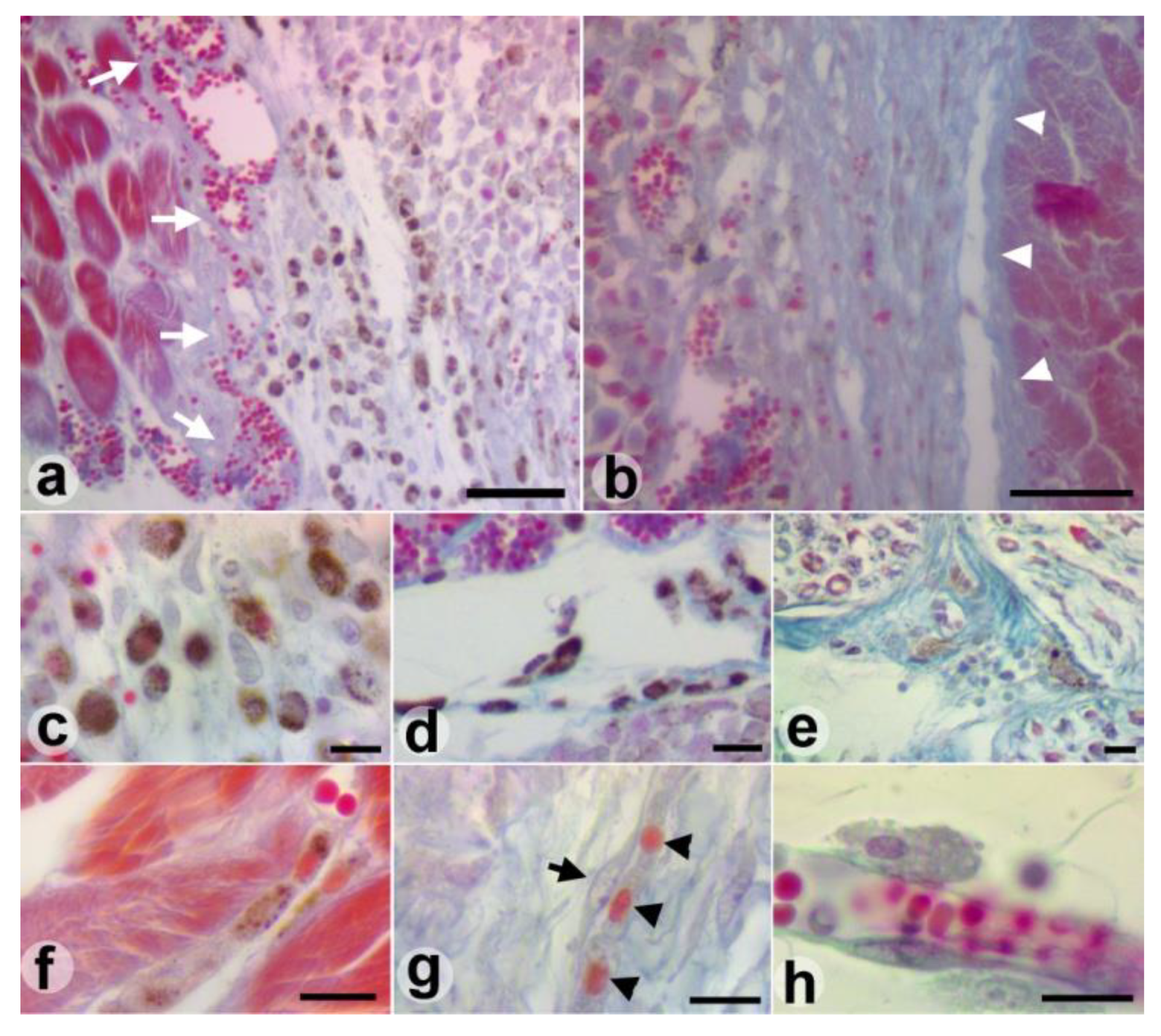
Several migration patterns of pigmented melanoma cells were identified in the peritumoral connective tissue and the muscles around the primary tumor. Changes in the shape and stiffness of the pigmented melanoma cells due to the interaction with the surrounding stromal elements can be observed during migration through the connective tissue. These changes in cell morphology are associated with a decrease in the pigment loading of melanoma cells. Individual low-pigmented melanoma cells with fibroblast-like spindle-shaped morphology and extension of the cell's leading edge attached to the matrix fibers of connective tissue were identified at a distance from the primary tumor. These so-called “path-generating cells” set the direction of migration and are followed by clusters of interconnected melanoma cells (Figure 2 a, b).
This is a way for pigmented cells to invade muscles that do not have direct contact with the primary tumor. Once these cells have set the migration route, a large number of pigment-loaded melanoma cells leave the primary tumor, undergo changes in shape and pigment content, and migrate through the peritumoral connective tissue to invade the distant muscle structure (Figure 2 c-g). Increased vascularity with large newly-formed perimysial vessels that dissociate muscle bundles were identified in the invasion front of the muscle by migratory melanoma cells (Figure 2 d). Only amelanic or low-level pigmented melanoma cells were identified in the lumen of these newly formed vessels (Figure 2 g).
The migration of pigmented melanoma cells through the muscle
Other patterns of migration related to the absence or presence of desmoplastic reaction in the invasion front (Figure 3 a, b) were also observed when the pigmented melanoma cells invade muscle bundles in direct contact with the primary tumor.
The newly formed perimysial and endomysial capillaries that dissociate the muscle fibers are used by chains or clusters of pigmented melanoma cells as an entry route when there is no desmoplastic reaction in the tumor-muscle interface (Figure 3 c, d).
Both “individual cell migration” and “collective migration” patterns were detected during the endomysial fibrous connective infiltration by the pigmented tumor cells. As the pigmented cells migrate, a depigmentation process takes place and the initial round cells become elongated (Figure 3 f). Chains or single melanoma cells with progressive depigmentation levels attached to the abluminal surface of vessels (angiotropism), the endomysium of muscle fibers (myotropism) or nerve fibers (neurotropism) could be highlighted in the tumor infiltrating muscle, away from the invasion front (Figure 3 e, f, h).
Areas with thickening and fibro sclerosis of the muscle perimysium containing fibroblast-like poorly loaded melanin pigment tumor cells were identified in the muscle- tumor contact sites with desmoplastic reaction. This fibro sclerotic reaction appears to prevent the infiltration of the muscle by the melanoma tumor cells and can be considered a barrier in front of the tumor invasion. However, fine networks of newly formed vessels could be identified on both sides of this fibro sclerotic barrier both in the desmoplastic tissue and the adjacent muscle fiber endomysium. Pigmented melanoma cells that migrate through the desmoplastic tissue undergo rapid depigmentation and fibroblast-like transformation. Cell groups formed by three depigmented melanoma cells flanked by a fibroblast were observed frequently during migration through desmoplastic tissue (Figure 3 g). No pigmented cell was observed in the lumen of the desmoplastic tissue vessels.
The migration of low-pigmented and amelanic melanoma cells through the muscle
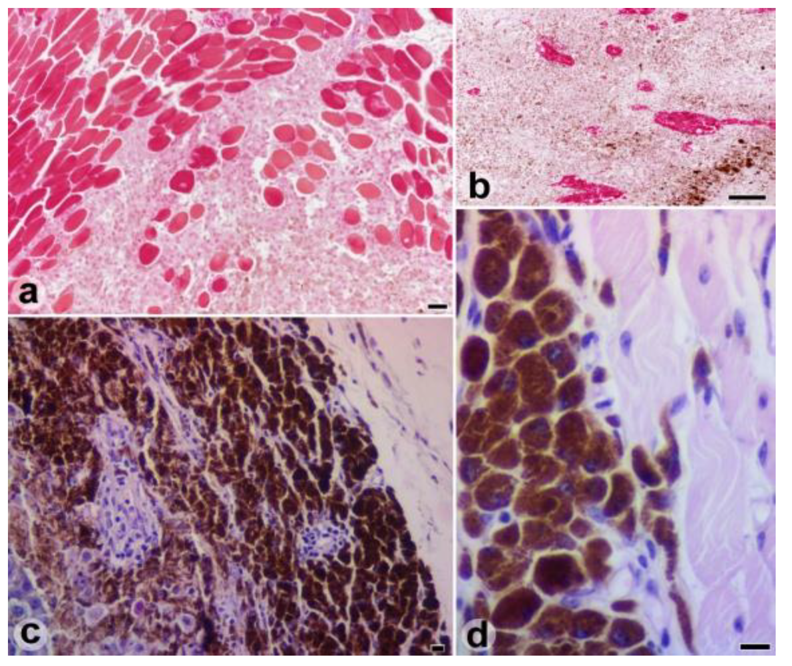
The replacement histopathological growth pattern was specific to muscle invasion by this type of melanoma cells (Figure 4 a).
The infiltration, dissociation, destruction, and replacement of muscle fibers by cancer cells, the lack of angiogenesis, and the ability to generate tumor emboli by rapidly invading the lumen of the lymphatic and blood vessels are the morphological aspects common in the case of invasion by amelanic or low pigmented melanoma cells. Finally, a tumor mass with numerous areas of necrosis and blood lacunae delimited by tumor cells replaced the initial muscle tissue (Figure 4 b).
The migration of heavily pigmented melanoma cells
A large subpopulation of heavily pigmented cells disposed at the periphery of the primary tumor was highlighted by the histological examination of the stopped growth tumor (Figure 4 c). Only a few heavily pigmented melanoma cells that migrated attached to the vessels or between neighboring muscle bundles were identified (Figure 4 d). The wedge-like clusters of slightly deformed cells that dissociated muscle bundles, the lack of melanoma cell depigmentation and the absence of host response were the histological aspects that characterize melanoma cell migration in this tumor.

Discussions

This study showed that the morphological features and tumor growth were similar in both groups of mice, even if the syngeneic transplant was different (cultured cells vs. tumor tissue). The difference in the mean volume of the tumors developed in the two groups of mice can be attributed to the different number of cells injected to produce the tumors, as no cell count could be made in the implanted tumor tissue. Unlike the allogeneic transplant with chemically-induced skin tumor tissues whose volumes started to decrease 8 days after inoculation [26], melanoma syngeneic transplanted tissues in mice showed a progressive volume increase due to the preservation of the tumor cell viability. This model allows the removal of the initial context that promotes tumor growth, created by the primary tumor interaction with the micro- and macro environment [27]. Therefore, when it is necessary to assess the melanoma cells that have undergone behavioral changes by applying therapy, the syngeneic tumor tissue transplantation may be a viable alternative. In addition, the ability to study the mechanisms of transmissible cancers [28] can be another facility offered by this experimental model.
In the case of the tumor with stopped growth, there are no signs of host response (replacement of melanoma cells with mononuclear infiltration and/or fibrosis with increased vascularity [29]) to consider it to be a histological regression. The peritumoral migration in small numbers of heavily pigmented melanoma cells and the presence of tumor emboli identified in the pulmonary arteries are elements that support the view that hyperpigmentation is likely the factor that slows tumor growth in this case than a sign of the spontaneous histological regression of the primary tumor.
A series of recent studies has shown that the level of pigment loading can affect the metastatic behavior of melanoma cells [23,24]. Melanin pigment synthesis and its loading into granule-like organelles called melanosomes are heterogeneous processes that can lead to different levels of melanoma cell pigmentation [30,31,32]. Due to the fact that loaded melanosomes are very stiff and hard to deform, the elasticity of the pigmented melanoma cells depends solely on the amount of endogenous pigment [23]. The cancer cells with high elasticity exhibited higher invasive potential (22).
Unlike heavily pigmented melanoma cells, the other pigmented melanoma cells exhibit more migratory abilities. Environmental properties directly influence the morphological changes of the pigmented cells during migration. Progressive depigmentation appears to be a necessity for adapting the shape of the cell and can approach complete depigmentation. Moreover, changes in pigment production are reversible, and melanoma cells could increase pigmentation when they reach metastatic locations [24].
The rapid depigmentation, the fibroblast-like transformation of melanoma cells, and the formation of cell groups consisting of a fibroblast and some melanoma cells, are morphological traits observed mainly in the melanoma that displays a variable pattern of desmoplasia. Melanoma– fibroblast interactions can be considered the cause of these morphological changes, knowing that melanoma cells can recruit and activate stromal fibroblasts which in turn provide the structural and chemical support for melanoma cell migration [33,34,35].
The lack of pigmented melanoma cells in the lumen of newly formed vessels could explain the local aggressiveness and low metastatic rate observed in these types of melanoma [36].
Whereas low-level pigmented or amelanotic melanoma cells can pass directly into the lumen of the primary tumor lymphatic vessels, it seems that pigmented melanoma cells need depigmentation to pass through the wall of the blood vessels, the ability of the melanoma cell to pass through a mechanical barrier being directly influenced by the level of pigmentation [37]. Therefore, it might be concluded that one of the causes of angiotropism is the pigmented melanoma cells’ inability to penetrate the vascular wall.
The invasion patterns of the muscles highlighted in the same tumor showed differences in the aggressiveness of various subpopulations of pigmented melanoma cells. While pigmented melanoma cells exhibited the ability to migrate with the induction of alterations in the muscle perimysium and endomysium, amelanotic and low- pigmented melanoma cells replaced muscle fibers with tumor tissue and rapidly entered the lymphatic vessels. The modulation of the migratory abilities by the pigment loading level could explain the possible relationship between the pigmentation of the melanoma cells and the patient’s survival, also highlighted in a series of recent studies [38,39].

Conclusions

This study showed that syngeneic B16F10 melanoma tissue transplantation in C57BL/6 mice was a viable technique for the study of melanoma cell behavior. The melanic pigment loading level and the ability to remove the pigment seem to be defining elements of the migratory behavior and aggressiveness of the B16F10 melanoma cells.

References

  1. Balch, C.M.; Soong, S.J.; Gershenwald, J.E.; et al. Prognostic factors analysis of 17,600 melanoma patients: Validation of the American Joint Committee on Cancer melanoma staging system. J Clin Oncol. 2001, 19, 3622–3634. [Google Scholar] [CrossRef]
  2. Anbari, K.K.; Schuchter, L.M.; Bucky, L.P.; et al. Melanoma of unknown primary site: presentation, treatment, and prognosis-a single institution study. University of Pennsylvania Pigmented Lesion Study Group. Cancer. 1997, 79, 1816–1821. [Google Scholar] [CrossRef]
  3. Fidler, I.J. Selection of successive tumour lines for metastasis. Nat New Biol. 1973, 242, 148–149. [Google Scholar] [CrossRef]
  4. Caisová, V.; Vieru, A.; Kumžáková, Z.; et al. Innate immunity based cancer immunotherapy: B16-F10 murine melanoma model. BMC Cancer. 2016, 16, 940. [Google Scholar] [CrossRef] [PubMed]
  5. Eberting, C.L.; Shrayer, D.P.; Butmarc, J.; Falanga, V. Histologic progression of B16 F10 metastatic melanoma in C57BL/6 mice over a six-week time period: distant metastases before local growth. J Dermatol. 2004, 31, 299–304. [Google Scholar] [CrossRef] [PubMed]
  6. Ishibashi, S.; Sonoda, K.; Fujii, K.; Ishikawa, K.; Shiraishi, N.; Kitano, S. A convenient murine model for the study of intra-abdominal lymph node metastasis. Oncol Rep. 2004, 12, 115–118. [Google Scholar] [CrossRef] [PubMed]
  7. Rozenberg, G.I.; Monahan, K.B.; Torrice, C.; Bear, J.E.; Sharpless, N.E. Metastasis in an orthotopic murine model of melanoma is independent of RAS/RAF mutation. Melanoma Res. 2010, 20, 361–371. [Google Scholar] [CrossRef]
  8. Lugassy, C.; Zadran, S.; Bentolila, L.A.; et al. Angiotropism, pericytic mimicry and extravascular migratory metastasis in melanoma: an alternative to intravascular cancer dissemination. Cancer Microenviron. 2014, 7, 139–152. [Google Scholar] [CrossRef]
  9. Bielenberg, D.R.; Zetter, B.R. The Contribution of Angiogenesis to the Process of Metastasis. Cancer J. 2015, 21, 267–273. [Google Scholar] [CrossRef]
  10. Bugyik, E.; Renyi-Vamos, F.; Szabo, V.; et al. Mechanisms of vascularization in murine models of primary and metastatic tumor growth. Chin J Cancer 2016, 35, 19. [Google Scholar] [CrossRef]
  11. Bentolila, L.A.; Prakash, R.; Mihic-Probst, D.; et al. Imaging of Angiotropism/Vascular Co-Option in a Murine Model of Brain Melanoma: Implications for Melanoma Progression along Extravascular Pathways. Sci Rep 2016, 6, 23834. [Google Scholar] [CrossRef] [PubMed]
  12. Kerbel, R.S. What is the optimal rodent model for anti- tumor drug testing? Cancer Metastasis Rev. 1998- 1999, 17, 301–304. [Google Scholar]
  13. Killion, J.J.; Radinsky, R.; Fidler, I.J. Orthotopic models are necessary to predict therapy of transplantable tumors in mice. Cancer Metastasis Rev. 1998- 1999, 17, 279–284. [Google Scholar] [CrossRef]
  14. Hoffman, R.M. Orthotropic metastatic mouse models for anticancer drug discovery and evaluation: a bridge to the clinic. Invest New Drugs. 1999, 17, 343–359. [Google Scholar] [PubMed]
  15. Fidler, I.J.; Nicolson, G.L. Organ selectivity for implantation survival and growth of B16 melanoma variant tumor lines. J Natl Cancer Inst. 1976, 57, 1199–1202. [Google Scholar]
  16. Herlyn, M.; Fukunaga-Kalabis, M. What is a good model for melanoma? J Invest Dermatol. 2010, 130, 911–912. [Google Scholar] [CrossRef]
  17. Zbytek, B.; Carlson, J.A.; Granese, J.; Ross, J.; Mihm, M.C.; Slominski, A. Current concepts of metastasis in melanoma. Expert Rev Dermatol. 2008, 3, 569–585. [Google Scholar]
  18. Valastyan, S.; Weinberg, R.A. Tumor metastasis: molecular insights and evolving paradigms. Cell. 2011, 147, 275–292. [Google Scholar]
  19. Mishra, H.; Mishra, P.K.; Ekielski, A.; Jaggi, M.; Iqbal, Z.; Talegaonkar, S. Melanoma treatment: from conventional to nanotechnology. J Cancer Res Clin Oncol. 2018, 144, 2283–2302. [Google Scholar]
  20. Mîndrilă, I.; Buteică, S.A.; Mihaiescu, D.E.; Badea, G.; Fudulu, A.; Mărgăritescu, D.N. Fe3O4/salicylic acid nanoparticles versatility in magnetic mediated vascular nanoblockage. J Nanopart Res. 2016, 18, 10. [Google Scholar]
  21. Zhou, Z.; Zheng, C.; Li, S.; et al. AFM nanoindentation detection of the elastic modulus of tongue squamous carcinoma cells with different metastatic potentials. Nanomedicine 2013, 9, 864–874. [Google Scholar] [CrossRef] [PubMed]
  22. Park, S. Nano-mechanical phenotype as a promising biomarker to evaluate cancer development, progression, and anti-cancer drug efficacy. J Cancer Prev. 2016, 21, 73–80. [Google Scholar] [CrossRef]
  23. Sarna, M.; Zadlo, A.; Czuba-Pelech, B.; Urbanska, K. Nanomechanical Phenotype of Melanoma Cells Depends Solely on the Amount of Endogenous Pigment in the Cells. Int J Mol Sci. 2018, 19, 607. [Google Scholar] [CrossRef]
  24. Pinner, S.; Jordan, P.; Sharrock, K.; et al. Intravital imaging reveals transient changes in pigment production and Brn2 expression during metastatic melanoma dissemination. Cancer Res. 2009, 69, 7969–7977. [Google Scholar] [CrossRef] [PubMed]
  25. Ogawa, Y.; Kawamura, T.; Furuhashi, M.; Tsukamoto, K.; Shimada, S. Improving chemotherapeutic drug penetration in melanoma by imatinib mesylate. J Dermatol Sci. 2008, 51, 190–199. [Google Scholar] [CrossRef]
  26. Zhang, Z.; Sun, H.; Zhang, J.; et al. Safety and Efficacy of Transplantation with Allogeneic Skin Tumors to Treat Chemically-Induced Skin Tumors in Mice. Med Sci Monit 2016, 22, 3113–3123. [Google Scholar] [CrossRef] [PubMed]
  27. Bissell, M.J.; Radisky, D. Putting tumours in context. Nat Rev Cancer. 2001, 1, 46–54. [Google Scholar] [CrossRef]
  28. Pye, R.J.; Pemberton, D.; Tovar, C.; et al. A second transmissible cancer in Tasmanian devils. Proc Natl Acad Sci U S A. 2016, 113, 374–379. [Google Scholar] [CrossRef]
  29. Aung, P.P.; Nagarajan, P.; Prieto, V.G. Regression in primary cutaneous melanoma: etiopathogenesis and clinical significance. Lab Invest 2017, 97, 657–668. [Google Scholar] [CrossRef]
  30. Lazova, R.; Pawelek, J.M. Why do melanomas get so dark? Exp Dermatol. 2009, 18, 934–938. [Google Scholar] [CrossRef]
  31. D’Ischia, M.; Wakamatsu, K.; Napolitano, A.; et al. Melanins and melanogenesis: Methods, standards, protocols. Pigment Cell Melanoma Res. 2013, 26, 616–633. [Google Scholar] [PubMed]
  32. Slominski, R.M.; Zmijewski, M.A.; Slominski, A.T. The role of melanin pigment in melanoma. Exp Dermatol. 2015, 24, 258–259. [Google Scholar] [CrossRef] [PubMed]
  33. Flach, E.H.; Rebecca, V.W.; Herlyn, M.; Smalley, K.S.; Anderson, A.R. Fibroblasts contribute to melanoma tumor growth and drug resistance. Mol Pharmacol. 2011, 8, 2039–2049. [Google Scholar]
  34. Zhou, L.; Yang, K.; Andl, T.; Wickett, R.R.; Zhang, Y. Perspective of Targeting Cancer-Associated Fibroblasts in Melanoma. J Cancer. 2015, 6, 717–726. [Google Scholar] [CrossRef]
  35. Löffek, S.; Zigrino, P.; Angel, P.; Anwald, B.; Krieg, T.; Mauch, C. High invasive melanoma cells induce matrix metalloproteinase-1 synthesis in fibroblasts by interleukin-1alpha and basic fibroblast growth factor- mediated mechanisms. J Invest Dermatol. 2005, 124, 638–643. [Google Scholar]
  36. Marques, P.C.; Diniz, L.M.; Spelta, K.; Nogueira, P.S.E. Desmoplastic melanoma: a rare variant with challenging diagnosis. A Bras Dermatol. 2019, 94, 82–85. [Google Scholar]
  37. Sarna, M.; Zadlo, A.; Hermanowicz, P.; Madeja, Z.; Burda, K.; Sarna, T. Cell elasticity is an important indicator of the metastatic phenotype of melanoma cells. Exp Dermatol. 2014, 23, 813–818. [Google Scholar]
  38. Thomas, N.E.; Kricker, A.; Waxweiler, W.T.; et al. Comparison of clinicopathologic features and survival of histopathologically amelanotic and pigmented melanomas: A population-based study. JAMA Dermatol. 2014, 150, 1306–1314. [Google Scholar] [CrossRef]
  39. Vernali, S.; Waxweiler, W.T.; Dillon, P.M.; et al. Association of Incident Amelanotic Melanoma with Phenotypic Characteristics, MC1R Status, and Prior Amelanotic Melanoma. JAMA Dermatol. 2017, 153, 1026–1031. [Google Scholar]
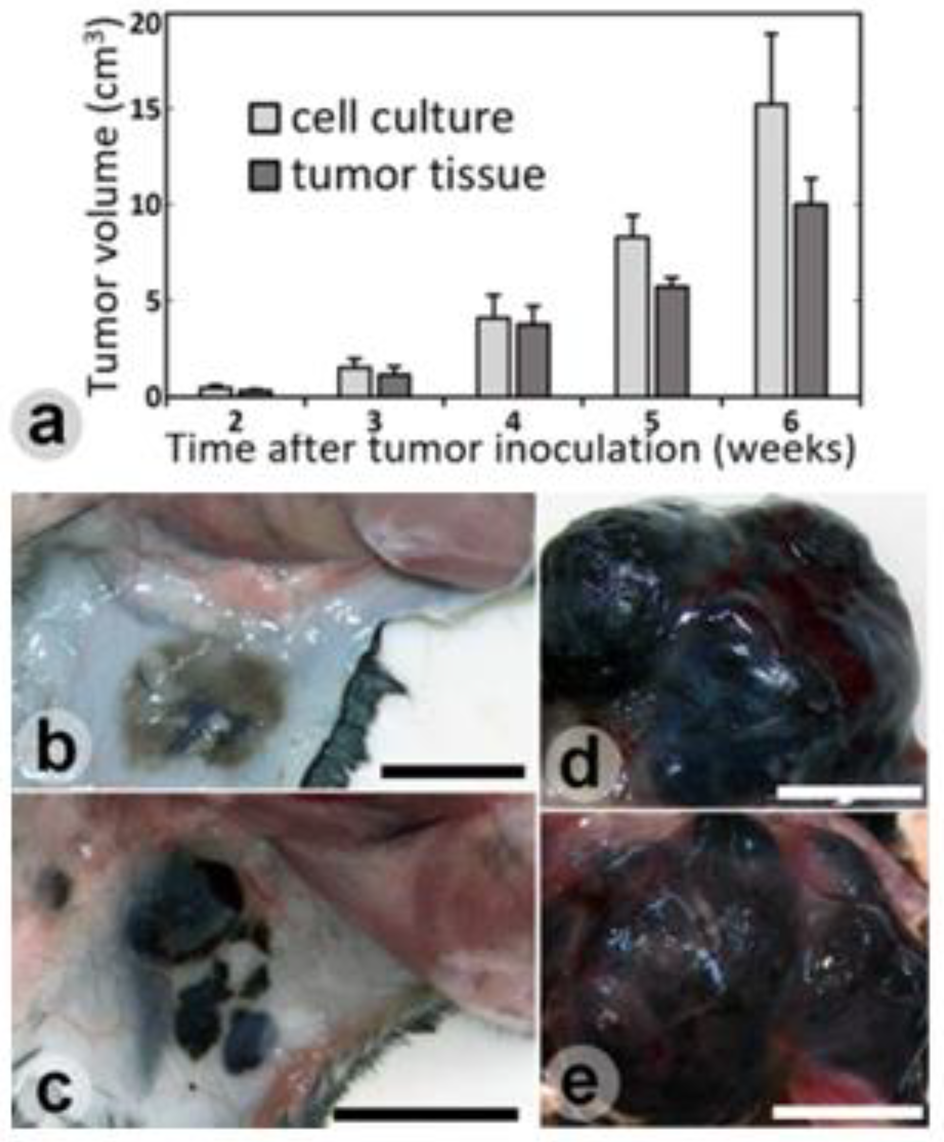
Figure 1. Growth assessment of syngeneic B16F10 melanoma cells or tissue transplanted into C57BL/6 mice (a). The implant site of non-developed tissue melanoma (b) and growth stopped melanoma (c). Six-week cells (d) and tissue (e) implanted melanoma. Bares = 1 mm.
Jmms 06 00045 g001
Figure 2. Cluster of migratory pigmented melanoma cells (a, b), and melanoma cell shape and pigment loading level changes (c-g) during peritumoral connective tissue migration. H&E (a-c, e, f) and azan trichrome (d, g) staining. Bare = 100 (a); 25 (b, c, d); 10 (e, f, g) μm.
Jmms 06 00045 g002
Figure 3. Interfaces of pigmented melanoma cell muscle migration with angiogenesis (a-white arrows), or desmoplastic reaction with strips of dense collagen (b-white arrowheads) that separates the muscle from the tumor. The migration patterns of the pigmented melanoma cells through muscle tissue: cluster of cells (c); Indian file pattern (d); neurotropism (e); endomysial (myotropism) (f); and angiotropism (h). Pack of fibroblast (black arrow) and depigmented melanoma cells (black arrowheads) in the desmoplastic area (g). Azan trichrome staining. Bare = 100 µm (a, b); 10 µm (c- h).
Jmms 06 00045 g003
Figure 4. The avascular infiltrative histopathological growth pattern of amelanotic and the low pigmented melanoma cells with progressive replacement of muscle fibers (a), extensive tumor necrosis and the occurrence of blood lacunae delimited by melanoma cells (b). The peripheral arrangement of heavily pigmented melanoma cells (c) and the invasion of the nearby muscle in the tumor with the stopped growth (d). Azan trichrome (a, b) and H&E (c, d) staining. Bare = 100 (a, b); 10 (c, d) µm.
Jmms 06 00045 g004