Next Article in Journal
Effects of the COVID-19 Pandemic on Clinical Manifestations and Therapeutic Outcomes in Acute Endophthalmitis
Previous Article in Journal
Assessment of the Bioaccumulation of Nicotine and Cotinine by the Crustacean Daphnia magna
 
 
Journal of Mind and Medical Sciences is published by MDPI from Volume 12 Issue 1 (2025). Previous articles were published by another publisher in Open Access under a CC-BY (or CC-BY-NC-ND) licence, and they are hosted by MDPI on mdpi.com as a courtesy and upon agreement with Valparaiso University (ValpoScholar).
Article

Infertility as a Possible Multifactorial Condition; The Experience of a Single Center

by
Cristina Diana Popescu
1,
Bashar Haj Hamoud
2,
Romina Marina Sima
3,4,
Anca Bobirca
3,5,
Oana Denisa Balalau
3,4,*,
Mihaela Amza
3,4,
Romeo Micu
6,
Gabriel Petre Gorecki
7 and
Liana Ples
3,4
1
Department PhD, IOSUD, Carol Davila University of Medicine and Pharmacy, Bucharest, Romania
2
Department for Gynecology, Obstetrics and Reproductive Medicine, Saarland University Hospital, Homburg, Germany
3
Carol Davila University of Medicine and Pharmacy, Bucharest, Romania
4
Department of Obstetrics and Gynecology, St John Hospital, Bucur Maternity, Bucharest, Romania
5
Department of Internal Medicine and Rheumatology, Dr. I. Cantacuzino Clinical Hospital, Bucharest, Romania
6
Iuliu Hatieganu University of Medicine and Pharmacy, Cluj-Napoca, Romania
7
Faculty of Medicine, Titu Maiorescu University, Bucharest, Romania
*
Author to whom correspondence should be addressed.
J. Mind Med. Sci. 2024, 11(2), 466-474; https://doi.org/10.22543/2392-7674.1535
Submission received: 19 March 2024 / Revised: 19 April 2024 / Accepted: 11 June 2024 / Published: 31 October 2024

Abstract

:
Objectives. Infertility is a topic of great interest around the world because it affects many couples at young ages. It can be caused by genetic background, associated with pathologies and/or external factors. The purpose of our study was to identify the causes of infertility of women presented in our clinic with this pathology. Materials and Methods. This retrospective study was performed on women with primary or secondary infertility. The analyzed data were age, weight, hereditary and personal pathological history, medication, menstrual cycle characteristics, standard blood tests, ultrasound, hysterosalpingography and hysteroscopy. Results. The study included 204 women with average age 35 years. The main diagnosis was primary infertility in 68.63% and secondary infertility in 31.37% cases. One of the most common diagnosed findings in ultrasound were uterine fibroids with an incidence of 6.86%, the incidence being higher among women with primary infertility than in women with secondary infertility. Regarding endometrial polyps, 96.15% of cases were observed ultrasonographical and the incidence of endometrial polyps was higher among women with primary infertility than in women with secondary infertility. Conclusions. This study identified that infertility is a multifactorial pathology, which requires multidisciplinary addressability. Gynecological pathology (such as tubal pathologies, uterine malformations, uterine fibroids, endometriosis, endometrial polyps, etc.) was very common among these patients, finding and treating the condition being the main objective of the study.

Introduction

Reproduction implies a multitude of biological processes from organ formation and development to neurological and endocrine regulation, hormone production, meiosis and mitosis [1]. Infertility is known as a major human reproductive disease and affects one in seven couples worldwide [2].
In recent decades genetics has progressed significantly bringing many benefits in the field of reproductive endocrinology and infertility studies. In this context pre-implantation genetic testing was considered one of the most important topics [3]; this method evaluates genetic status of the embryos prior to transfer for in vitro fertilization. Technological advances such as next-generation sequencing have increased efficiency and accuracy [4].
Infertility was defined as the inability to achieve pregnancy after 6 or 12 months of unprotected sexual contact, depending on the age group. A complete and correctly performed medical anamnesis plays an important role, as well as a clinical examination [5]. The causes of infertility in women have been defined as uterine, tubal, ovarian, hormonal pathology, but external factors such as alcohol or illicit substances, smoking, diet, weight or psycho-emotional factors should not be neglected [6].
A review (2023) of the literature by the International Federation of Gynecologists and Obstetricians (FIGO) has supported that weight loss in obese patients has a beneficial effect on fertility [7]. Another study published (January 2023) showed that women with polycystic ovary syndrome and high body mass index were less successful in getting pregnant [8]. Weight loss was associated with positive results compared to maintaining a stable weight or increasing weight. According to a World Health Organization publication it has been stated that obesity rates among young women are increasing worldwide and it is estimated that 20% of women will be obese by 2025 [9].
There are numerous studies in the literature about the types of infertility, primary or secondary, and their associated causes. A recent published study evaluating the role of hysteroscopy on endometrial pathology claimed that transvaginal ultrasound was easily correlated with hysteroscopy and the hysteroscopic diagnostic and treatment techniques significantly increased the chances of achieving pregnancy if the cause was endometrial [10].
Studies have shown that endometrial polyps are involved in 8-12% of infertility cases [11] in women of childbearing age, adenomyosis in another 35% of cases [12] and uterine fibroids affect more than 70% of women over the age of 40 [13]. A study involving 215 patients diagnosed with endometrial polyps was published in the literature [14,15]; of these patients those who underwent hysteroscopic polypectomy were twice as successful in becoming pregnant than those that have not been operated [16]. In terms of the relationship between endometrial polyps and infertility, the data showed that potential mechanisms include both mechanical action and the release of molecules that negatively affect sperm transport or embryo implantation; there was evidence of increased levels of glycodelin [17], aromatase [18], inflammatory markers [19] and reduced levels of HOXA-10 and -11 messenger RNA [20]. Adenomyosis has also been shown to be a factor in infertility by altering the architecture and function of the myometrium, altering uterine peristalsis and sperm transport [21]. Adenomyosis may manifest itself as reduced endometrial receptivity with defects or abnormalities of implantation markers [22].
Regarding fibroids, the mechanisms involved in infertility impairment have been described as abnormal uterine contractility, abnormal endometrial cytokine expression, abnormal vascularity and chronic endometrial inflammation [23]. There was evidence that hysteroscopic myomectomy improved spontaneous pregnancy rates by 21%-39% [24,25].
A study published in 2021 claimed that synechia was mainly responsible for hypomenorrhea even amenorrhea and could be associated with chronic pelvic pain or dysmenorrhea—it could be diagnosed during a systematic infertility evaluation. The main hypothesis was that obliteration of the cavity limits sperm migration, especially if adhesions involved the tubal ostia and cervix. The goal of treating synechia is to restore a normal-sized cavity with a functional endometrium [26].
Despite de advances in diagnosis there are still cases where infertility is not explained; about 15% of couples diagnosed with infertility have “unexplained infertility” [27].

Material and Methods
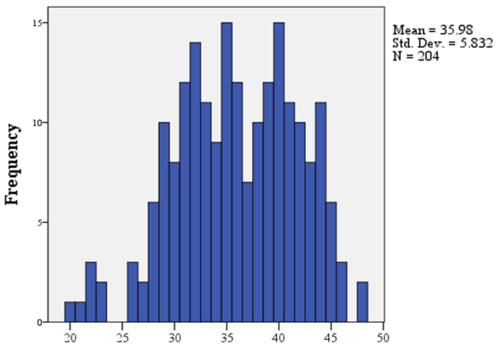
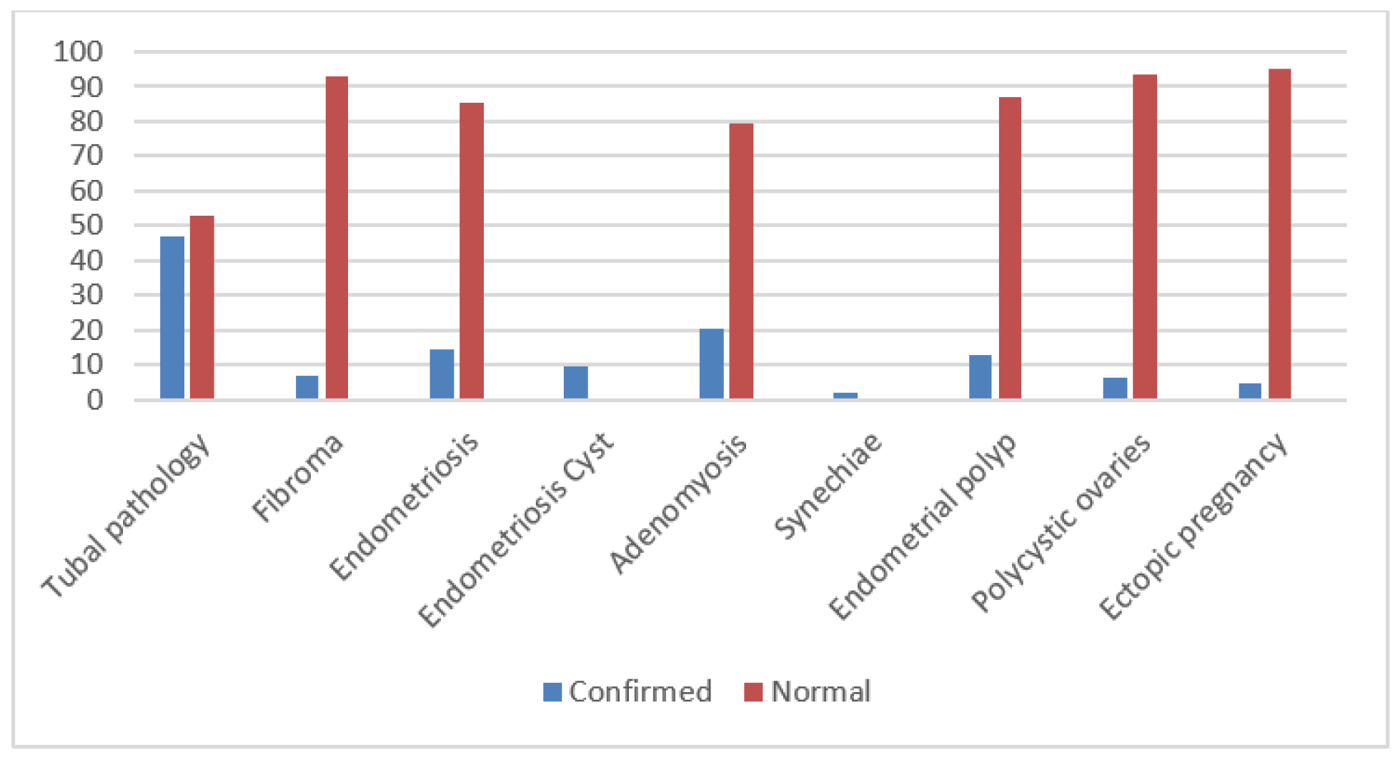
This retrospective study was performed in our clinic and included women with primary or secondary infertility investigates over 12 months in 2022. The following parameters were collected: personal data (age, weight, hereditary and personal pathological history, medication), menstrual cycle characteristics, blood tests, ultrasound, hysterosalpingography and hysteroscopy data. All patients included in the study were over 18 years old and had a minimum of one year of unprotected sexual contact without pregnancy; all of them wanted to get pregnant.
Inclusion criteria were formulated according to the diagnosis of infertility, all included patients having this diagnosis established, but also transvaginal ultrasound as the main method of examination—all patients had at least one transvaginal ultrasound before being included in the study. The exclusion criteria consisted of partial or total lack of data to complete the parameters as well as patients who had completed all the needed investigations but only wanted to preserve oocytes (either did not want a pregnancy at the moment or did not have a partner).
We wanted to include in this group of patients those at fertile age, even extreme age, the age limit being between 20 and 48 years, all patients having menstruation and already established the diagnosis of infertility. The body mass index, associated factors such as smoking, but also the hereditary or personal pathological history of each patient—both medical and surgical—were analyzed; patients with polycystic ovary syndrome or low ovarian reserve (the lowest value was 0.04 ng/ml) were also included in the study.
All patients underwent transvaginal ultrasonography and hysterosalpingography or hysteroscopy. The study was approved by the Ethics Committee (12.04.2023, number 9082).
The parameters were evaluated and processed in IBM SPSS Statistics version 21. The statistical tests used in the analysis were Chi-Square Test used to test whether two normal or dichotomous variables are associated, Mann-Whitney U Test to determine whether there were differences between two independent groups when the dependent variable was either ordinal or continuous but not normally distributed and Spearman Correlation Indicator measured the strength and direction of the link between two variables and analyses the relationship between them.
The purpose of our study was to identify the causes of infertility of women with this pathology presented in our clinic.

Results

The study included 204 women aged 20 to 48 years, half of them over 36 years (50%), the average age of the patients analyzed was 35.98 years (Histogram 1). Of the total patients 68.63% were diagnosed with primary infertility (n = 140) and 31.37% with secondary infertility (n = 64).
Figure 1. Histogram no. 1, with age distribution of the patients included in the study.
Jmms 11 00059 g001
The patients included in the study had a Body Mass Index (BMI) between 16.71 (underweight) and 46.98 (morbidly obese) with a mean BMI of 24.11 (normal weight). The percentage of normal weight patients was 60.83% (n = 73) and underweight was only 5% (n = 6) (Table 1).
Non-smoking patients predominated in the analyzed group represented 81.86% of cases (n = 167), while 18.14% of patients were smokers (n = 37); the incidence of smoking was higher among patients with secondary infertility compared to those with primary infertility (21.87% vs. 16.43%). Only 6.37% of women had a family history such as hypertension, diabetes, heart attack, stroke, etc. (n = 13), while the rest denied it (93.63%, n = 191). Half of the patients had a personal pathological history such as hypothyroidism, peptic ulcer, appendectomy, cholecystectomy, varicose veins in the lower limbs, hypertension, thrombophilia, endometriosis, etc. (50%, n = 102), the incidence of which was significantly higher among women with secondary infertility (n = 43) than those with primary infertility (n = 59) (67.19% vs. 42.14%). Of the patients with a personal pathological history 0.98% of them had a history of Tuberculosis (n = 2). 1.96% patients stated that they were in menopause or perimenopause (n = 4) and 1. 47% had undergone tubal permeabilization procedures (n = 3); 7.35% were on medication (most commonly Euthyrox) (n = 15), 4.9% had hypothyroidism (n = 10). Regarding arterial hypertension, 1.96% (n = 4) of the patients included in the study were diagnosed with this pathology.
The patients included in the group had AMH (Anti-Müllerian Hormone) test values ranging from 0.04 ng/ml to 17.80 ng/ml, with a mean value of 2.42 ng/ml: 10.87% of the patients had low ovarian reserve (n = 20) with values between 1 and 1.5 ng/ml, 19.57% of patients had very low AMH values indicating menopausal approach (n = 36) with values between 0,5 and 1 ng/ml and 17.93% of the patients indicated values attesting the onset of menopause (n = 33) with values bellow 0,5 ng/ml (Histogram 2). Patients with normal ovarian reserve were aged between 23 and 45 years, with a mean age of 34.42 years and those with ovarian reserve corresponding to menopause were aged between 29 and 45 years, with a mean age of 39 years.
In 6.37% of patients polycystic ovary syndrome (PCOS) was diagnosed (n = 13): 7.14% of women with primary infertility (n = 10) and 4.69% of women with secondary infertility (n = 3). Patients with this diagnosis were between 22 and 39 years old, with an average age of 30.23 years.
The patients included in the group had Thyroid Stimulating Hormone (TSH) values ranging from 0.24 μIU/mL to 5.21 μIU/mL, with a mean value of 1.80 μIU/mL; 96.18% of patients had normal TSH values (n = 126), only 1.53% had low values (n = 2) and 2.29% had high TSH values (n = 3). Prolactin values in most patients were within normal limits (79.55%, n = 35), but elevated values were also found (15.91%, n = 7); 89.52% had normal CA125 values (n = 111) and 10.48% had elevated values (n = 13).
Figure 2. Histogram no. 2 with AMH value distribution.
Jmms 11 00059 g002
Regarding ectopic pregnancies, 4.90% of the women in the group had at least one ectopic pregnancy in their history (n = 10); most of them had no ectopic pregnancies (95.10%, n = 194), but 2.45% (n = 5) of the women had a previous ectopic pregnancy, 1.96% (n = 2) had two ectopic pregnancies, and 0.49% (n = 1) had 3 ectopic pregnancies in the past.
The patients included in the group had menarche between 10 and 17 years, half of them earlier than 12 years (50%). The mean age of menarche onset was 12.5 years. Only 2.5% of them had irregular menstrual cycles (n = 5), with the group predominantly composed of patients with regular period. Dysmenorrhea was frequently reported, 60.29% of patients were affected (n = 123), while 39.71% of women denied dysmenorrhea symptoms (n = 81). A large proportion of women in the group experienced dyspareunia, i.e., 43.14% of patients were affected (n = 88). The incidence of HBS, HCV, HIV or VDRL infections was 1.96% in the studied group (n = 4); 34.31% of patients were diagnosed with thrombophilia (n = 70).
In terms of HPV infection, 13 of the 204 patients included in the group had HPV testing, 23.08% of them were infected with strain 16 (n = 3), 53.85% of negative cases (n = 7) and 15.38% of positive cases but without known strain (n = 2). Of all patients, 91,67% (n = 187) had normal results on Pap Cytology, but in 7.35% of cases inflammation was detected (n = 15) and in 0.98% of cases an altered result (LSIL – low grade intraepithelial lesion or ASCUS—atypical squamous cells of undetermined significance) was obtained (n = 2).
Of the total of 204 patients, 134 performed the hysterosalpingography (HSG), the results obtained indicating that 47.01% (n = 63) of them had non-permeable fallopian tubes. In women with non-permeable fallopian tubes 54.55% of the cases showed bilateral non-permeability (n = 30) (Figure 3) and in 15.87% of patients with altered HSG result only narrowed tubal was present (n = 10); another 2 of the patients had both non-permeable (bilateral) and narrowed tubal evidence.
All patients included in the study group underwent transvaginal ultrasound; 1 out of 3 patients showed modified ultrasound—36.76% of them showed modified results (n = 75). Out of the total patients 158 underwent hysteroscopy, 82.28% of them had normal results (n = 130).
Investigations confirmed the presence of uterine fibroids in 6.86% of patients (n = 14); in all 14 cases the fibroids were G3-G4 and one case G0. Women with fibroids ranged in age from 33 to 44 years, with a mean age of 39 years; the incidence of fibroids was higher among women with primary infertility (n = 12 - 8.57%). It was found that 14.71% of the patients in the group had endometriosis (n = 30), the incidence of endometriotic cyst was 9.80%—20 of the patients. The incidence of adenomyosis was 20.59% in the group of patients studied, affecting 42 women (Figure 4).
Synechiae were diagnosed in 4 patients, representing 1.96% of the patients in the group. Regarding annexes, 88.73% had normal adnexa (n = 181), but 10.29% of them had ovarian cysts (n = 21) and 0.98% had hydrosalpinx (n = 2) (Figure 5). In terms of tubal ligation in the antecedent, it was identified in 2.45% of patients (n = 5).
The incidence of endometrial polyp in the analyzed group was 12.75% being diagnosed in 26 patients; 33.33% of patients with modified ultrasound showed endometrial polyp (n = 25) and 75% of patients with modified hysteroscopy showed endometrial polyp (n = 21). 15% of primary infertility patients were diagnosed with endometrial polyp (n = 21) and 7.81% of secondary infertility patients (n = 5).

Discussions

According to the World Health Organization, infertility has been studied as a health problem affecting 48 million couples and 186 million people worldwide. Endocrine factors were defined as one of the many important causes and the association with societal progress was not to be overlooked. Many of the chemicals used in the food industry directly affected endocrine function [28]. Some of these substances were strongly associated with diseases attributed to female infertility such as polycystic ovary syndrome, endometriosis, irregular menstrual cycle, development of ovarian follicles [29].
Malnutrition was also a very important factor already proven. It was known that over 98% of female survivors of Auschwitz had amenorrhoea immediately after their arrival in the concentration camps [30]. Further research had shown that exogenous hormones administered without the women’s consent played an important role [31]. They influenced the amenorrhoea induction as well as subsequent primary and secondary infertility [32].
Our study based on STROBE criteria indicated that infertility is as reported in the literature a plurifactorial condition [33].
The type of infertility depends on the age intervals of women at reproductive age. In our study the age limits varied between 20 and 48 years with an average age of 35.98 years old. Patients with primary infertility were slightly younger ranged in age from 20 to 48 years, with a mean age of 35.45 years and patients with secondary infertility ranged in age from 28 to 48 years, with a mean age of 37.13 years. Patients with secondary infertility were slightly older, but the difference is not significant. Analysis of data from a study called the Global Burden of Disease Study found that women aged 20-24 had the lowest infertility rates (3%) compared to the group of women aged 35-39 which had higher infertility rates (5.5%) [34]. The prevalence of infertility increases with age [35]; 1 in 8 women of childbearing age between 15 and 49 received investigation and treatment for infertility diagnosis worldwide. Success rates were variable depending on the age of the patient and the cause of infertility [36].
In terms of body mass index most patients were normal weight (3 out of 5 patients) and overweight (1 out of 5), but rarely obese (1 out of 10) or underweight (less than 1 out of 10). There were no significant differences in BMI between women with primary infertility and those with secondary infertility and no statistically significant association between patient age and body mass index. Obesity in women of reproductive age was known to be an increasingly common problem worldwide, reduced fertility and increased the time it takes to get pregnant [37,38].
In this study there were no significant differences between smoking or non-smoking patients and type of infertility (primary or secondary) among the patients analyzed, although the incidence of smoking was little higher among patients with secondary infertility. Data from the literature described a study conducted in the US in which 3665 women aged 18 to 45 from the National Health and Nutrition Examination Survey were included; the study was conducted over 5 years [39]. Women smokers included in this study were more frequently associated with infertility and the findings revealed that stopping smoking could reduce the risk of infertility [40].
Half of the patients included in the study related the personal pathological history (PPA) and statistical analysis showed a slight tendency for a direct association between PPA and type of infertility, in that women with secondary infertility tended to have more frequent personal pathological history than those with primary infertility. No statistically significant associations were found between the presence of PPA, BMI, smoking or age.
Hypothyroidism was found to be significantly more common in women with secondary infertility (2.86% among women with primary infertility and 9.38% among those with secondary infertility). Thyroid pathology had a significant influence on the processes of folliculogenesis, fertilization and implantation [41]. Women presenting for thyroid dysfunction should be evaluated with special care because fertility whether we referred to natural conception or through assisted reproductive technologies could be strongly influenced by thyroid function [42]. Normal TSH values were found in most patients regardless of infertility type (96.59% in those with primary infertility vs. 95.35% in those with secondary infertility), but low TSH values were found exclusively in patients with secondary infertility (4.65%) and high TSH values were found exclusively in patients with primary infertility (3.41%).
In terms of ovarian reserve there were no significant differences between the primary infertility group and the secondary infertility group. It was found a significant decrease in ovarian reserve with increasing age, so that in the group of patients studied an increased AMH value was associated with younger patients, while a decreased AMH value was associated with older age. Concerning dysmenorrhea, it was frequently reported among the patients enrolled, 3 out of 5 patients reported having the symptoms; patients with normal AMH value reported dysmenorrhea in 71.67% of cases, while patients with low ovarian reserve reported it in 47.22% of cases. There was a slight tendency of association between the presence of dysmenorrhea and ovarian reserve, in the meaning that women with low ovarian reserve or those at perimenopause tended to have dysmenorrhea less often compared to women in other fertility groups. There were studies in the literature that have reported no association between thyroid function and anti-Müllerian hormone (AMH) levels [43]; these results had suggested that there was no causal association between genetically predicted thyroid function and AMH levels in the European population [44].
It was noticed that patients with micropolycystic ovaries were significantly younger than the rest of the women included in the group. Women with this diagnosis were more frequently on medication (e.g., Euthyrox), with a slight tendency of association between the presence of micropolycystic ovaries and the fact that the patient was on medication; 20% of hypothyroid patients had micropolycystic ovaries. A significantly higher incidence of irregular menstrual cycles was noted among women diagnosed with PCOS, 23.08% of whom experienced irregular menstrual periods (n = 3), while only 1.05% of patients not experiencing PCOS had irregular menstrual cycles (n = 2). Statistical analysis suggested that patients with PCOS had a moderate tendency to develop irregular menstrual cycles. The direct cause of infertility in PCOS patients was influenced by ovulation and implantation disorders which were caused by inflammation of the ovarian tissue and endometrium and by disorders of the immune and metabolic systems [45]. Systemic inflammatory response associated with metabolic disorders, insulin resistance, hyperadrenalism, insufficient progesterone secretion and oxidative stress facilitated the onset of disease and infertility [46]. Depending on genetic background and environmental factors, some diseases may appear earlier, while others may appear years after a diagnosis of infertility [47].
Evaluation and treatment for infertility were recommended and evaluation included assessment of the uterus and fallopian tubes with ultrasonography or hysterosalpingography when needed [48]. Laparoscopy, hysteroscopy or magnetic resonance imaging could be required in patients with endometriosis, leiomyomas or evidence of previous pelvic infection [49]. Stopping or limiting alcohol consumption, avoiding tobacco and illicit drug use, as well as weight loss when necessary and healthy diet could improve success rates [50].
Of the patients who underwent hysterosalpingography, the results obtained indicating that half of them had non-permeable fallopian tubes; in women with non-permeable fallopian tubes more than half of the cases showed bilateral non-permeability. A study published in 2021 of 199 patients diagnosed with infertility who underwent hysterosalpingography showed that 21% of them had unilateral and 7% bilateral non permeable fallopian tubes [51]. In the present study the percentage of altered hysterosalpingography was higher than already published in the literature. Compared to literature data which showed that 36.9% of hysteroscopies were unmodified in women with infertility [52], our study found that 82.28% were normal.
All patients included in the study group had at least one transvaginal ultrasound, most of them with normal results. One of the most commonly diagnosed was uterine fibroids with an incidence of 6.86% of all patients; 17.33% of patients with altered ultrasound showed fibroids. Next 46.15% of fibroid cases resulted in modified hysteroscopies (n = 6). A doubling of the incidence of increased CRP among fibroid patients was noted, so that more than half of fibroid patients had increased CRP values (55.56%), practically more than 1 in 2 fibroid patients had increased CRP, which indicated that fibroid patients had a tendency to have elevated CRP values. It was observed that in patients with uterine fibroids the CA125 marker was more frequently elevated, i.e., one third of fibroid patients had elevated CA125 values (33.33%), while normal values were found in 66.67% of them. Practically 3 out of 10 fibroid patients had elevated CA 125 values, indicated that fibroid patients had a tendency to have elevated CA 125 values. A higher incidence of fibroids was observed among patients with endometriosis, 16.67% of patients with endometriosis had fibroids (n = 5), while among patients without endometriosis only 5.17% had fibroids, which indicated that patients with endometriosis had a tendency to have fibroids. Concerning uterine fibroids, it was noted that its incidence was higher among women with primary infertility than in women with secondary infertility (8.57% vs. 3.13%), patients with fibroids had more frequent endometriotic cysts than patients without fibroids (21.43% vs. 8.95%) and patients with fibroids had more frequent endometrial polyps than patients without fibroids (28.57% vs. 11.58%). Another study in the literature published in 2023 showed that of the 1024 patients who were included in the study 66% were examined ultrasonographically and of these 10.51% were diagnosed with uterine fibroids. The incidence of uterine fibroids was similar to our study [53].
There were studies in the literature suggesting that subserosal leiomyomas and intramyometrial tumors ≤4 cm in diameter did not influence achieving pregnancy [54], but intramural tumors >4 cm in diameter or intracavitary tumors have been associated with negative outcomes [55,56]. These data confirmed that the position of the fibroid plays an important role in infertility [57], as well as the importance of fibroid removal when appropriate to increase the chances of achieving pregnancy [58].
Regarding endometrial polyps, 96.15% of cases were observed ultrasonographical and 91.30% of endometrial polyp cases were associated with modified hysteroscopy. It was noted that the incidence of endometrial polyps was higher among women with primary infertility than in women with secondary infertility (15% vs. 7.81%), women with endometrial polyps had increased CA 125 values more frequently compared to those without polyps (23.53% vs. 8.41%) and they had a slightly older age compared to those without polyps (mean age 37.52 years vs. 35.69 years).
This study aimed to evaluate if the data concerning the infertility causes and risk factors provided by the literature can be superposed in our infertile population. In many cases our study did not confirm positive correlations for some factors as high BMI, smoking and infertility.
The limitations were the available clinical and paraclinical data for each patient with the infertility diagnosis. The available data of patients who had done all the required investigations for oocyte collection were also studied; they either did not want a pregnancy in the near future or did not have a partner. All these patients were over 35 years old and had a low AMH value. This subgroup of patients was later excluded as they are not the subject of this study.
It is necessary to point out that the unrealized desire to have children is a challenge and there are many interrelated factors, so that the success of fertility treatment does not depend only on endometrial pathology. Our results showed that further research is needed in this sector.

Conclusions

The present study showed that infertility is a multifactorial pathology that requires multidisciplinary addressability. Gynecological pathology is very common among these patients, whether primary or secondary infertility, and finding the cause and treating it is the main objective.
Ultrasound is one of the most important diagnostic methods for many gynecological pathologies. All patients included in the group underwent transvaginal ultrasound; this was complemented by hysteroscopy or hysterosalpingography where necessary. The most frequent associated gynecological pathologies in our cohort population were G0-G1 fibroids and endometrial polyps.

Author Contributions

Conceptualization, L.P. and R.M.S.; methodology, R.M.S. and B.H.H.; software, R.M.S. and A.B.; validation, L.P., C.D.P. and R.M.S.; formal analysis, R.M. and O.D.B.; investigation, C.D.P.; resources, C.D.P., M.A. and G.P.G.; data curation, C.D.P. and M.A.; writing—original draft preparation, C.D.P.; writing—review and editing, R.M.S., L.P. and B.H.H.; visualization, C.D.P., R.M. and O.D.B.; supervision, L.P.; project administration, R.M.S. and A.B.; funding acquisition, none. All authors have read and agreed to the published version of the manuscript.

Institutional Review Board Statement

Any aspect of the work covered in this manuscript has been conducted with the ethical approval of all relevant bodies and that such approvals are acknowledged within the manuscript.

Informed Consent Statement

Informed consent was obtained from all subjects involved in the study. The study was approved by the Ethics Committee (9082/12.04.2023).

Conflicts of Interest

There are no known conflicts of interest in the publication of this article. The manuscript was read and approved by all authors.

References

  1. Van Der Kelen, A.; Okutman, Ö.; Javey, E.; et al. A systematic review and evidence assessment of monogenic gene-disease relationships in human female infertility and differences in sex development. Hum Reprod Update. 2023, 29, 218–232. [Google Scholar] [CrossRef] [PubMed]
  2. Sang, Q.; Ray, P.F.; Wang, L. Understanding the genetics of human infertility. Science. 2023 Apr 14, 380, 158–163. [Google Scholar] [CrossRef]
  3. Brezina, P.R.; Kutteh, W.H. Clinical applications of preimplantation genetic testing. BMJ. 2015, 350, g7611, Published 2015 Feb 19. [Google Scholar] [CrossRef] [PubMed]
  4. Lee, I.T.; Kappy, M.; Forman, E.J.; Dokras, A. Genetics in reproductive endocrinology and infertility. Fertil Steril 2023, 120(3 Pt 1), 521–527. [Google Scholar] [CrossRef]
  5. Hwang, S.I.; Yoon, Y.J.; Sung, S.H.; Cho, S.J.; Park, J.K. Acupuncture Treatment for Emotional Problems in Women with Infertility: A Systematic Review and Meta-Analysis. Healthcare (Basel). 2023, 11, 2704. [Google Scholar] [CrossRef] [PubMed]
  6. Phillips, K.; Olanrewaju, R.A.; Omole, F. Infertility: Evaluation and Management. Am Fam Physician. 2023, 107, 623–630. [Google Scholar]
  7. Ennab, F.; Atiomo, W. Obesity and female infertility. Best Pract Res Clin Obstet Gynaecol. 2023, 89, 102336. [Google Scholar] [CrossRef]
  8. Gautam, D.; Purandare, N.; Maxwell, C.V.; et al. The challenges of obesity for fertility: A FIGO literature review. Int J Gynaecol Obstet 2023, 160 Suppl 1(Suppl 1), 50–55. [Google Scholar] [CrossRef]
  9. Haase, C.L.; Varbo, A.; Laursen, P.N.; Schnecke, V.; Balen, A.H. Association between body mass index, weight loss and the chance of pregnancy in women with polycystic ovary syndrome and overweight or obesity: A retrospective cohort study in the UK. Hum Reprod. 2023, 38, 471–481. [Google Scholar] [CrossRef]
  10. Popescu, C.D.; Sima, R.M.; Amza, M.; et al. Hysteroscopy for Infertility in Young Women—Our Experience. Maedica (Bucur). 2023, 18, 631–638. [Google Scholar] [CrossRef]
  11. Dreisler, E.; Stampe Sorensen, S.; Ibsen, P.H.; Lose, G. Prevalence of endometrial polyps and abnormal uterine bleeding in a Danish population aged 20-74 years. Ultrasound Obstet Gynecol 2009, 33, 102–108. [Google Scholar] [CrossRef] [PubMed]
  12. Pinzauti, S.; Lazzeri, L.; Tosti, C.; et al. Transvaginal sonographic features of diffuse adenomyosis in 18-30-year-old nulligravid women without endometriosis: Association with symptoms. Ultrasound Obstet Gynecol. 2015, 46, 730–736. [Google Scholar] [CrossRef] [PubMed]
  13. Baird, D.D.; Dunson, D.B.; Hill, M.C.; et al. High cumulative incidence of uterine leiomyoma in black and white women: Ultrasound evidence. Am J Obstet Gynecol. 2003, 188, 100–107. [Google Scholar] [CrossRef]
  14. Pérez-Medina, T.; Bajo-Arenas, J.; Salazar, F.; et al. Endometrial polyps and their implication in the pregnancy rates of patients undergoing intrauterine insemination: A prospective, randomized study. Hum Reprod. 2005, 20, 1632–1635. [Google Scholar] [CrossRef] [PubMed]
  15. Bosteels, J.; Kasius, J.; Weyers, S.; Broekmans, F.J.; Mol, B.W.; D’Hooghe, T.M. Hysteroscopy for treating subfertility associated with suspected major uterine cavity abnormalities. Cochrane Database Syst Rev 2015, CD009461. [Google Scholar] [CrossRef]
  16. Kalampokas, T.; Tzanakaki, D.; Konidaris, S.; Iavazzo, C.; Kalampokas, E.; Gregoriou, O. Endometrial polyps and their relationship in the pregnancy rates of patients undergoing intrauterine insemination. Clin Exp Obstet Gynecol. 2012, 39, 299–302. [Google Scholar]
  17. Richlin, S.S.; Ramachandran, S.; Shanti, A.; Murphy, A.A.; Parthasarathy, S. Glycodelin levels in uterine flushings and in plasma of patients with leiomyomas and polyps: Implications for implantation. Hum Reprod. 2002, 17, 2742–2747. [Google Scholar] [CrossRef]
  18. Maia, H., Jr.; Pimentel, K.; Silva, T.M.; et al. Aromatase and cyclooxygenase-2 expression in endometrial polyps during the menstrual cycle. Gynecol Endocrinol. 2006, 22, 219–224. [Google Scholar] [CrossRef]
  19. Ben-Nagi, J.; Miell, J.; Yazbek, J.; Holland, T.; Jurkovic, D. The effect of hysteroscopic polypectomy on the concentrations of endometrial implantation factors in uterine flushings. Reprod Biomed Online. 2009, 19, 737–744. [Google Scholar] [CrossRef]
  20. Rackow, B.W.; Jorgensen, E.; Taylor, H.S. Endometrial polyps affect uterine receptivity. Fertil Steril. 2011, 95, 2690–2692. [Google Scholar] [CrossRef]
  21. Sunkara, S.K.; Khan, K.S. Adenomyosis and female fertility: A critical review of the evidence. J Obstet Gynaecol. 2012, 32, 113–116. [Google Scholar] [CrossRef]
  22. Vercellini, P.; Consonni, D.; Dridi, D.; Bracco, B.; Frattaruolo, M.P.; Somigliana, E. Uterine adenomyosis and in vitro fertilization outcome: A systematic review and meta-analysis. Hum Reprod. 2014, 29, 964–977. [Google Scholar] [CrossRef] [PubMed]
  23. Miura, S.; Khan, K.N.; Kitajima, M.; et al. Differential infiltration of macrophages and prostaglandin production by different uterine leiomyomas. Hum Reprod. 2006, 21, 2545–2554. [Google Scholar] [CrossRef] [PubMed]
  24. Goldberg, J.; Pereira, L. Pregnancy outcomes following treatment for fibroids: Uterine fibroid embolization versus laparoscopic myomectomy. Curr Opin Obstet Gynecol. 2006, 18, 402–406. [Google Scholar] [CrossRef] [PubMed]
  25. Keltz, J.; Levie, M.; Chudnoff, S. Pregnancy Outcomes After Direct Uterine Myoma Thermal Ablation: Review of the Literature. J Minim Invasive Gynecol. 2017, 24, 538–545. [Google Scholar] [CrossRef]
  26. Jegaden, M.; Capmas, P.; Debras, E.; et al. Treatment of synechiae related to infertility. Gynecol Obstet Fertil Senol. 2021, 49, 930–935. [Google Scholar] [CrossRef]
  27. Carson, S.A.; Kallen, A.N. Diagnosis and Management of Infertility: A Review. JAMA. 2021, 326, 65–76. [Google Scholar] [CrossRef]
  28. Solecki, R.; Kortenkamp, A.; Bergman, Å.; et al. Scientific principles for the identification of endocrine-disrupting chemicals: A consensus statement. Arch Toxicol. 2017, 91, 1001–1006. [Google Scholar] [CrossRef]
  29. Silva, A.B.P.; Carreiró, F.; Ramos, F.; Sanches-Silva, A. The role of endocrine disruptors in female infertility. Mol Biol Rep 2023, 50, 7069–7088. [Google Scholar] [CrossRef]
  30. Kleinplatz, P.J.; Weindling, P. Women’s experiences of infertility after the Holocaust. Soc Sci Med. 2022, 309, 115250. [Google Scholar] [CrossRef]
  31. Pasternak, A.; Brooks, P.G. The long-term effects of the Holocaust on the reproductive function of female survivors. J Minim Invasive Gynecol. 2007, 14, 211–217. [Google Scholar] [CrossRef]
  32. Gordon, EG. A Medical Education Recommendation for Improving Sexual Health and Humanism and Professionalism. Sex Med Rev. 2021, 9, 23–35. [Google Scholar] [CrossRef] [PubMed]
  33. Koroma, L.; Stewart, L. Infertility: Evaluation and initial management. J Midwifery Womens Health. 2012, 57, 614–621. [Google Scholar] [CrossRef] [PubMed]
  34. Sun, H.; Gong, T.T.; Jiang, Y.T.; Zhang, S.; Zhao, Y.H.; Wu, Q.J. Global, regional, and national prevalence and disability-adjusted life-years for infertility in 195 countries and territories, 1990-2017: Results from a global burden of disease study, 2017. Aging (Albany NY). 2019, 11, 10952–10991. [Google Scholar] [CrossRef]
  35. Snow, M.; Vranich, T.M.; Perin, J.; Trent, M. Estimates of infertility in the United States: 1995-2019. Fertil Steril. 2022, 118, 560–567. [Google Scholar] [CrossRef]
  36. Case, A.M. Infertility evaluation and management. Strategies for family physicians. Can Fam Physician. 2003, 49, 1465–1472. [Google Scholar]
  37. Poston, L.; Caleyachetty, R.; Cnattingius, S.; et al. Preconceptional and maternal obesity: Epidemiology and health consequences. Lancet Diabetes Endocrinol. 2016, 4, 1025–1036. [Google Scholar] [CrossRef]
  38. Hanson, M.; Gluckman, P.; Bustreo, F. Obesity and the health of future generations. Lancet Diabetes Endocrinol. 2016, 4, 966–967. [Google Scholar] [CrossRef] [PubMed]
  39. He, S.; Wan, L. Associations between smoking status and infertility: A cross-sectional analysis among USA women aged 18-45 years. Front Endocrinol (Lausanne). 2023, 14, 1140739, Published 2023 Apr 19. [Google Scholar] [CrossRef]
  40. Macaluso, M.; Wright-Schnapp, T.J.; Chandra, A.; et al. A public health focus on infertility prevention, detection, and management. Fertil Steril. 2010, 93, 16.e1-10. [Google Scholar] [CrossRef]
  41. Vannucchi, G.; Persani, L.; Fugazzola, L. Thyroid pathology and female fertility: Myth or reality? Ann Endocrinol (Paris). 2022, 83, 168–171. [Google Scholar] [CrossRef] [PubMed]
  42. Concepción-Zavaleta, M.J.; Coronado-Arroyo, J.C.; Quiroz-Aldave, J.E.; Concepción-Urteaga, L.A.; Paz-Ibarra, J. Thyroid dysfunction and female infertility. A comprehensive review. Diabetes Metab Syndr. 2023, 17, 102876. [Google Scholar] [CrossRef]
  43. Practice Committee of the American Society for Reproductive Medicine. Testing and interpreting measures of ovarian reserve: A committee opinion. Fertil Steril. 2015, 103, e9–e17. [Google Scholar] [CrossRef] [PubMed]
  44. Liang, Z.; Xu, Z.; Liu, J. Mendelian randomization study of thyroid function and anti-Müllerian hormone levels. Front Endocrinol (Lausanne). 2023, 14, 1188284. [Google Scholar] [CrossRef] [PubMed]
  45. Mascarenhas, M.N.; Flaxman, S.R.; Boerma, T.; Vanderpoel, S.; Stevens, G.A. National, regional, and global trends in infertility prevalence since 1990: A systematic analysis of 277 health surveys. PLoS Med. 2012, 9, e1001356. [Google Scholar] [CrossRef]
  46. Islam, H.; Masud, J.; Islam, Y.N.; Haque, F.K.M. An update on polycystic ovary syndrome: A review of the current state of knowledge in diagnosis, genetic etiology, and emerging treatment options. Womens Health (Lond). 2022, 18, 17455057221117966. [Google Scholar] [CrossRef]
  47. Kicińska, A.M.; Maksym, R.B.; Zabielska-Kaczorowska, M.A.; Stachowska, A.; Babińska, A. Immunological and Metabolic Causes of Infertility in Polycystic Ovary Syndrome. Biomedicines. 2023, 11, 1567. [Google Scholar] [CrossRef]
  48. Koniares, K.G.; Patel, K.; Baecher-Lind, L. Evaluation and Management of Infertility for Patients Without Insurance Coverage. Clin Obstet Gynecol. 2022, 65, 739–752. [Google Scholar] [CrossRef]
  49. Butts, S.F. Health disparities of African Americans in reproductive medicine. Fertil Steril. 2021, 116, 287–291. [Google Scholar] [CrossRef]
  50. Infertility Workup for the Women’s Health Specialist: ACOG Committee Opinion, Number 781. Obstet Gynecol. 2019, 133, e377–e384. [CrossRef]
  51. Antonisamy, N.; Reddy, N.S.; Chinta, P.; et al. Role of Hysterosalpingography in Diagnosing Tubal Blockage—A Prospective Diagnostic Study. J Hum Reprod Sci. 2021, 14, 386–391. [Google Scholar] [CrossRef] [PubMed]
  52. Citu, C.; Gorun, F.; Motoc, A.; et al. Hysteroscopy as a Primary Tool in Exploration and Treatment of Infertility: Single Center Experience in Western Romania. Diagnostics (Basel). 2021, 11, 1917, Published 2021 Oct 16. [Google Scholar] [CrossRef] [PubMed]
  53. Kaseso, D.; Valyananzighu, J.; Mulisya, O.; et al. Hysterosalpingographic, ultrasonographic and clinical profile of infertile women in Butembo, Eastern Democratic Republic of Congo. Pan Afr Med J. 2023, 46, 105, Published 2023 Dec 14. [Google Scholar] [CrossRef]
  54. Casini, M.L.; Rossi, F.; Agostini, R.; Unfer, V. Effects of the position of fibroids on fertility. Gynecol Endocrinol. 2006, 22, 106–109. [Google Scholar] [CrossRef] [PubMed]
  55. Munro, M.G. Uterine polyps, adenomyosis, leiomyomas, and endometrial receptivity. Fertil Steril. 2019, 111, 629–640. [Google Scholar] [CrossRef]
  56. Klatsky, P.C.; Tran, N.D.; Caughey, A.B.; Fujimoto, V.Y. Fibroids and reproductive outcomes: A systematic literature review from conception to delivery. Am J Obstet Gynecol. 2008, 198, 357–366. [Google Scholar] [CrossRef]
  57. Giatras, K.; Berkeley, A.S.; Noyes, N.; Licciardi, F.; Lolis, D.; Grifo, J.A. Fertility after hysteroscopic resection of submucous myomas. J Am Assoc Gynecol Laparosc. 1999, 6, 155–158. [Google Scholar] [CrossRef]
  58. Eldar-Geva, T.; Meagher, S.; Healy, D.L.; et al. Effect of intramural, subserosal, and submucosal uterine fibroids on the outcome of assisted reproductive technology treatment. Fertil Steril. 1998, 70, 687–691. [Google Scholar] [CrossRef]
Figure 3. Hysterosalpingography results in patients with non-permeable fallopian tubes.
Jmms 11 00059 g003
Figure 4. Distribution of gynecological pathology.
Jmms 11 00059 g004
Figure 5. Adnexal pathology among patients included in the study (88,73%normal, 10,29% ovarian cyst and 0,98% hydrosalpinx).
Jmms 11 00059 g005
Table 1. Body weight distribution according to BMI (Body Mass Index).
FrequencyPercentValid PercentCumulative Percent
ValidUnderweight62.95.05.0
Normal weight7335.860.865.8
Overweight2813.723.389.2
Obesity grade I115.49.298.3
Obesity grade II1.5.899.2
Morbid obesity1.5.8100.0
Total12058.8100.0
MissingSystem8441.2
Total204100.0

