
Treatment of Anomalous Coronary Arteries—Surgical Revascularisation Using the Pure Internal Thoracic Artery Technique


Abstract
1. Introduction
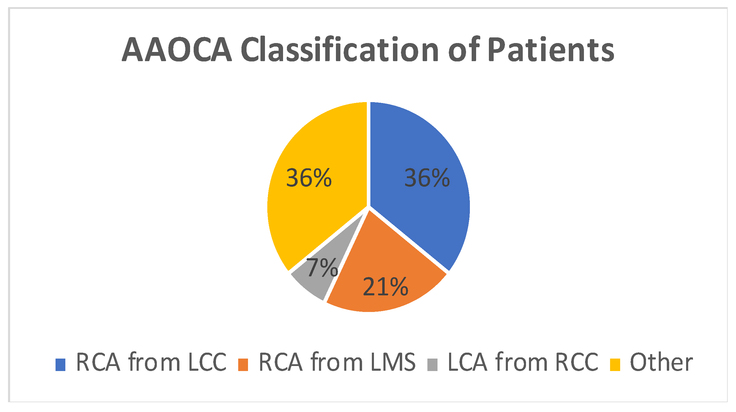
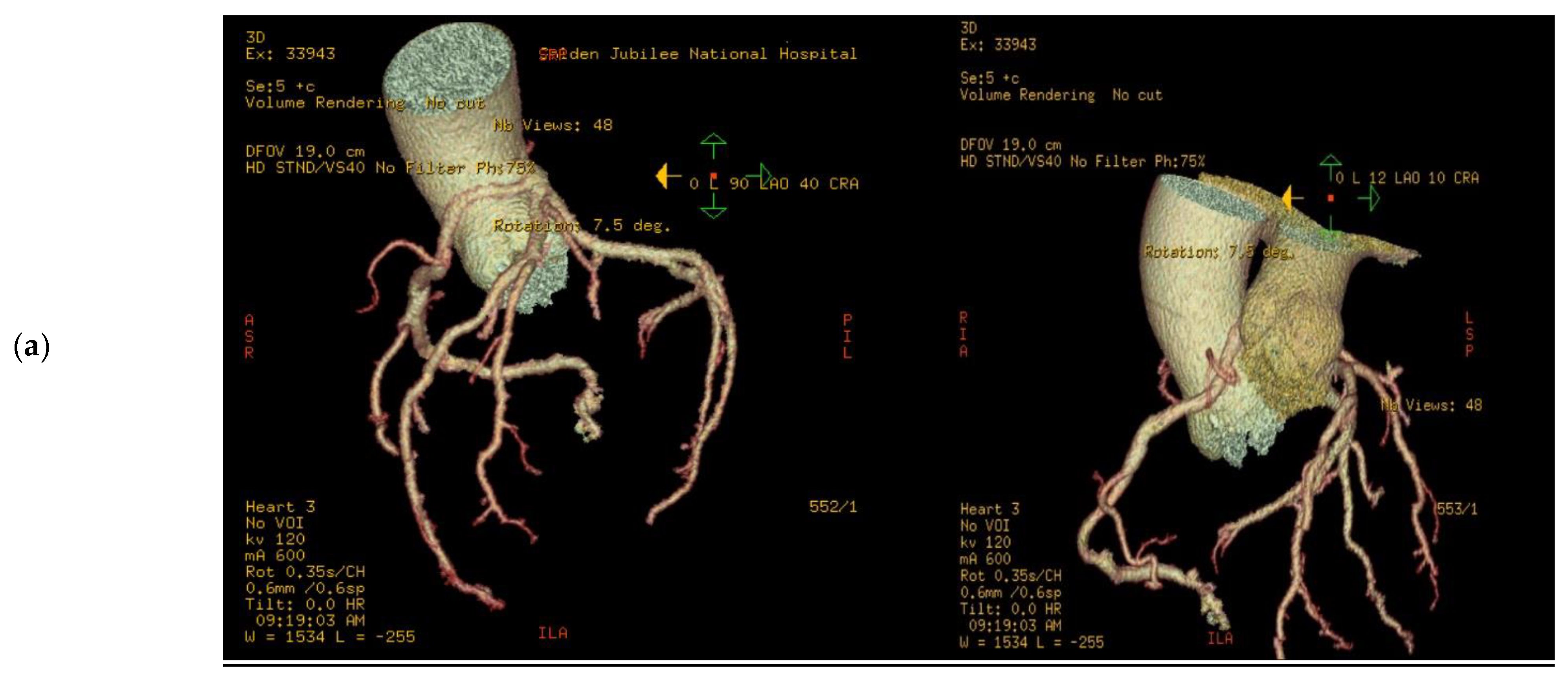
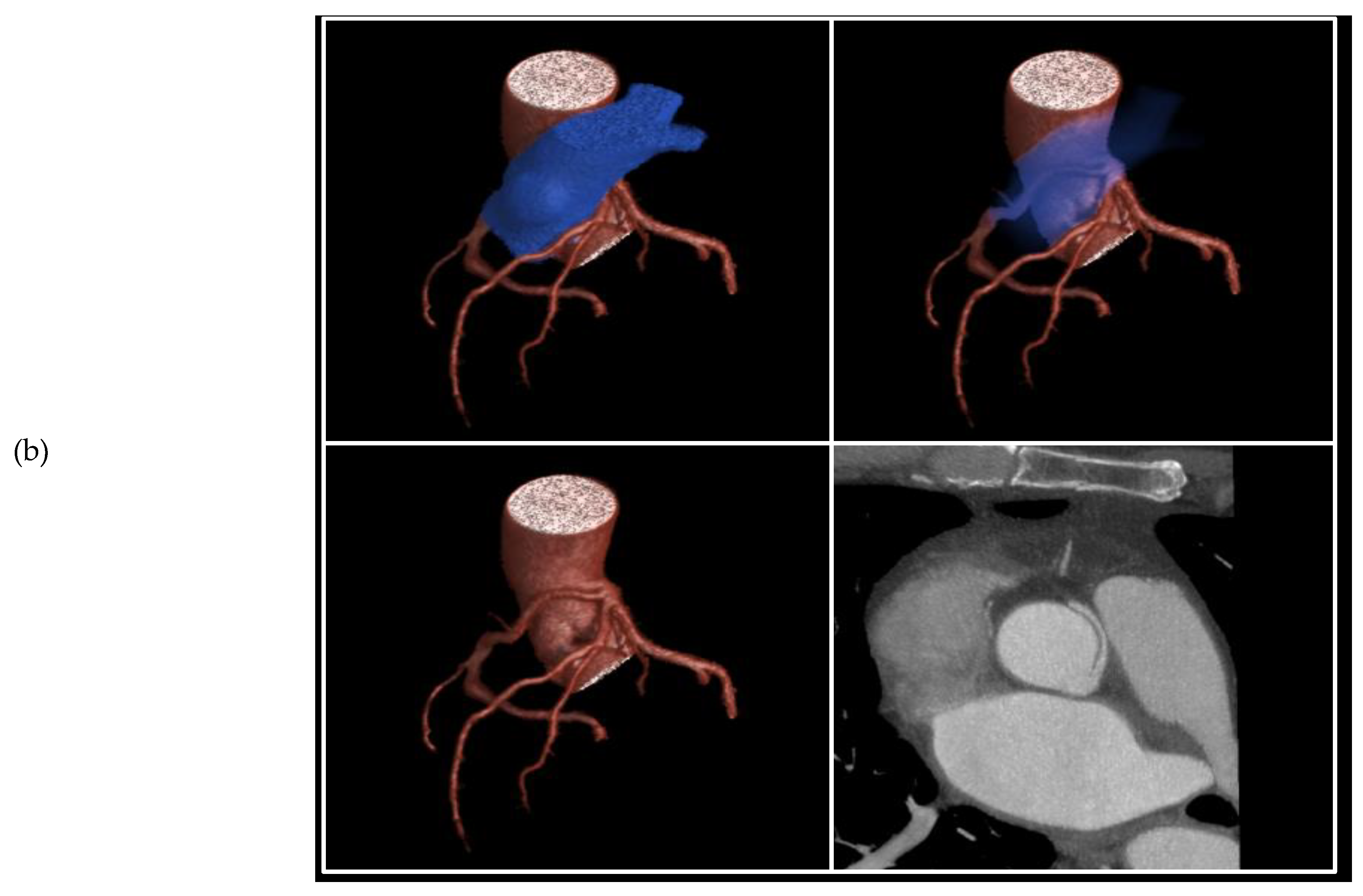
2. Results
3. Discussion
- Non-Surgical: Exercise (high-intensity) restriction
- Surgical:
- Unroofing of coronary artery;
- Patch arterioplasty;
- Coronary re-implantation;
- Coronary artery bypass grafting.
- Circumflex artery from the right sinus of Valsalva;
- A single coronary artery from the left sinus of Valsalva;
- Both coronary arteries from the right sinus of Valsalva;
- Left anterior descending artery (LAD) from the right sinus of Valsalva;
- Right coronary artery (RCA) arising from the left sinus of Valsalva;
- Left main coronary artery arising from the right sinus of Valsalva.
4. Methods
5. Conclusions
Author Contributions
Funding
Institutional Review Board Statement
Informed Consent Statement
Data Availability Statement
Conflicts of Interest
Abbreviations
| CABG | Coronary artery bypass grafting |
| AAOCA | Anomalous aortic origin of coronary artery |
| SCD | Sudden Cardiac Death |
| RITA | Right internal thoracic artery |
| LITA | Left internal thoracic artery |
| PITA | Pure internal thoracic artery |
| IQR | Interquartile range |
| RCA | Right coronary artery |
| LMS | Left main stem |
| LCA | Left coronary artery |
| RCS | Right coronary sinus |
| LCS | Left coronary sinus |
| AVR | Aortic valve replacement |
| SoV | Sinus of Valsalva |
| ACC/AHA | American College of Cardiology/American Heart Association |
| AAOLCA | Anomalous aortic origin of left coronary artery |
| ACAOS | Anomalous coronary arteries with the origin of the anomalous vessel from the opposite sinus of Valsalva |
| LAD | Left anterior descending artery |
| ITA | Internal thoracic artery |
| Cx | Circumflex artery |
| OM1 | 1st Obtuse marginal artery |
| PDA | Posterior descending artery |
| m-AVR | Mechanical aortic valve replacement |
| t-AVR | Tissue/bioprosthetic aortic valve replacement |
References
- Angelini, P. Coronary artery anomalies–current clinical issues: Definitions, classification, incidence, clinical relevance, and treatment guidelines. Tex. Heart Inst. J. 2002, 29, 271–278. [Google Scholar]
- Yamanaka, O.; Hobbs, R.E. Coronary artery anomalies in 126,595 patients undergoing coronary arteriography. Catheter. Cardiovasc. Diagn. 1990, 21, 28–40. [Google Scholar] [CrossRef]
- Tuo, G.; Marasini, M.; Brunelli, C.; Zannini, L.; Balbi, M. Incidence and clinical relevance of primary congenital coronary artery anomalies of the coronary arteries in children and adults. Cardiol. Young 2013, 23, 381–386. [Google Scholar] [CrossRef] [PubMed]
- Labombarda, F.; Coutance, G.; Pellissier, A.; Mery-Alexandre, C.; Roule, V.; Maragnes, P.; Milliez, P.; Saloux, E. Major congenital coronary artery anomalies in a paediatric and adult population: A prospective echocardiographic study. Eur. Heart J.-Cardiovasc. Imaging 2014, 15, 761–768. [Google Scholar] [CrossRef]
- Pelliccia, A.; Spataro, A.; Maron, B.J. Prospective echocardiographic screening for coronary artery anomalies in 1,360 elite competitive athletes. Am. J. Cardiol. 1993, 72, 978–979. [Google Scholar] [CrossRef]
- Davis, J.A.; Cecchin, F.; Jones, T.K.; Portman, M.A. Major coronary artery anomalies in a pediatric population: Incidence and clinical importance. J. Am. Coll. Cardiol. 1993, 37, 593–597. [Google Scholar] [CrossRef] [PubMed]
- Maron, B.J.; Shirani, J.; Poliac, L.C.; Mathenge, R.; Roberts, W.; Mueller, F.O. Sudden Death in Young Competitive Athletes Clinical, Demographic, and Pathological Profiles. JAMA 1996, 276, 199–204. [Google Scholar] [CrossRef]
- Angelini, P. Novel imaging of coronary artery anomalies to assess their prevalence, the causes of clinical symptoms, and the risk of sudden cardiac death. Circ. Cardiovasc. Imaging 2014, 7, 747–754. [Google Scholar] [CrossRef] [PubMed]
- Maron, B.; Doerer, J.; Haas, T.; Tierney, D. Sudden deaths in young competitive athletes: Analysis of 1866 deaths in the United States, 1980–2006. Circulation 2009, 119, 1085–1092. [Google Scholar] [CrossRef]
- Stecker, E.C.; Reinier, K.; Marijon, E.; Narayanan, K. Public Health Burden of Sudden Cardiac Death in the United States. Circ. Arrhythmia Electrophysiol. 2014, 7, 212–217. [Google Scholar] [CrossRef]
- Basso, C.; Maron, B.J.; Corrado, D.; Thiene, G. Clinical profile of congenital coronary artery anomalies with origin from the wrong aortic sinus leading to sudden death in young competitive athletes. J. Am. Coll. Cardiol. 2000, 35, 1493–1501. [Google Scholar] [CrossRef] [PubMed]
- Tweddell, J.A. Expert consensus guidelines: Anomalous aortic origin of a coronary artery. J. Thorac. Cardiovasc. Surg. 2017, 153, 1440–1457. [Google Scholar] [CrossRef]
- Mustafa, I.; Gula, G.; Radley-Smith, R.; Durrer, S.; Yacoub, M. Anomalous origin of the left coronary artery from the anterior aortic sinus: A potential cause of sudden death. J. Thorac. Cardiovasc. Surg. 1981, 82, 297–300. [Google Scholar] [CrossRef] [PubMed]
- Romp, R.; Helong, R.; Landolfo, C.; Sanders, S.; Miller, C. Outcome of unroofing procedure for repair of anomalous aortic origin of left or right coronary artery. Ann. Thorac. Surg. 2003, 76, 589–596. [Google Scholar] [CrossRef]
- van Son, J.; Mohr, F. Modified unroofing procedure in anomalous aortic origin of left or right coronary artery. Ann. Thorac. Surg. 1997, 64, 568–569. [Google Scholar] [CrossRef]
- Furukawa, K.; Itoh, T. Direct Coronary Reimplantation for Repair of Anomalous Aortic Origin of Left or Right Coronary Artery. Ann. Thorac. Surg. 2005, 79, 389–390. [Google Scholar] [CrossRef]
- Fernandes, E.D.; Kadivar, H.; Hallman, G.L.; Reul, G.J.; Ott, D.A.; Cooley, D.A. Congenital malformations of the coronary arteries: The Texas Heart Institute experience. Ann. Thorac. Surg. 1992, 54, 732–740. [Google Scholar] [CrossRef]
- Fedoruk, L.; Kern, J.; Peeler, B.; Kron, I. Anomalous origin of the right coronary artery. Right internal thoracic artery to right coronary artery bypass graft is not an answer. J. Thorac. Cardiovasc. Surg. 2007, 133, 456–460. [Google Scholar] [CrossRef]
- Tavaf-Motamen, H.; Bannister, S.P.; Corcoran, P.C.; Stewart, R.W.; Mulligan, C.R.; DeVries, W.C. Repair of Anomalous Origin of Right Coronary Artery From the Left Sinus of Valsalva. Ann. Thorac. Surg. 2008, 85, 2135–2136. [Google Scholar] [CrossRef]
- Taylor, A.J.; Rogan, K.M.; Virmani, R. Sudden cardiac death associated with isolated congenital coronary artery anomalies. J. Am. Coll. Cardiol. 1992, 20, 640–647. [Google Scholar] [CrossRef]
- Brothers, J.; Gaynor, J.W.; Paridon, S.; Lorber, R.; Jacobs, M. Anomalous Aortic Origin of a Coronary Artery with an Interarterial Course: Understanding Current Management Strategies in Children and Young Adults. Pediatr. Cardiol. 2009, 30, 911–921. [Google Scholar] [CrossRef]
- Cheezum, M.K.; Liberthson, R.R.; Shah, N.R.; Villines, T.C.; O’Gara, P.T.; Landzberg, M.J.; Blankstein, R. Anomalous Aortic Origin of a Coronary Artery From the Inappropriate Sinus of Valsalva. J. Am. Coll. Cardiol. 2017, 69, 1592–1608. [Google Scholar] [CrossRef]
- Gräni, C.; Benz, D.; Schmied, C.; Vontobel, J.; Possner, M.; Clerc, O.; Mikulicic, F.; Stehli, J.; Fuchs, T.; Pazhenkottil, A.; et al. Prevalence and characteristics of coronary artery anomalies detected by coronary computed tomography angiography in 5 634 consecutive patients in a single centre in Switzerland. Swiss Med. Wkly. 2016, 146, w14294. [Google Scholar] [CrossRef] [PubMed]
- Angelini, P.; Velasco, J.A.; Ott, D.; Khoshnevis, G.R. Anomalous coronary artery arising from the opposite sinus: Descriptive features and pathophysiologic mechanisms, as documented by intravascular ultrasonography. J. Invasive Cardiol. 2003, 15, 507–514. [Google Scholar] [PubMed]
- Jegatheeswaran, A.; Devlin, P.; McCrindle, B.; Williams, W.; Jacobs, M.; Blackstone, E. Features associated with myocardial is-chemia in anomalous aortic origin of a coronary artery: A congenital heart surgeons’ society study. J. Thorac. Cardiovasc. Surg. 2019, 158, 822–834. [Google Scholar] [CrossRef]
- Peñalver, J.M.; Mosca, R.S.; Weitz, D.; Phoon, C.K.L. Anomalous aortic origin of coronary arteries from the opposite sinus: A critical appraisal of risk. BMC Cardiovasc. Disord. 2012, 12, 83. [Google Scholar] [CrossRef] [PubMed]
- Warnes, C.; Williams, R.; Bashore, T.; Child, J.; Connolly, H.; Dearani, J. ACC/AHA 2008 Guidelines fro management of Adults with Congenital Heart Disease: A report of the American College of Cardiology/ American Heart Association Task Forceon Practice Guidelines. Circulation 2008, 118, e714–e833. [Google Scholar]
- Cohen, A.J.; Grishkin, B.A.; Helsel, R.A.; Head, H.D. Surgical therapy in the management of coronary anomalies: Emphasis on utility of internal mammary artery grafts. Ann. Thorac. Surg. 1989, 47, 630–637. [Google Scholar] [CrossRef]
- Sabik, J.; Lytle, B.; Blackstone, E.; Khan, M.; Houghtaling, P.; Cosgrove, D. Does competitive flow reduce internal thoracic artery graft patency? Ann. Thorac. Surg. 2003, 76, 1490–1497. [Google Scholar] [CrossRef]
- Navia, D.; Cosgrove, D.; Lytle, B.; Taylor, P.; McCarthy, P.; Stewart, R. Is the internal thoracic artery the conduit of choice to replace a stenotic vein graft? Ann. Thorac. Surg. 1994, 57, 40–44. [Google Scholar] [CrossRef]
- Sharma, V.; Burkhart, H.; Dearani, J.; Suri, R.; Daly, R.; Park, S. Surgical unroofing of anomalous aortic origin of a coronary artery: A single-center experience. Ann. Thorac. Surg. 2014, 98, 941–945. [Google Scholar] [CrossRef] [PubMed]
- Ibraheem, W.; Abass, O.; Toema, A.; Yehia, A. Coronary artery bypass grafting experience in the setting of an anomalous origin of the right coronary artery from the left sinus of Valsalva: Midterm results. J. Card. Surg. 2019, 34, 1162–1171. [Google Scholar] [CrossRef] [PubMed]


Disclaimer/Publisher’s Note: The statements, opinions and data contained in all publications are solely those of the individual author(s) and contributor(s) and not of MDPI and/or the editor(s). MDPI and/or the editor(s) disclaim responsibility for any injury to people or property resulting from any ideas, methods, instructions or products referred to in the content. |
© 2023 by the authors. Licensee MDPI, Basel, Switzerland. This article is an open access article distributed under the terms and conditions of the Creative Commons Attribution (CC BY) license (https://creativecommons.org/licenses/by/4.0/).
Share and Cite
James, R.L.; Das De, S.; Singh Avtaar Singh, S.; Dreisbach, J.; Watkins, S.; Al-Attar, N. Treatment of Anomalous Coronary Arteries—Surgical Revascularisation Using the Pure Internal Thoracic Artery Technique. J. Cardiovasc. Dev. Dis. 2023, 10, 155. https://doi.org/10.3390/jcdd10040155
James RL, Das De S, Singh Avtaar Singh S, Dreisbach J, Watkins S, Al-Attar N. Treatment of Anomalous Coronary Arteries—Surgical Revascularisation Using the Pure Internal Thoracic Artery Technique. Journal of Cardiovascular Development and Disease. 2023; 10(4):155. https://doi.org/10.3390/jcdd10040155
Chicago/Turabian StyleJames, Ramon L., Sudeep Das De, Sanjeet Singh Avtaar Singh, John Dreisbach, Stuart Watkins, and Nawwar Al-Attar. 2023. "Treatment of Anomalous Coronary Arteries—Surgical Revascularisation Using the Pure Internal Thoracic Artery Technique" Journal of Cardiovascular Development and Disease 10, no. 4: 155. https://doi.org/10.3390/jcdd10040155
APA StyleJames, R. L., Das De, S., Singh Avtaar Singh, S., Dreisbach, J., Watkins, S., & Al-Attar, N. (2023). Treatment of Anomalous Coronary Arteries—Surgical Revascularisation Using the Pure Internal Thoracic Artery Technique. Journal of Cardiovascular Development and Disease, 10(4), 155. https://doi.org/10.3390/jcdd10040155