

REHABS: An Innovative and User-Friendly Device for Rehabilitation

 ,
,  ,
,  ,
,  , and
, and
Abstract
:1. Introduction
2. Materials and Methods
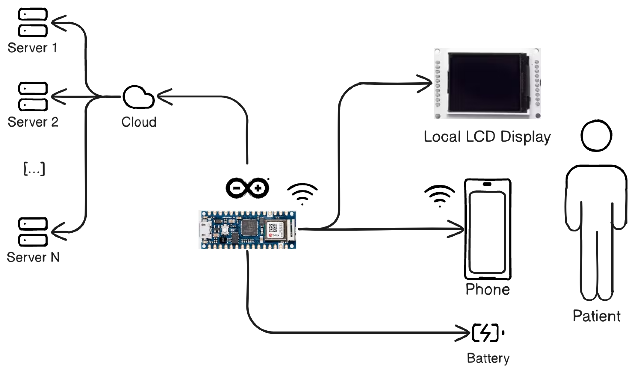
2.1. System Architecture
| Algorithm 1 Object detection algorithm |
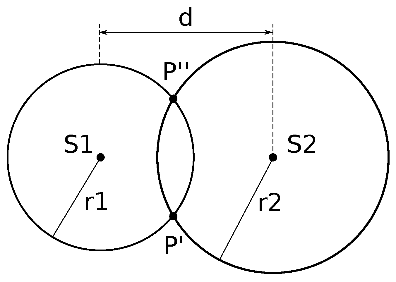
| Require: d distance between the two sensors, time of flight from sensor S1, time of flight from sensor S2 1: /* Convert time of flight from sensors to distances */ 2: 3: 4: /* Combine to calculate object coordinates */ 5: 6: 7: return |
2.2. Device Use Configuration
2.3. Rehabilitation Exercises
3. Results
4. Discussion
5. Conclusions
Author Contributions
Funding
Institutional Review Board Statement
Informed Consent Statement
Data Availability Statement
Acknowledgments
Conflicts of Interest
References
- O’Sullivan, S.B.; Schmitz, T.J.; Fulk, G. Physical Rehabilitation; FA Davis: Philadelphia, PA, USA, 2019. [Google Scholar]
- Gilmour, G.S.; Nielsen, G.; Teodoro, T.; Yogarajah, M.; Coebergh, J.A.; Dilley, M.D.; Martino, D.; Edwards, M.J. Management of functional neurological disorder. J. Neurol. 2020, 267, 2164–2172. [Google Scholar] [CrossRef] [PubMed]
- Kolar, P.; Calta, J.; Kriz, J.; Lewit, K.; Dyrhonová, O.; Bitnar, P.; Valouchová, P.; Čech, Z.; Kobesová, A.; Smékal, D.; et al. Clinical Rehabilitation; Alena Kobesová: Prague, Czechoslovakia, 2014. [Google Scholar]
- Canino, G.; Guzzi, P.H.; Tradigo, G.; Zhang, A.; Veltri, P. On the analysis of diseases and their related geographical data. IEEE J. Biomed. Health Inform. 2015, 21, 228–237. [Google Scholar] [CrossRef] [PubMed]
- Morera-Balaguer, J.; Botella-Rico, J.M.; Martínez-González, M.C.; Medina-Mirapeix, F.; Rodríguez-Nogueira, Ó. Physical therapists’ perceptions and experiences about barriers and facilitators of therapeutic patient-centred relationships during outpatient rehabilitation: A qualitative study. Braz. J. Phys. Ther. 2018, 22, 484–492. [Google Scholar] [CrossRef] [PubMed]
- Carter, R.; Lubinsky, J. Rehabilitation Research: Principles and Applications; Elsevier Health Sciences: Amsterdam, The Netherlands, 2015. [Google Scholar]
- Ellis, T.; Motl, R.W. Physical activity behavior change in persons with neurologic disorders: Overview and examples from Parkinson disease and multiple sclerosis. J. Neurol. Phys. Ther. 2013, 37, 85–90. [Google Scholar] [CrossRef] [PubMed]
- Succurro, E.; Pedace, E.; Andreozzi, F.; Papa, A.; Vizza, P.; Fiorentino, T.V.; Perticone, F.; Veltri, P.; Cascini, G.L.; Sesti, G. Reduction in global myocardial glucose metabolism in subjects with 1-hour postload hyperglycemia and impaired glucose tolerance. Diabetes Care 2020, 43, 669–676. [Google Scholar] [CrossRef] [PubMed]
- Mercatelli, D.; Pedace, E.; Veltri, P.; Giorgi, F.M.; Guzzi, P.H. Exploiting the molecular basis of age and gender differences in outcomes of SARS-CoV-2 infections. Comput. Struct. Biotechnol. J. 2021, 19, 4092–4100. [Google Scholar] [CrossRef] [PubMed]
- Espay, A.J.; Aybek, S.; Carson, A.; Edwards, M.J.; Goldstein, L.H.; Hallett, M.; LaFaver, K.; LaFrance, W.C.; Lang, A.E.; Nicholson, T.; et al. Current concepts in diagnosis and treatment of functional neurological disorders. JAMA Neurol. 2018, 75, 1132–1141. [Google Scholar] [CrossRef] [PubMed]
- Barnes, M.P.; Good, D.C. Neurological Rehabilitation; Elsevier: Amsterdam, The Netherlands, 2013. [Google Scholar]
- Vizza, P.; Mirarchi, D.; Tradigo, G.; Redavide, M.; Bossio, R.B.; Veltri, P. Vocal signal analysis in patients affected by Multiple Sclerosis. Procedia Comput. Sci. 2017, 108, 1205–1214. [Google Scholar] [CrossRef]
- Mirarchi, D.; Vizza, P.; Tradigo, G.; Lombardo, N.; Arabia, G.; Veltri, P. Signal analysis for voice evaluation in Parkinson’s disease. In Proceedings of the 2017 IEEE International Conference on Healthcare Informatics (ICHI), Park City, UT, USA, 23–26 August 2017; pp. 530–535. [Google Scholar]
- Amato, F.; Cannataro, M.; Manfredi, C.; Garozzo, A.; Lombardo, N.; Cosentino, C. Early detection of voice diseases via a web-based system. In Proceedings of the Models and Analysis of Vocal Emissions for Biomedical Applications: 5th International Workshop, Firenze, Italy, 13–15 December 2007; pp. 1000–1004. [Google Scholar]
- Fickling, S.D.; Greene, T.; Greene, D.; Frehlick, Z.; Campbell, N.; Etheridge, T.; Smith, C.J.; Bollinger, F.; Danilov, Y.; Rizzotti, R.; et al. Brain vital signs detect cognitive improvements during combined physical therapy and neuromodulation in rehabilitation from severe traumatic brain injury: A case report. Front. Hum. Neurosci. 2020, 14, 347. [Google Scholar] [CrossRef]
- Mak, M.K.; Wong-Yu, I.S.; Shen, X.; Chung, C.L. Long-term effects of exercise and physical therapy in people with Parkinson disease. Nat. Rev. Neurol. 2017, 13, 689–703. [Google Scholar] [CrossRef]
- Radder, D.L.; Sturkenboom, I.H.; van Nimwegen, M.; Keus, S.H.; Bloem, B.R.; de Vries, N.M. Physical therapy and occupational therapy in Parkinson’s disease. Int. J. Neurosci. 2017, 127, 930–943. [Google Scholar] [CrossRef] [PubMed]
- Prasetyo, Y.T. Factors Affecting Gross Manual Dexterity: A Structural Equation Modeling Approach. In Proceedings of the 2020 IEEE 7th International Conference on Industrial Engineering and Applications (ICIEA), Bangkok, Thailand, 16–18 April 2020. [Google Scholar]
- Mousavi Hondori, H.; Khademi, M. A review on technical and clinical impact of microsoft kinect on physical therapy and rehabilitation. J. Med. Eng. 2014, 2014, 846514. [Google Scholar] [CrossRef] [PubMed]
- Porciuncula, F.; Roto, A.V.; Kumar, D.; Davis, I.; Roy, S.; Walsh, C.J.; Awad, L.N. Wearable movement sensors for rehabilitation: A focused review of technological and clinical advances. PM&R 2018, 10, S220–S232. [Google Scholar]
- Palumbo, A.; Vizza, P.; Calabrese, B.; Ielpo, N. Biopotential signal monitoring systems in rehabilitation: A review. Sensors 2021, 21, 7172. [Google Scholar] [CrossRef] [PubMed]
- Tradigo, G.; Vizza, P.; Guzzi, P.H.; Fragomeni, G.; Ammendolia, A.; Veltri, P. A programmable device to guide rehabilitation patients: Design, testing and data collection. In Proceedings of the 2020 IEEE International Conference on Bioinformatics and Biomedicine (BIBM), Seoul, Republic of Korea, 16–19 December 2020; pp. 1487–1491. [Google Scholar]
- Chu, C.Y.; Patterson, R.M. Soft robotic devices for hand rehabilitation and assistance: A narrative review. J. Neuroeng. Rehabil. 2018, 15, 1–14. [Google Scholar] [CrossRef] [PubMed]
- Castelli, L.; Iacovelli, C.; Loreti, C.; Malizia, A.M.; Barone Ricciardelli, I.; Tomaino, A.; Fusco, A.; Biscotti, L.; Padua, L.; Giovannini, S. Robotic-assisted rehabilitation for balance in stroke patients (ROAR-S): Effects of cognitive, motor and functional outcomes. Eur. Rev. Med. Pharmacol. Sci. 2023, 27, 8198–8211. [Google Scholar]
- Carrillo, C.; Tilley, D.; Horn, K.; Gonzalez, M.; Coffman, C.; Hilton, C.; Mani, K. Effectiveness of robotics in stroke rehabilitation to accelerate upper extremity function: Systematic review. Occup. Ther. Int. 2023, 2023, 7991765. [Google Scholar] [CrossRef]
- Tenforde, A.S.; Hefner, J.E.; Kodish-Wachs, J.E.; Iaccarino, M.A.; Paganoni, S. Telehealth in physical medicine and rehabilitation: A narrative review. PM&R 2017, 9, S51–S58. [Google Scholar]
- Domingues, V.L.; de Freitas, T.B.; Palma, G.d.S.; Makhoul, M.P.; Torriani-Pasin, C. Physical exercise program via telemonitoring to individuals with Parkinson’s disease during COVID-19 pandemic: Phase I clinical trial. Braz. J. Mot. Behav. 2022, 16, 17. [Google Scholar]
- Antoniello, A.; Sabatelli, A.; Valenti, S.; Di Tillo, M.; Pepa, L.; Spalazzi, L.; Andrenelli, E.; Capecci, M.; Ceravolo, M.G. A low-cost telerehabilitation and telemonitoring system for people with Parkinson’s disease: The architecture. In Proceedings of the 2022 IEEE 12th International Conference on Consumer Electronics (ICCE-Berlin), Berlin, Germany, 2–6 September 2022; pp. 1–2. [Google Scholar]
- Borghese, N.A.; Essenziale, J.; Mainetti, R.; Mancon, E.; Pagliaro, R.; Pajardi, G. Hand rehabilitation and telemonitoring through smart toys. Sensors 2019, 19, 5517. [Google Scholar] [CrossRef]
- Peretti, A.; Amenta, F.; Tayebati, S.K.; Nittari, G.; Mahdi, S.S. Telerehabilitation: Review of the state-of-the-art and areas of application. JMIR Rehabil. Assist. Technol. 2017, 4, e7511. [Google Scholar] [CrossRef] [PubMed]
- Sapanel, Y.; Tadeo, X.; Brenna, C.T.; Remus, A.; Koerber, F.; Cloutier, L.M.; Tremblay, G.; Blasiak, A.; Hardesty, C.L.; Yoong, J.; et al. Economic Evaluation Associated With Clinical-Grade Mobile App–Based Digital Therapeutic Interventions: Systematic Review. J. Med. Internet Res. 2023, 25, e47094. [Google Scholar] [CrossRef] [PubMed]
- Arntz, A.; Weber, F.; Handgraaf, M.; Lällä, K.; Korniloff, K.; Murtonen, K.P.; Chichaeva, J.; Kidritsch, A.; Heller, M.; Sakellari, E.; et al. Technologies in Home-Based Digital Rehabilitation: Scoping Review. JMIR Rehabil. Assist. Technol. 2023, 10, e43615. [Google Scholar] [CrossRef] [PubMed]
- Aggogeri, F.; Mikolajczyk, T.; O’Kane, J. Robotics for rehabilitation of hand movement in stroke survivors. Adv. Mech. Eng. 2019, 11, 1687814019841921. [Google Scholar] [CrossRef]
- Herrera-Luna, I.; Rechy-Ramirez, E.J.; Rios-Figueroa, H.V.; Marin-Hernandez, A. Sensor fusion used in applications for hand rehabilitation: A systematic review. IEEE Sens. J. 2019, 10, 3581–3592. [Google Scholar] [CrossRef]
- Bernocchi, P.; Mulè, C.; Vanoglio, F.; Taveggia, G.; Luisa, A.; Scalvini, S. Home-based hand rehabilitation with a robotic glove in hemiplegic patients after stroke: A pilot feasibility study. Top. Stroke Rehabil. 2018, 25, 114–119. [Google Scholar] [CrossRef]
- Jo, I.; Park, Y.; Lee, J.; Bae, J. A portable and spring-guided hand exoskeleton for exercising flexion/extension of the fingers. Mech. Mach. Theory 2019, 135, 176–191. [Google Scholar] [CrossRef]
- Sandison, M.; Phan, K.; Casas, R.; Nguyen, L.; Lum, M.; Pergami-Peries, M.; Lum, P.S. HandMATE: Wearable robotic hand exoskeleton and integrated android app for at home stroke rehabilitation. In Proceedings of the 2020 42nd Annual International Conference of the IEEE Engineering in Medicine & Biology Society (EMBC), Montreal, QC, Canada, 20–24 July 2020; pp. 4867–4872. [Google Scholar]
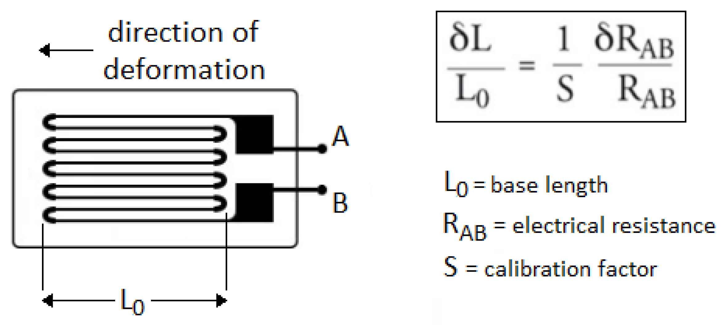
- Keil, S. Technology and Practical Use of Strain Gages with Particular Consideration of Stress Analysis Using Strain Gages; Wiley: Hoboken, NJ, USA, 2017. [Google Scholar]
- Mathiowetz, V.; Weber, K.; Kashman, N.; Volland, G. Adult norms for the nine hole peg test of finger dexterity. Occup. Ther. J. Res. 1985, 5, 24–38. [Google Scholar] [CrossRef]


| Number of Measure | Detected Value for S1 | Detected Value for S2 | Detected Value for S3 |
|---|---|---|---|
| 1 | 20 | 34 | 50 |
| 2 | 21 | 35 | 52 |
| 3 | 25 | 36 | 54 |
| 4 | 26 | 41 | 68 |
| 5 | 27 | 42 | 70 |
| 6 | 28 | 43 | 71 |
| 7 | 29 | 44 | 72 |
| 8 | 30 | 45 | 74 |
| Mean | 25.75 | 40 | 63.87 |
| Disease | Description |
|---|---|
| Loeys-Dietz Syndrome | Rare genetic connective tissue disease |
| Multiple sclerosis | Chronic demyelinating neurodegenerative disease affecting any type of nerve |
| Omarthrosis | Degenerative disease affecting the shoulder joint, causing the wear of the articular cartilage |
| Parkinson | Neurodegenerative disese with mobility impairment |
| Rhizoarthrosis | Arthrosis localized at the level of the trapezio-metacarpal joint |
| Cerebellar Ataxia | Cerebellum damage causing impairment in motor skills |
| Limb-girdle muscular dystrophy | Diseases characterized by weakness and wasting of the muscles in the arms and legs |
| Stroke | Cerebrovascular event associated with different types of movement disorders |
| Hirayama Syndrome | Cervical myelopathy presenting spinal muscular atrophy of the distal upper limbs |
| Experiments | Control | Study | p-Value |
|---|---|---|---|
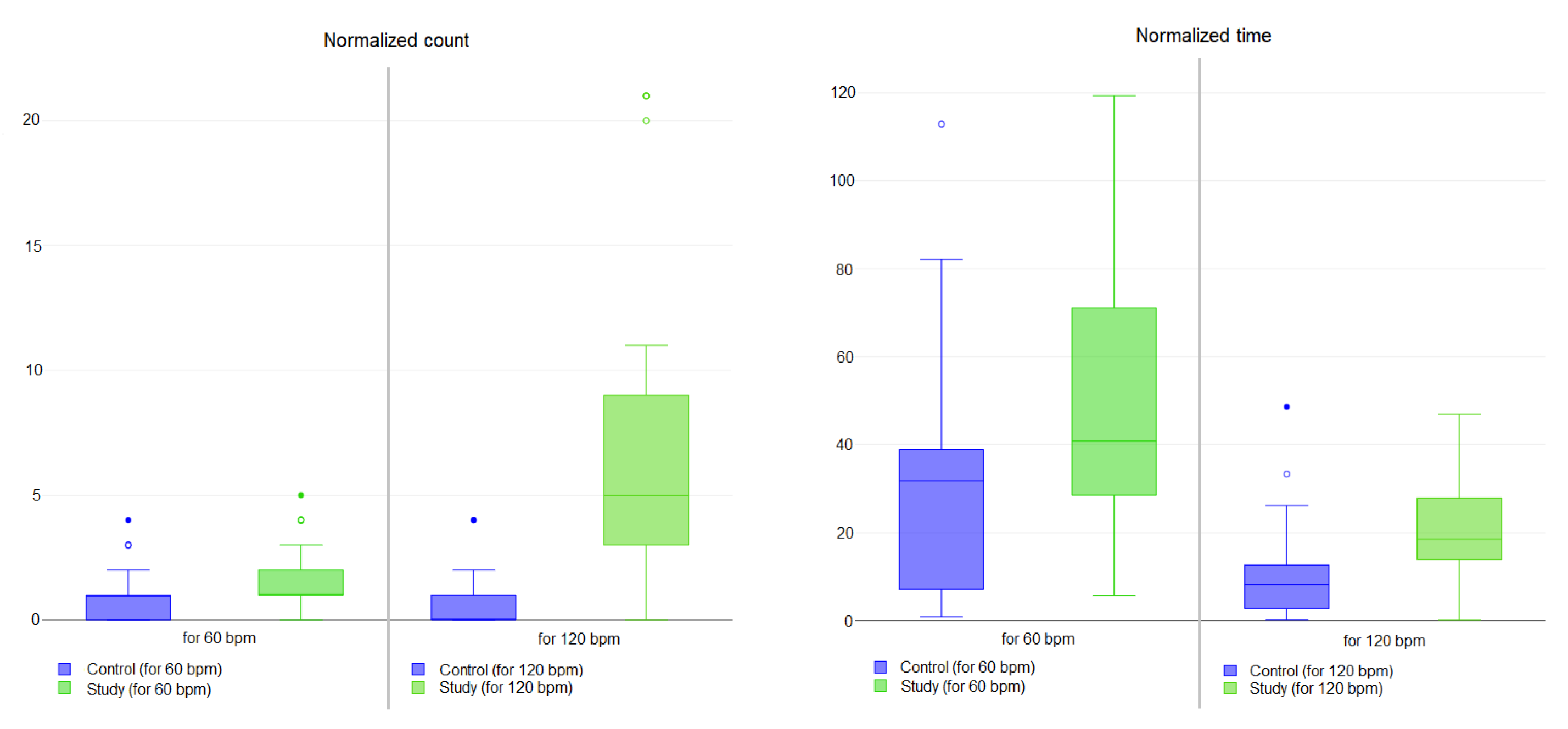
| Normalized count at 60 bpm | 0.72 ± 0.93 | 1.58 ± 1.22 | <0.01 |
| Normalized time at 60 bpm | 30.16 ± 25.82 | 47.96 ± 29.15 | <0.01 |
| Normalized count at 120 bpm | 0.67 ± 1.00 | 6.50 ± 5.84 | <0.01 |
| Normalized time at 120 bpm | 9.89 ± 10.00 | 21.57 ± 12.81 | <0.01 |
| Experiments | Control | Study | p-Value |
|---|---|---|---|
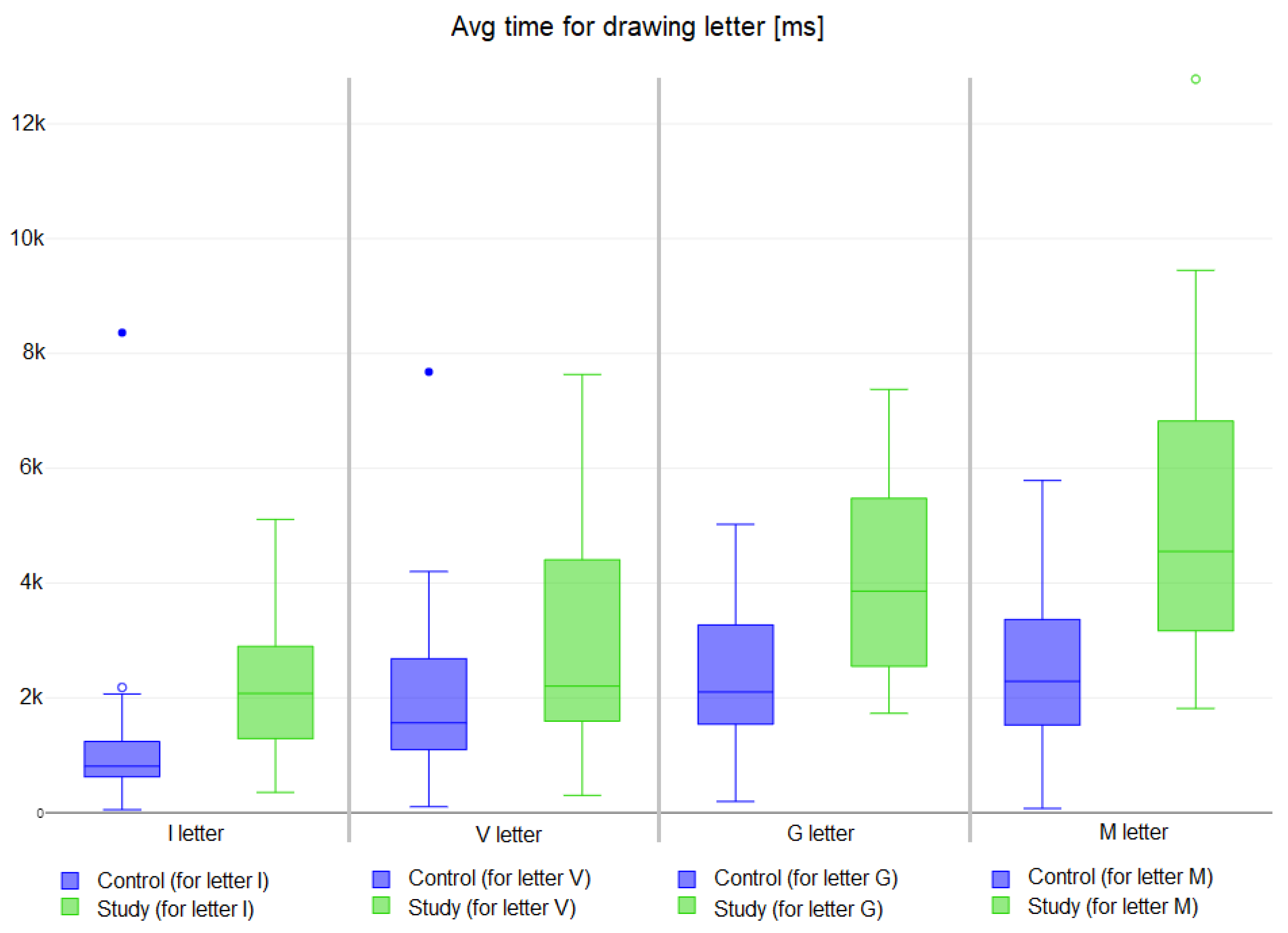
| Letter I | 1127.18 ± 1286.60 | 2109.98 ± 977.15 | <0.01 |
| Letter V | 2056.48 ± 1398.00 | 2954.70 ± 1915.17 | <0.01 |
| Letter G | 2426.98 ± 1284.80 | 4130.80 ± 1763.47 | <0.01 |
| Letter M | 2522.55 ± 1388.21 | 5245.03 ± 2421.81 | <0.01 |
| Experiments | Control | Study | p-Value |
|---|---|---|---|
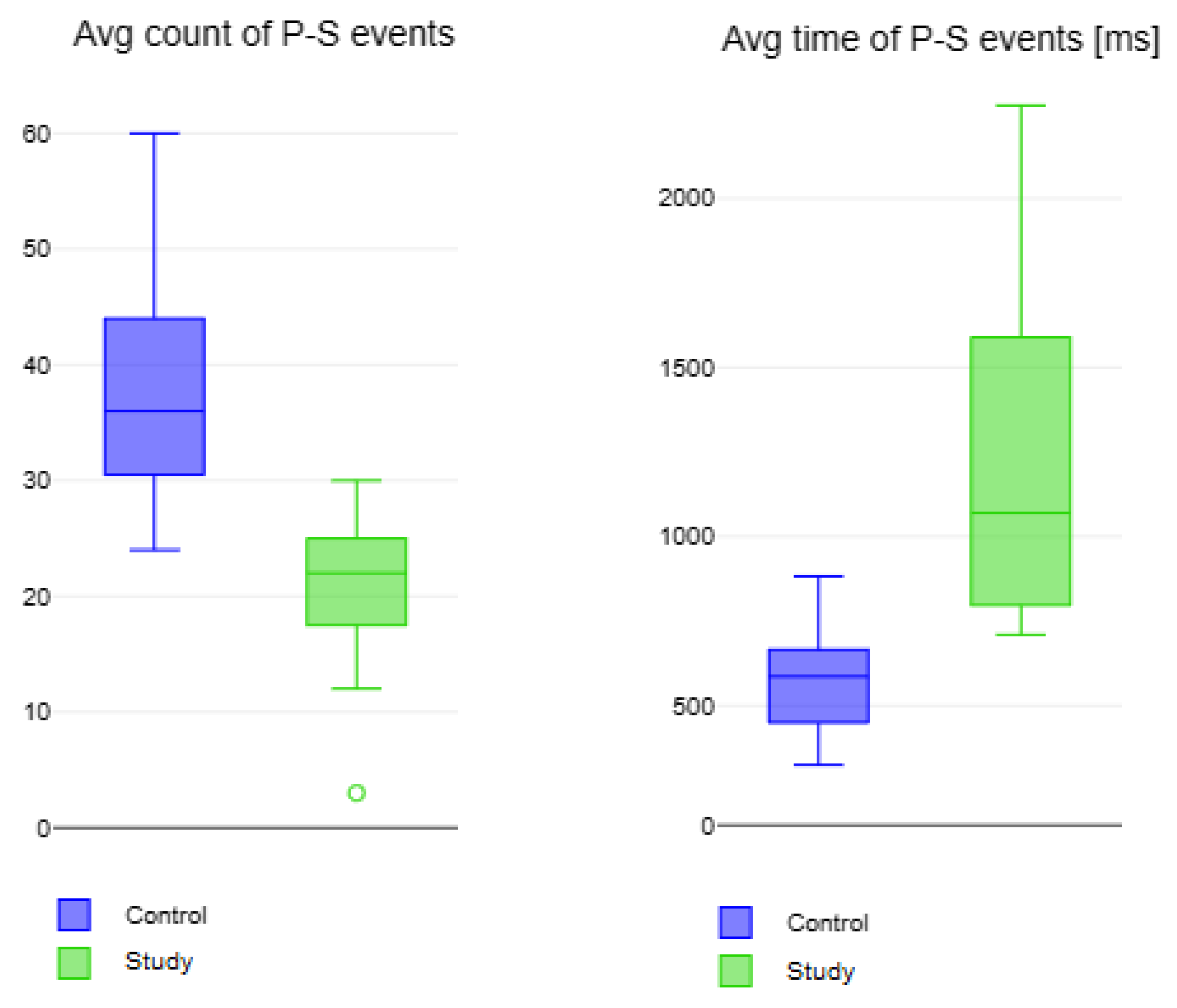
| Avg count of P-S events | 37.60 ± 8.81 | 20.73 ± 5.56 | <0.01 |
| Avg Time for a P-S event [ms] | 575.08 ± 131.59 | 1203.00 ± 433.73 | <0.01 |
Disclaimer/Publisher’s Note: The statements, opinions and data contained in all publications are solely those of the individual author(s) and contributor(s) and not of MDPI and/or the editor(s). MDPI and/or the editor(s) disclaim responsibility for any injury to people or property resulting from any ideas, methods, instructions or products referred to in the content. |
© 2023 by the authors. Licensee MDPI, Basel, Switzerland. This article is an open access article distributed under the terms and conditions of the Creative Commons Attribution (CC BY) license (https://creativecommons.org/licenses/by/4.0/).
Share and Cite
Vizza, P.; Marotta, N.; Ammendolia, A.; Guzzi, P.H.; Veltri, P.; Tradigo, G. REHABS: An Innovative and User-Friendly Device for Rehabilitation. Bioengineering 2024, 11, 5. https://doi.org/10.3390/bioengineering11010005
Vizza P, Marotta N, Ammendolia A, Guzzi PH, Veltri P, Tradigo G. REHABS: An Innovative and User-Friendly Device for Rehabilitation. Bioengineering. 2024; 11(1):5. https://doi.org/10.3390/bioengineering11010005
Chicago/Turabian StyleVizza, Patrizia, Nicola Marotta, Antonio Ammendolia, Pietro Hiram Guzzi, Pierangelo Veltri, and Giuseppe Tradigo. 2024. "REHABS: An Innovative and User-Friendly Device for Rehabilitation" Bioengineering 11, no. 1: 5. https://doi.org/10.3390/bioengineering11010005
APA StyleVizza, P., Marotta, N., Ammendolia, A., Guzzi, P. H., Veltri, P., & Tradigo, G. (2024). REHABS: An Innovative and User-Friendly Device for Rehabilitation. Bioengineering, 11(1), 5. https://doi.org/10.3390/bioengineering11010005