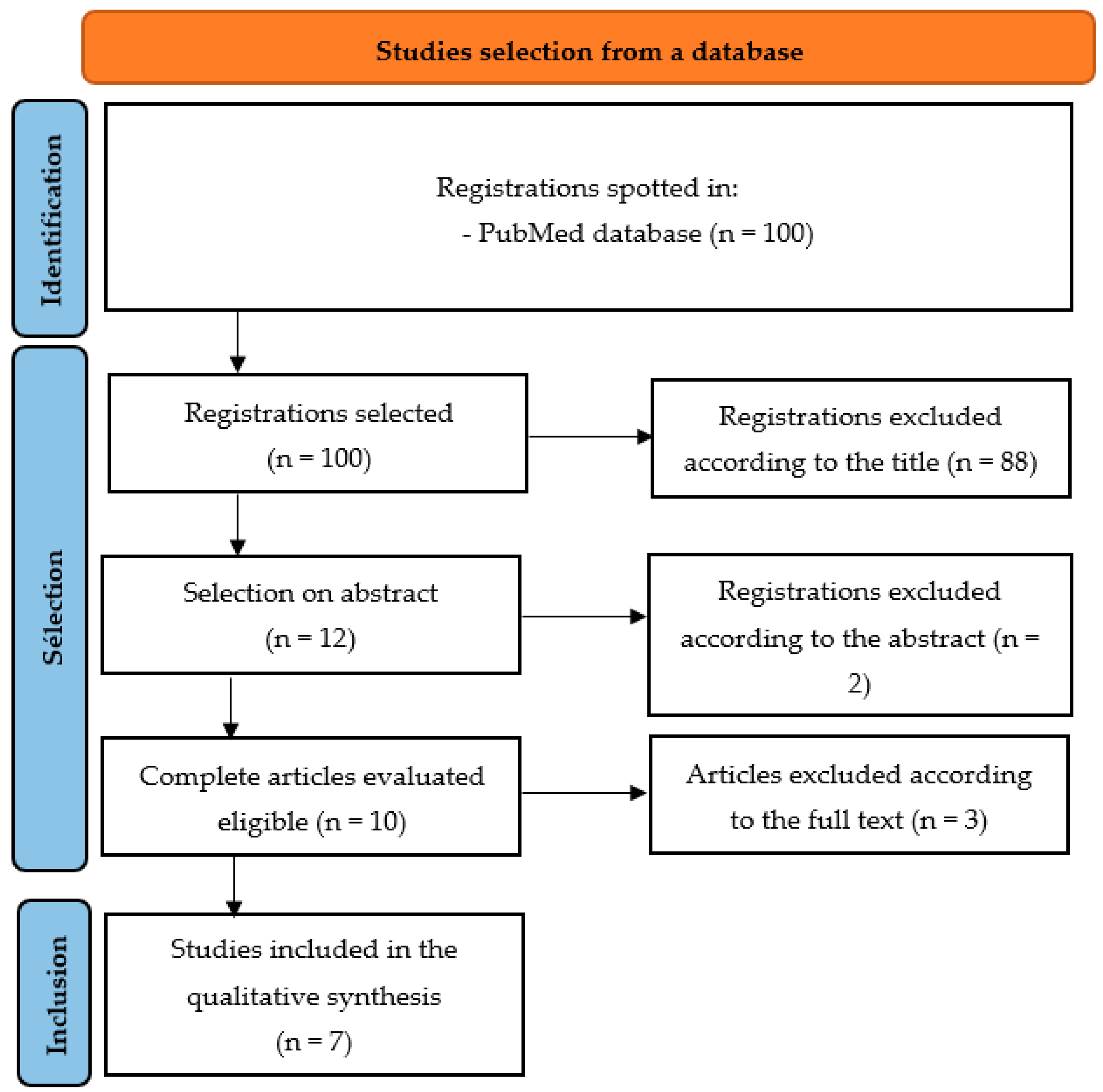
This systematic review, based on any prospective or retrospective study published after 2021 (date of the last search in a published systematic review of the literature on the subject [
17]), included seven articles to answer to the following research question: “Is orthodontic-surgical treatment a risk factor for temporomandibular disorders (TMDs)?” Concerning the diagnosis of TMDs and disc displacement, all included articles were based on the diagnostic algorithm of the “Diagnostic Criteria for Temporomandibular Disorders (DC/TMDs)”, as this is the consensus standardized evaluation protocol for TMDs [
3] and/or on the magnetic resonance imaging (MRI), recognized as the reference standard diagnostic method [
27].
4.1. Population Studied
The seven studies included 570 patients, 529 of whom were undergoing orthodontic-surgical treatment (
Table 2). The study by Madhan et al. [
26] included a control group of 30 patients. The variables relating to the demographics of the different studies included, according to the data available, are detailed in
Table 2. The study by Castro et al. [
20] included a group of 19 patients off-topic, who underwent orthodontic-surgical treatments with surgical repositioning of the TMJ disc.
No study specifies the conservative treatments possibly implemented for the management of TMDs before or after OS. This constitutes a bias for the interpretation of the impact of the OS on the signs and symptoms of TMDs.
The studies were carried out in the following locations: Brazil (two studies, Bergamaschi et al. [
23] and Castro et al. [
20]); China (Toh et al. [
21]); France (Roland-Billecart et al. [
22]); India (two studies, Sahu et al. [
24] and Kaur et al. [
25]); and Denmark (Madhan et al. [
26]). The French (Roland-Billecart et al. [
22]) and Chinese (Toh et al. [
21]) translations have been validated (respectively, since 2018 and 2016), but the inclusion of the French study took place prior to validation (2013 to 2015). The Danish (Madhan et al. [
26]) and Indian (Sahu et al. [
24] et Kaur et al. [
25]) versions are currently being validated. The Portuguese (Brazilian) version has been available since 2019 but the inclusions of the two Brazilian studies (Castro et al. [
20] and Bergamaschi et al. [
23]) were made before this date. The version of DC/TMDs used was validated in terms of translation quality in only one study (Toh et al. [
21]). This constitutes a bias in the interpretation of the results.
4.2. Joint Noises
Three studies [
20,
24,
25] involving a total of one hundred and eleven patients considered the presence of preoperative joint noise and whether it persisted after surgery (
Table 5). Combining these three studies, the mean percentage of patients with initial joint noise was 48.7%, whereas after surgery, the percentage was significantly reduced to 17.1% (
p < 0.001).
The studies by Castro et al. [
20] and Sahu et al. [
24] also showed a significant difference in the presence of joint noise before and after OS. However, it was necessary to distinguish between the proportion of joint noises detected before the operation that persisted after the operation and the proportion that appeared after the operation. In the study by Castro et al. [
20], out of the 11 patients with joint noise, no patient had noise that persisted after the operation (assessed at 16.2 months on average; between 6 and 51 months). In the study by Sahu et al. [
24], out of the 28 patients with joint noise, 5 retained their clicking (at 6 months). Finally, in the study by Kaur et al. [
25], out of the 15 patients with TMJ noise, 6 patients retained it after surgery (at 6 months). This means that 64.8% of the patients with a joint noise before surgery no longer had it after surgery (at 6 months), and the impact of OS on this symptom was significant according to these three studies.
Conversely, out of the eighteen patients in the study by Castro et al. [
20], eight had no joint noise before surgery, and only one of these patients (12.5%) developed joint noise at more than 6 months. In the study by Sahu et al. [
24], out of the 56 patients in the study, 28 had no joint noise before the operation, and 5 patients (17.85%) had a postoperative noise at 6 months. Of the 17 patients in the study by Kaur et al. [
25] who did not experience a clicking sound, 2 (11.76%) did at 6 months post-op. Combining these results, out of the 57 patients with no initial clicking, 8 developed clicking after the procedure (15%;
p = 0.55). Furthermore, the onset or resolution of joint clicking was not correlated with the patient’s initial skeletal class, nor was it correlated with the surgical technique, according to the study by Kaur et al. [
25]. Elimination of clicking after surgery may be the result of non-reduction of the discs. MRI is necessary to objectively determine the position of the disc.
4.3. Disc Displacement
Four studies [
20,
21,
22,
26] assessed the presence of disc displacement before and after OS. Four studies [
21,
22,
23,
26] based their diagnosis of disc displacement on a set of interview and clinical examination criteria. Specificity (sp) and sensitivity (se) of these criteria (DC/TMDs) for the diagnostic of the different types of disc displacement were measured (Schiffman) (total disc displacement with reduction: se = 0.34 and sp = 0.92; total disc displacement with reduction and intermittent blocking: se = 0.38 and sp = 0.98; non-reducible disc displacement with opening limitation: se = 0.80 and sp = 0.97; and non-reducible disc displacement without opening limitation: se =0.54 and sp = 0.79). In addition to the interview and clinical examination, MRI is considered the reference standard for the diagnosis of disc displacements [
3]. One of the five studies that looked at disc displacement used MRI (
Table 6) [
20].
Although the materials and methods of the study by Castro et al. [
20] specified the absence of TMD symptoms in the preoperative phase, only 36% of the TMJs had a disc in a normal position, 41.5% had a reducible disc displacement, and 22% had an irreducible disc displacement (
Table 7). This can be explained by the fact that the presence of clicking, although a sign of TMD, was not taken into account in the formation of this group; moreover, because the clicking was isolated, without other signs and symptoms, it did not justify a necessity for intervention. According to the results of this single study, which was based on MRI and a small sample size (18 patients, 36 TMJs), 38.4% of the discs in the normal pre-surgical position would shift (30.7% with reduction and 7.7% without reduction), and 26.7% of the discs shifting with reduction would reposition, while 33.33% would worsen to a shift without reduction. In other words, a reducible disc displacement before surgery was maintained in 40% of the cases after surgery. Irreducible displacements were maintained in 75% of the cases after surgery, and in the remaining 25% of the cases, the displacement became reducible. Overall, in the pre-surgery phase, 63.88% of TMJs had a displaced disc, compared with 66.66% in the post-surgery phase. This gives the impression of stability; however, in reality, of the entire group of 36 TMJs, only 20 were in the same condition after OS, and of the 23 TMJs whose discs were initially displaced, 10 improved (43.5%) and 6 worsened (26%). It is important to note at this stage that the importance of TMD symptoms (pain and functional limitation) are not always correlated with the extent of the damage (disc displacement as degenerative damage). This has led to the concept that pain in some patients with TMDs may result from altered central nervous system pain processing [
5]. Studies based only on MRI will identify variable disc positions, not necessarily related to TMD symptoms [
28].
The results of the studies which based their diagnosis of joint disorder on DC/TMDs are shown in
Table 7. The study by Roland-Billecart et al. [
22] focused on reducible disc displacements in particular and showed no significant difference before (43 out of 183 patients; 23.5%) and one year after OS (27 out of 183 patients; 14.8%). The study did, however, distinguish between the reducible displacements resolved after surgery (24 out of 43 patients: 56%; thus, 44% of the reducible displacements remained) and those that would have appeared after surgery (8 out of 138 patients: 5.8%). The only other study to have measured these proportions was that of Castro et al. [
20] (
Table 6), in which the reducible displacements resolved after OS represented 26.7% of the cases (which is two times less), and the disc displacements that appeared after OS represented 30.7% of the cases (which is five times more). For disc displacements, even if MRI interpretation is sometimes limited [
28], the reference standard for diagnosis is MRI, and the credibility of the study by Castro et al. [
20] seems superior.
In the four studies in which the diagnosis was based on the DC/TMDs [
21,
22,
23,
26], the formulated diagnostic titles were heterogeneous and often grouped a number of disorders together.
The study by Bergamaschi et al. [
23] measured the number of patients with disc displacement without differentiating according to the type of displacement and revealed no significant difference before (33.33%) and after OS (26.33%).
The studies by Toh et al. [
21] and Madhan et al. [
26] measured the number of patients with joint disorders, including different types of disc displacement, degenerative damage, and dislocation, before OS and at 1 and 2 years after OS, respectively. The study by Toh et al. [
21] followed the same group of patients before and after surgery and showed a significant reduction in these disorders (20%;
p = 0.011), with no significant difference according to type of procedure (as detailed in
Table 7), extent of surgical movement, skeletal class (I, II, or III), or presence of asymmetry. The study by Madhan et al. [
26], which involved separate groups of patients, showed no significant difference in the prevalence of joint disorder before and after OS. The latter study is the only one that attempted to measure degenerative TMJ damage, and its results (with a very small sample) showed no difference before and after OS.
4.4. Arthralgia
Five studies [
22,
23,
24,
25,
26] used the DC/TMD criteria to measure the number of patients presenting with arthralgia during orthodontic-surgical treatment, and one study [
20] measured the intensity of joint pain using a visual analog scale (score from 0 to 10) (
Table 8). These six studies involved 427 patients. The studies by Bergamaschi et al. [
4] and Sahu et al. [
24], which did not distinguish between persistent arthralgia and arthralgia that appeared after OS, nevertheless showed a significant overall reduction in the number of patients with arthralgia after surgery (
p = 0.016 and 0.001, respectively). Three other studies [
22,
25,
26] did not show a significant difference before and after surgery, but they all included elements pointing toward an improvement. Indeed, the study by Madhan et al. [
26] tended toward an overall reduction in the prevalence of arthralgia in patients as early as 4 months after OS, and the studies by Roland et al. [
22] and Kaur et al. [
25] also showed, respectively, that of the 26 (14.2%) and 9 (24.3%) patients with preoperative arthralgia, 11 patients (6%) and 2 patients (5.4%) remained in the same conditions for more than 6 months, which correspond to the resolution of 57 to 77% of arthralgia after OS. Longer-term follow-up would be necessary to be able to confirm and determine the cause of the improvement in joint condition: reduction in overloads inherent to the postoperative condition, better neuromuscular coordination, good intercuspation and/or improved function. The positive psychosocial impact (axis II; paragraph 4.8) of orthodontic-surgical treatment may also have an influence on patient behavior and the progression of pain.
The study by Castro et al. [
20] showed a reduction in the average intensity of joint pain using a visual analog scale in patients after the operation (at an average of 16.2 months), but without revealing any significant difference between the two measures (
Table 8).
After compiling the five studies [
22,
23,
24,
25,
26], which measured the prevalence of patients with arthralgia before and after OS, the mean percentage of patients with arthralgia before OS was 21.5%, whereas at more than 6 months after the intervention, this percentage was significantly reduced to 10.9% (
p < 0.001).
Only the studies by Roland-Billecart et al. [
22] and Kaur et al. [
25] measured the number of patients who were free of preoperative arthralgia (185 patients in the two studies combined) and who reported arthralgia after OS (16 patients); the percentage was 8.6%.
Regarding the influence of the osteosynthesis system on the occurrence of arthralgia after OS, the study by Roland-Billecart et al. [
22] did not reveal any difference linked to the rigidity of the system. The research by Kaur et al. [
25] did not show any influence of skeletal class or type of procedure on the occurrence of arthralgia after OS.
4.5. Myalgia
Five studies [
22,
23,
24,
25,
26] used the DC/TMD criteria to measure the number of patients presenting with myalgia (
Table 9) during orthodontic-surgical treatment, and one study (Castro et al. [
20]) measured the intensity of myalgia using a visual analog scale (score from 0 to 10). These six studies involved 427 patients (
Table 9).
The study by Sahu et al. [
24] showed a significant overall reduction in the number of patients with myalgia after surgery (
p = 0.036). The other five studies [
20,
22,
23,
25,
26] did not show a significant difference before and after surgery, but they all showed evidence of improvement.
The study by Castro et al. [
20] showed a reduction in the average intensity of muscle pain in patients after the operation (at an average of 16.2 months), but without revealing any significant difference between the two measures (
Table 9).
On the other hand, the studies by Madhan et al. [
26] and Berghamaschi et al. [
23], which did not discriminate between persistent myalgia and myalgia that appeared after OS, nevertheless tended toward a gradual overall decrease in the prevalence of patients with myalgia after OS. The studies by Roland et al. [
22] and Kaur et al. [
25], which did make this distinction, also showed that of the thirty (16%) and eight (21.6%) patients with preoperative myalgia, eight (4.4%) and zero patients, respectively, remained in the same condition for more than 6 months, which correspond to the resolution of 73 to 100% of myalgia after OS.
After compiling the five studies [
22,
23,
24,
25,
26] that measured the prevalence of patients with myalgia before and after OS, the mean percentage of patients with myalgia before OS was 23.8%, whereas at more than 6 months after the procedure, this percentage was significantly reduced to 11.7% (
p < 0.001).
As for articular symptoms, longer-term follow-up would be necessary to be able to confirm and determine the cause of the improvement in muscular condition: reduction in overloads inherent to the postoperative condition, better neuromuscular coordination, good intercuspation and/or improved function.
Only the studies by Roland-Billecart et al. [
22] and Kaur et al. [
25] measured the number of patients who were free of preoperative myalgia (182 patients in the two studies combined) and who reported myalgia after OS (14 patients); thus, this percentage was 7.7%.
Regarding the influence of the osteosynthesis system on the occurrence of myalgia after OS, the study by Roland-Billecart et al. [
22] did not show any difference linked to the rigidity of the system. The research by Kaur et al. [
25] did not show any influence of skeletal class or type of procedure on the occurrence of post-OS myalgia.
4.6. Headaches
Four studies [
22,
24,
25,
26] measured the number of patients presenting with headaches (
Table 10) during orthodontic-surgical treatment using the DC/TMD criteria (DC/TMD symptom questionnaire), and one study (Castro et al., [
20]) measured the headache intensity using a visual analog scale (score from 0 to 10). These five studies involved 384 patients (
Table 10).
The studies by Roland-Billecart et al. [
22] and Sahu et al. [
24] showed a significant overall reduction in the number of patients presenting with headaches after surgery (
p = 0.005 and
p < 0.001, respectively). The other three studies [
20,
25,
26] did not show a significant difference before and after surgery, but they all showed evidence of improvement.
The study by Castro et al. [
20] showed a reduction in the average intensity of the patients’ headaches after the operation (at an average of 16.2 months), but without revealing any significant difference between the two measures (
Table 10).
The study by Madhan et al. [
26] showed a greater reduction in the prevalence of headaches at 4 months post-OS than at 24 months.
The studies by Roland-Billecart et al. [
22] and Kaur et al. [
25], which differentiated between persistent headaches and headaches that appeared after OS, showed that of the eleven (6%) and eight (21.6%) patients with headaches before the operation, only one (0.55%) and zero, respectively, had them for more than 6 months, which in this small sample correspond to the resolution of 91 to 100% of the headaches. The headaches were diagnosed only using the DC/TMD symptom questionnaire; the type of headache was therefore not specified.
After compiling the four studies [
22,
24,
25,
26] which measured the prevalence of patients with headaches before and after OS, the mean percentage of patients with headaches before OS was 17.6%, whereas at more than 6 months after the intervention, this percentage had been reduced to 5.55%, with no significant difference.
Only the studies by Roland-Billecart et al. [
22] and Kaur et al. [
25] measured the number of patients who were free of preoperative headaches (201 patients in the two studies combined) and who reported headaches after OS (two patients); the percentage was therefore 1%.
Regarding the influence of the osteosynthesis system on the occurrence of myalgia after OS, the study by Roland-Billecart et al. [
22] did not show any difference related to the rigidity of the system. The research by Kaur et al. [
25] showed no influence of skeletal class or type of procedure on the occurrence of post-OS headaches.
4.7. Maximum Mouth Opening
Five studies measured the mean unassisted maximum mouth opening (MMO) of patients before and after surgery (
Table 11). Only one study (Toh et al. [
21], 64 patients) measured OBM at 4 months post-surgery and showed a significant decrease in OBM (
p < 0.001; about 14.61 mm). There were two studies (Toh et al. [
21] and Sahu et al. [
24], 120 patients in total) that measured OBM at 6 months post-surgery and showed a significant decrease in OBM (from 2.27 to 7.58 mm). There were two studies (Toh et al. [
21] and Bergamaschi et al. [
23], 107 patients in total) that measured OBM at 1 year post-surgery and showed a significant decrease in OBM (from 4.64 to 7 mm). Conversely, the studies by Kaur et al. [
25] and Castro et al. [
20] showed no significant difference in OBM, respectively, at 6 months post-op and at more than 6 months up to 51 months (16.2 months on average) post-op, even though the trend was toward a decrease in amplitude (from 1.11 to 1.35 mm).
The research by Kaur et al. [
25] and Toh et al. [
21] did not show any influence of skeletal class and/or asymmetry or of the type of intervention on mouth opening amplitude.
4.9. Evolution of Mandibular Function and Psychosial Characteristics
Three studies measured the impact of orthodontic-surgical treatment on mandibular function (
Table 13). The study by Castro et al. [
20] assessed mandibular function using a visual analog scale, with 0 corresponding to no functional discomfort and 10 to maximum functional discomfort. Before surgery, i.e., during orthodontic treatment, the average functional discomfort (18 patients) was 1.22 ± 2.10, which decreased non-significantly to 0.22 ± 0.64 6 months after surgery.
The study by Bergamaschi et al. [
23] used the OHIP-14 questionnaire to assess oral health-related quality of life. It comprised 14 questions (score out of 56) grouped into seven domains: functional limitation, physical pain, psychological discomfort, physical disability, psychological disability, social disability, and disability. The higher the score, the greater the impact on quality of life. The improvement in quality of life was significant 1 year after surgery compared with before surgery (i.e., during orthodontic treatment) (
p < 0.001). Bergamaschi et al. [
23] also studied non-specific physical symptoms including pain (NSPSIP), and non-specific physical symptoms excluding pain (NSPSEP); these symptoms were classified into three degrees (normal, moderate, and severe), then dichotomized according to the presence of suffering (moderate and severe) and the absence of suffering (normal). Compared with the pre-surgery phase, the scores improved 1 year after surgery, and they improved significantly (
p = 0.013) for the NSPSEP. The study by Bergamaschi et al. [
23] also measured depression and chronic pain before and after orthodontic-surgical treatment. The authors found that the OHIP-14 score was elevated in patients with chronic pain (
p < 0.001), depression (
p < 0.001), and moderate or severe NSPSIP (
p = 0.025). Depression was associated with all domains of the OHIP-14, demonstrating its importance with regard to perceived quality of life. In brackets, some articles emphasize the importance of the patient’s psychological state before starting orthodontic-surgical treatment [
29].
Madhan et al. [
26] used the JFLS-8 functional limitation scale (score out of 80) which assessed mastication, mandibular mobility, and verbal and non-verbal communication in eight criteria scored from 0 (no limitation) to 10 (severe limitation). Before orthodontic treatment, the mean score was 8 ± 9. It fell to 6 ± 8 during orthodontic treatment, just before surgery, then rose to 8 ± 8 at 4 months and fell again to 3 ± 5 2 years after surgery. The improvement in function, assessed using the JFLS-8 scale, was significant between the pre-orthodontic phase and 2 years after surgery. It is probably more interesting to analyze the impact of OS on mandibular function by comparing the patient’s condition before orthodontic treatment (rather than during orthodontic treatment) with that after completion of orthodontic-surgical treatment.
To conclude this section, it is imperative to recognize the limitations inherent in this systematic literature review. First, each included study has limitations, which the authors mentioned at the end of their Discussion section. Indeed, we can cite as an example a short follow-up, a non-homogeneous distribution of patients in the different groups, statistical analysis which did not consider inter-examiner variability, etc. It should also be noted that, while most studies were prospective, two of the seven included were retrospective, and not all included a control group, which may have implications for the strength of the evidence presented.
Furthermore, the variability between studies is notable, particularly regarding the diversity of materials and methods used. Not all included studies examined each of the variables described below, and follow-up periods can be quite disparate across studies. The types of surgery performed in the studies are disparate, which makes the interpretation of the results complex. The considerable heterogeneity in sample sizes, ranging from 37 to 183 patients, with a predominance of woman participants (on average 66% woman), warrants careful consideration. Although there is some consistency in the mean age of included patients, generally between 20 and 30 years, this population is not representative of the overall population.
Given these limitations, the results of this systematic review, while offering interesting insights, should be interpreted in the context of the mentioned limitations. Addressing these limitations through further research is essential to improve knowledge in this area.


 ,
, 
