
GLUT1 Expression in Cutaneous Sebaceous Lesions Determined by Immunohistochemical Staining Patterns


Abstract
1. Introduction
2. Materials and Methods
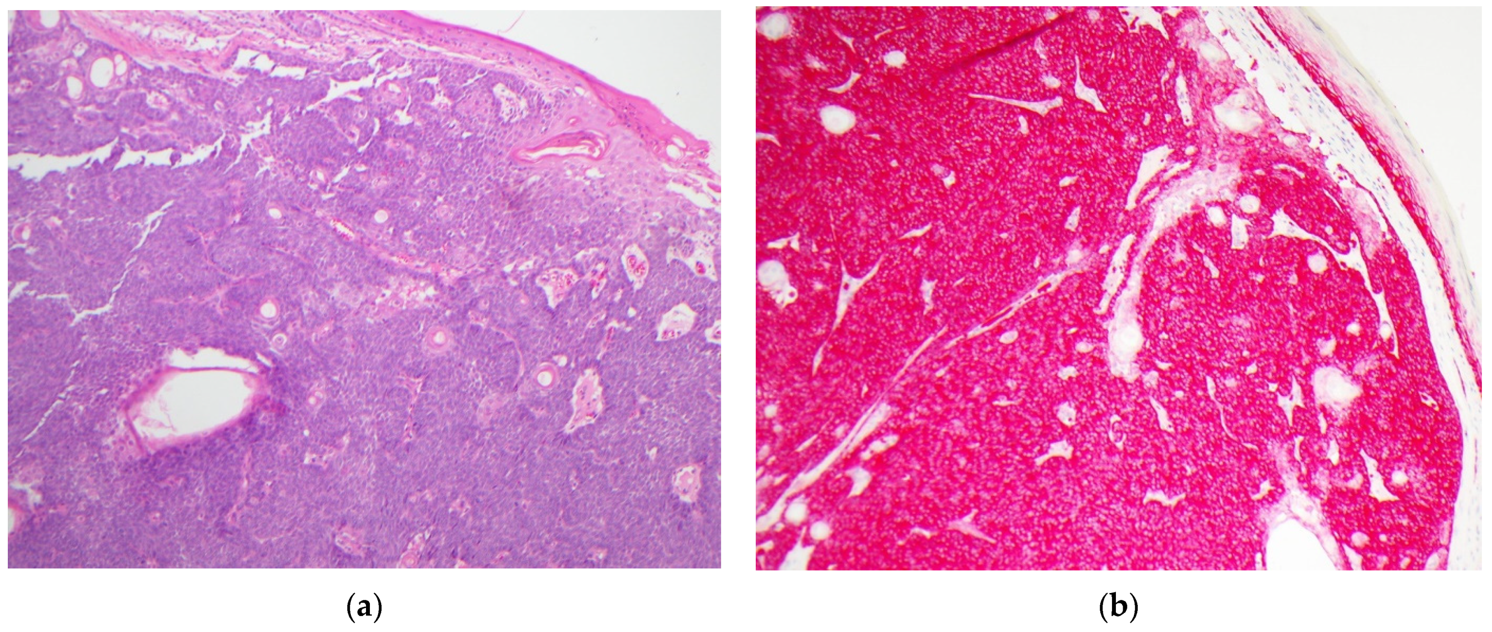
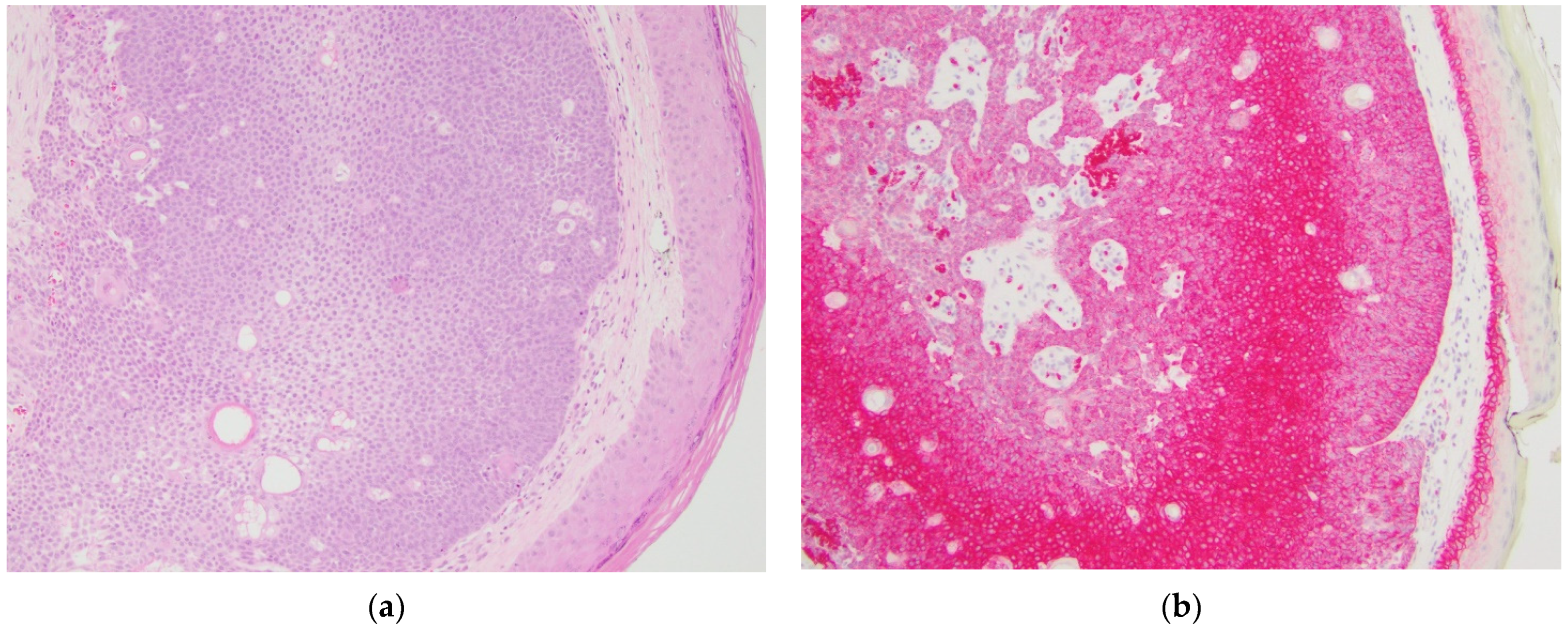
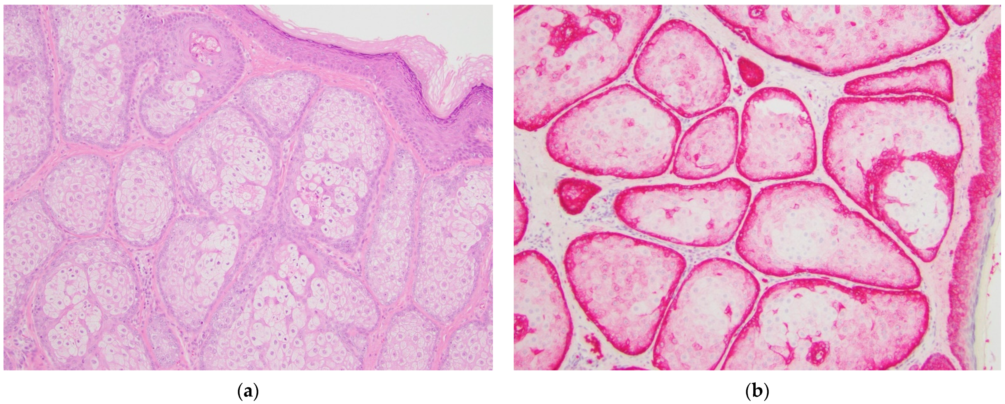
3. Results
4. Discussion
Author Contributions
Funding
Institutional Review Board Statement
Informed Consent Statement
Conflicts of Interest
References
- Iacobelli, J.; Harvey, N.T.; Wood, B.A. Sebaceous lesions of the skin. Pathology 2017, 49, 688–697. [Google Scholar] [CrossRef] [PubMed]
- John, A.M.; Schwartz, R.A. Muir-Torre syndrome (MTS): An update and approach to diagnosis and management. J. Am. Acad. Dermatol. 2016, 74, 558–566. [Google Scholar] [CrossRef] [PubMed]
- Harvey, N.T.; Budgeon, C.A.; Leecy, T.; Beer, T.W.; Kattampallil, J.; Yu, L.; Van Vliet, C.; Muirhead, R.; Sparrow, S.; Swarbrick, N.; et al. Interobserver variability in the diagnosis of circumscribed sebaceous neoplasms of the skin. Pathology 2013, 45, 581–586. [Google Scholar] [CrossRef] [PubMed]
- Zhang, Z.; Zi, Z.; Lee, E.E.; Zhao, J.; Contreras, D.C.; South, A.P.; Abel, E.D.; Chong, B.F.; Vandergriff, T.; Hosler, G.A.; et al. Differential glucose requirement in skin homeostasis and injury identifies a therapeutic target for psoriasis. Nat. Med. 2018, 24, 617–627. [Google Scholar] [CrossRef] [PubMed]
- Liberti, M.V.; Locasale, J.W. The Warburg Effect: How Does it Benefit Cancer Cells? Trends Biochem. Sci. 2016, 41, 211–218. [Google Scholar] [CrossRef] [PubMed]
- Pragallapati, S.; Manyam, R. Glucose transporter 1 in health and disease. J. Oral Maxillofac. Pathol. 2019, 23, 443–449. [Google Scholar] [CrossRef] [PubMed]
- Nakazato, K.; Mogushi, K.; Kayamori, K.; Tsuchiya, M.; Takahashi, K.I.; Sumino, J.; Michi, Y.; Yoda, T.; Uzawa, N. Glucose metabolism changes during the development and progression of oral tongue squamous cell carcinomas. Oncol. Lett. 2019, 18, 1372–1380. [Google Scholar] [CrossRef] [PubMed]
- Rudlowski, C.; Becker, A.J.; Schroder, W.; Rath, W.; Buttner, R.; Moser, M. GLUT1 messenger RNA and protein induction relates to the malignant transformation of cervical cancer. Am. J. Clin. Pathol. 2003, 120, 691–698. [Google Scholar] [CrossRef] [PubMed][Green Version]
- Koch, A.; Lang, S.A.; Wild, P.J.; Gantner, S.; Mahli, A.; Spanier, G.; Berneburg, M.; Muller, M.; Bosserhoff, A.K.; Hellerbrand, C. Glucose transporter isoform 1 expression enhances metastasis of malignant melanoma cells. Oncotarget 2015, 6, 32748–32760. [Google Scholar] [CrossRef] [PubMed]
- Yan, S.; Coffing, B.N.; Li, Z.; Xie, H.; Brennick, J.B.; Beg, H.A.; Froehlich, H.M.; Wells, W.A. Diagnostic and Prognostic Value of ProEx C and GLUT1 in Melanocytic Lesions. Anticancer Res. 2016, 36, 2871–2880. [Google Scholar] [PubMed]
- Pinheiro, C.; Miranda-Goncalves, V.; Longatto-Filho, A.; Vicente, A.L.; Berardinelli, G.N.; Scapulatempo-Neto, C.; Costa, R.F.; Viana, C.R.; Reis, R.M.; Baltazar, F.; et al. The metabolic microenvironment of melanomas: Prognostic value of MCT1 and MCT4. Cell Cycle 2016, 15, 1462–1470. [Google Scholar] [CrossRef] [PubMed]
- Uhlenhake, E.E.; Clark, L.N.; Smoller, B.R.; Shalin, S.C.; Gardner, J.M. Nuclear factor XIIIa staining (clone AC-1A1 mouse monoclonal) is a sensitive and specific marker to discriminate sebaceous proliferations from other cutaneous clear cell neoplasms. J. Cutan. Pathol. 2016, 43, 649–656. [Google Scholar] [CrossRef] [PubMed]
- Tjarks, B.J.; Pownell, B.R.; Evans, C.; Thompson, P.A.; Kerkvliet, A.M.; Koch, M.R.D.; Jassim, A.D. Evaluation and comparison of staining patterns of factor XIIIa (AC-1A1), adipophilin and GATA3 in sebaceous neoplasia. J. Cutan. Pathol. 2018, 45, 1–7. [Google Scholar] [CrossRef] [PubMed]
- Plaza, J.A.; Mackinnon, A.; Carrillo, L.; Prieto, V.G.; Sangueza, M.; Suster, S. Role of immunohistochemistry in the diagnosis of sebaceous carcinoma: A clinicopathologic and immunohistochemical study. Am. J. Dermatopathol. 2015, 37, 809–821. [Google Scholar] [CrossRef] [PubMed]
- Mulay, K.; White, V.A.; Shah, S.J.; Honavar, S.G. Sebaceous carcinoma: Clinicopathologic features and diagnostic role of immunohistochemistry (including androgen receptor). Can. J. Ophthalmol. 2014, 49, 326–332. [Google Scholar] [CrossRef] [PubMed]
- Bayer-Garner, I.B.; Givens, V.; Smoller, B. Immunohistochemical staining for androgen receptors: A sensitive marker of sebaceous differentiation. Am. J. Dermatopathol. 1999, 21, 426–431. [Google Scholar] [CrossRef] [PubMed]
- Li, F.Z.; Ye, Q.; Ran, L.W.; Fang, S. Adipophilin expression in skin lesions with clear cell histology. J. Clin. Pathol. 2021. [Google Scholar] [CrossRef] [PubMed]
- Yang, H.M.; Cabral, E.; Dadras, S.S.; Cassarino, D.S. Immunohistochemical expression of D2-40 in benign and malignant sebaceous tumors and comparison to basal and squamous cell carcinomas. Am. J. Dermatopathol. 2008, 30, 549–554. [Google Scholar] [CrossRef] [PubMed]
- Calder, K.B.; Khalil, F.K.; Schlauder, S.; Cualing, H.D.; Morgan, M.B. Immunohistochemical expression of survivin in cutaneous sebaceous lesions. Am. J. Dermatopathol. 2008, 30, 545–548. [Google Scholar] [CrossRef] [PubMed]
- Jessup, C.J.; Redston, M.; Tilton, E.; Reimann, J.D. Importance of universal mismatch repair protein immunohistochemistry in patients with sebaceous neoplasia as an initial screening tool for Muir-Torre syndrome. Hum. Pathol. 2016, 49, 1–9. [Google Scholar] [CrossRef] [PubMed]


| Diagnosis | Sebaceous Carcinoma | Sebaceoma | Sebaceous Adenoma | Sebaceous Hyperplasia |
|---|---|---|---|---|
| # of cases | 10 | 9 | 10 | 10 |
| Age (years) | ||||
| Average | 64 | 67 | 74 | 58 |
| Range | 47–89 | 46–82 | 68–88 | 40–84 |
| Gender (male) | 60% | 56% | 90% | 70% |
| Sites | Eyelid, nose, face (other), scalp, neck, shoulder, chest, flank, thigh | Nose, ear, face (other), back | Nose, face (other), scalp, back | Nose, face (other), chest, thigh |
| GLUT1 Staining Pattern | Diffuse cytoplasmic and membranous staining in basaloid cells (variable) | >50% Diffuse cytoplasmic and membranous staining | <50% Diffuse cytoplasmic and membranous staining in greater than 1 layer | Only single layer of basaloid cells highlighted by GLUT1 |
| MMR 1 deficiency | ||||
| All retained | 1/10 | 3/9 | 6/10 | |
| MLH1/PMS2 | 1/10 | 0/9 | 0/10 | |
| MSH2/MSH6 | 6/10 | 1/9 | 2/10 | |
| MSH6 only | 1/10 | 0/9 | 0/10 | |
| not done | 1/10 | 5/9 | 2/10 | 10/10 |
Publisher’s Note: MDPI stays neutral with regard to jurisdictional claims in published maps and institutional affiliations. |
© 2021 by the authors. Licensee MDPI, Basel, Switzerland. This article is an open access article distributed under the terms and conditions of the Creative Commons Attribution (CC BY) license (https://creativecommons.org/licenses/by/4.0/).
Share and Cite
Barron, C.R.; Smoller, B.R. GLUT1 Expression in Cutaneous Sebaceous Lesions Determined by Immunohistochemical Staining Patterns. Dermatopathology 2021, 8, 258-264. https://doi.org/10.3390/dermatopathology8030031
Barron CR, Smoller BR. GLUT1 Expression in Cutaneous Sebaceous Lesions Determined by Immunohistochemical Staining Patterns. Dermatopathology. 2021; 8(3):258-264. https://doi.org/10.3390/dermatopathology8030031
Chicago/Turabian StyleBarron, Cynthia Reyes, and Bruce R. Smoller. 2021. "GLUT1 Expression in Cutaneous Sebaceous Lesions Determined by Immunohistochemical Staining Patterns" Dermatopathology 8, no. 3: 258-264. https://doi.org/10.3390/dermatopathology8030031
APA StyleBarron, C. R., & Smoller, B. R. (2021). GLUT1 Expression in Cutaneous Sebaceous Lesions Determined by Immunohistochemical Staining Patterns. Dermatopathology, 8(3), 258-264. https://doi.org/10.3390/dermatopathology8030031