Type I Neurofibromatosis: Case Report and Review of the Literature Focused on Oral and Cutaneous Lesions


Abstract
1. Introduction
2. Background
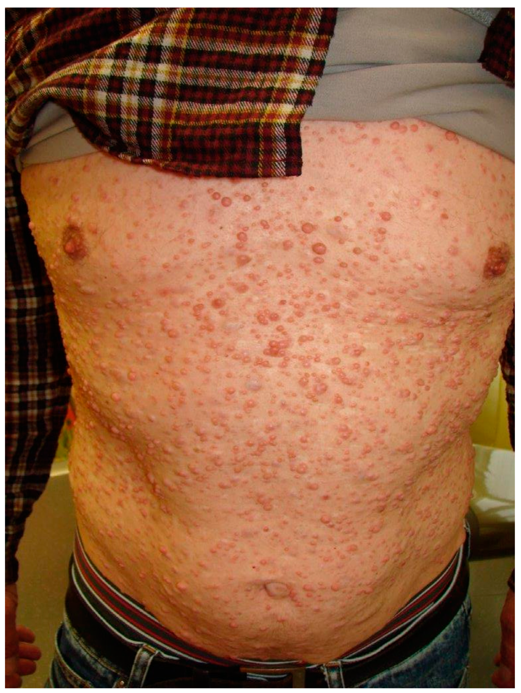
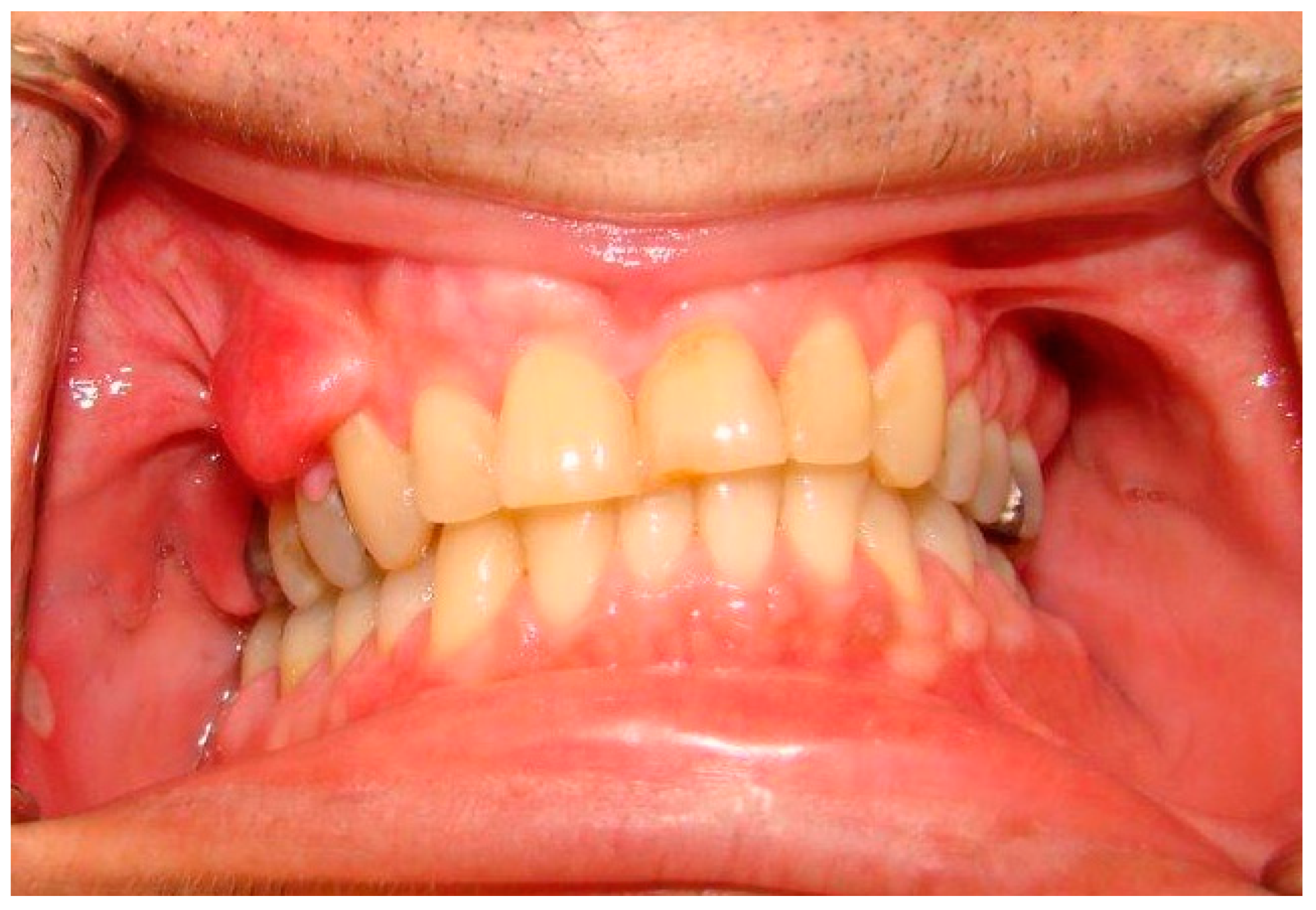
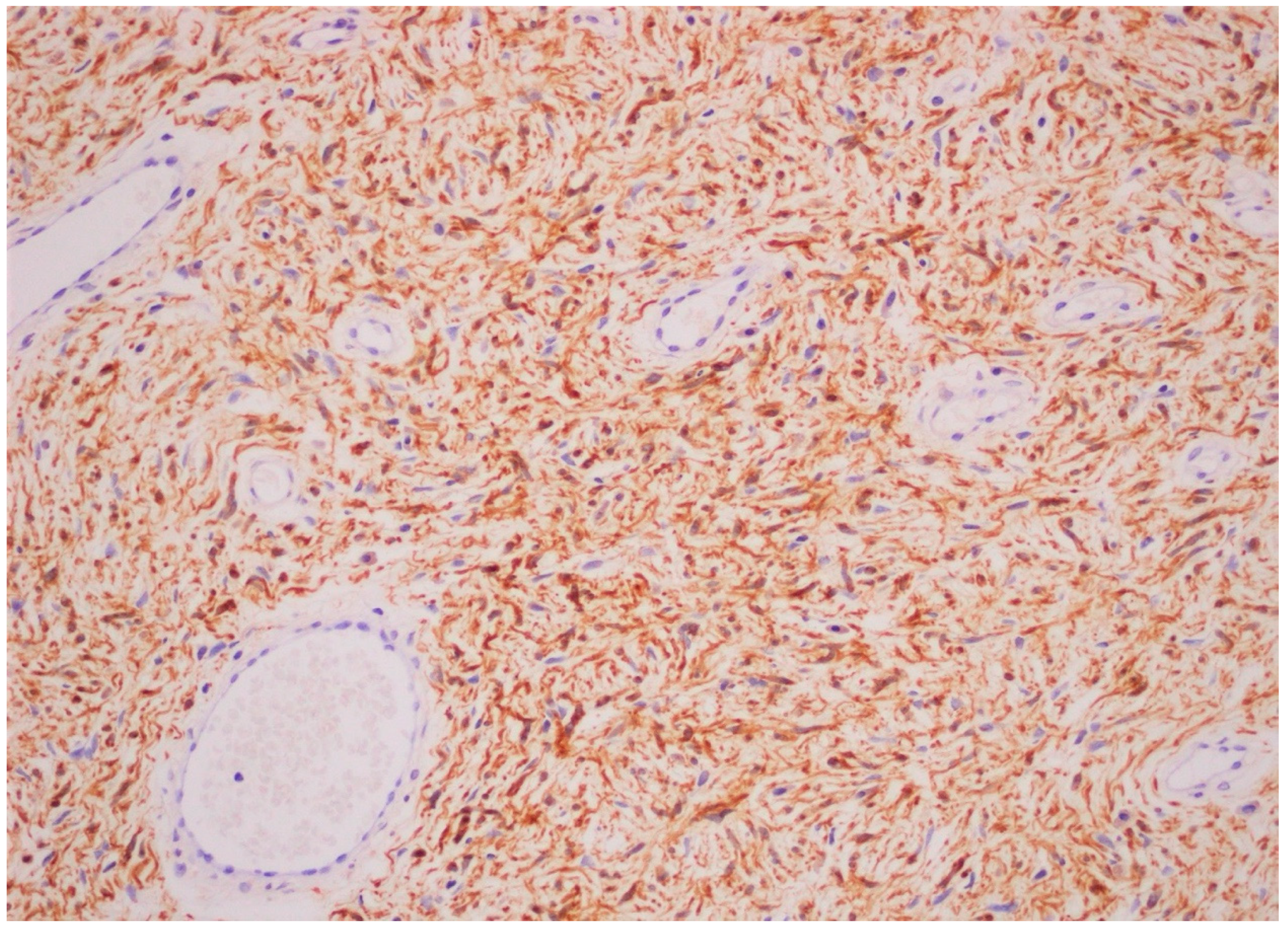
3. Case Report
4. Discussion
5. Conclusions
Author Contributions
Funding
Institutional Review Board Statement
Informed Consent Statement
Data Availability Statement
Conflicts of Interest
References
- Bachelet, J.T.; Combemale, P.; Devic, C.; Foray, N.; Jouanneau, E.; Breton, P. Prise en charge des atteintes craniofaciales de la neurofibromatose de type 1 [Management of craniofacial type 1 neurofibromatosis]. Rev. Stomatol. Chir. Maxillofac. Chir. Orale 2015, 116, 209–214. [Google Scholar] [PubMed]
- Shetty, B.; Umesh, Y.; Kranti, K.; Seshan, H. Periodontal manifestations of von Recklinghausen neuro fibromatosis. J. Indian Soc. Periodontol. 2013, 17, 253–256. [Google Scholar] [CrossRef]
- Visnapuu, V.; Peltonen, S.; Alivuotila, L.; Happonen, R.-P.; Peltonen, J. Craniofacial and oral alterations in patients with Neurofibromatosis 1. Orphanet J. Rare Dis. 2018, 13, 1–9. [Google Scholar] [CrossRef] [PubMed]
- Javed, F.; Ramalingam, S.; Ahmed, H.B.; Gupta, B.; Sundar, C.; Qadri, T.; Al-Hezaimi, K.; Romanos, G.E. Oral manifestations in patients with neurofibromatosis type-1: A comprehensive literature review. Crit. Rev. Oncol. 2014, 91, 123–129. [Google Scholar] [CrossRef] [PubMed]
- Bongiorno, M.; Pistone, G.; Arico, M. Manifestations of the tongue in Neurofibromatosis type 1. Oral Dis. 2006, 12, 125–129. [Google Scholar] [CrossRef]
- Santoro, R.; Santoro, C.; Loffredo, F.; Romano, A.; Perrotta, S.; Serpico, R.; Lauritano, D.; Lucchese, A. Oral Clinical Manifestations of Neurofibromatosis Type 1 in Children and Adolescents. Appl. Sci. 2020, 10, 4687. [Google Scholar] [CrossRef]
- Visnapuu, V.; Peltonen, S.; Tammisalo, T.; Peltonen, J.; Happonen, R.-P. Radiographic Findings in the Jaws of Patients with Neurofibromatosis 1. J. Oral Maxillofac. Surg. 2012, 70, 1351–1357. [Google Scholar] [CrossRef] [PubMed]
- Ferrari, F.; Masurel, A.; Olivier-Faivre, L.; Vabres, P. Juvenile Xanthogranuloma and Nevus Anemicus in the Diagnosis of Neurofibromatosis Type 1. JAMA Dermatol. 2014, 150, 42–46. [Google Scholar] [CrossRef]
- Cunha, K.S.; Barboza, E.P.; Dias, E.P.; Oliveira, F.M. Neurofibromatosis type I with periodontal manifestation. A case report and literature review. Br. Dent. J. 2004, 196, 457–460. [Google Scholar] [CrossRef]
- Bergqvist, C.; Network, N.F.; Servy, A.; Valeyrie-Allanore, L.; Ferkal, S.; Combemale, P.; Wolkenstein, P. Neurofibromatosis 1 French national guidelines based on an extensive literature review since 1966. Orphanet J. Rare Dis. 2020, 15, 1–23. [Google Scholar] [CrossRef]
- Jouhilahti, E.-M.; Visnapuu, V.; Soukka, T.; Aho, H.; Peltonen, S.; Happonen, R.-P.; Peltonen, J. Oral soft tissue alterations in patients with neurofibromatosis. Clin. Oral Investig. 2011, 16, 551–558. [Google Scholar] [CrossRef] [PubMed]
- Christen-Zaech, S.; Vernez, M. Syndromes néoplasiques héréditaires avec atteinte cutanée. Rev. Med. Suisse. 2008, 4, 1095–1102. [Google Scholar] [PubMed]
- García de Marcos, J.A.; Dean Ferrer, A.; Alamillos Granados, F.; Ruiz Masera, J.J.; García de Marcos, M.J.; Vidal Jiménez, A.; Valenzuela Salas, B.; García Lainez, A. Gingival neurofibroma in a neurofibromatosis type 1 patient. Med. Oral Patol. Oral Cir. Bucal. 2007, 12, E287–E291. [Google Scholar] [PubMed]
- Thompson, L.D.R.; Koh, S.S.; Lau, S.K. Sporadic Neurofibroma of the Tongue Unassociated with Neurofibromatosis Type I: A Clinicopathologic Study of Ten Cases. Head Neck Pathol. 2020, 14, 374–380. [Google Scholar] [CrossRef] [PubMed]
- Bouimetarhan, L.; Bellamlih, H.; En-Nafaa, I.; Fenni, J.E.; Amil, T.; Radouane, B. Neurofibfrome plexiforme cervical: À propos d’un cas [Plexiform cervical neurofibroma: About a case]. Pan Afr. Med. J. 2018, 30, 41. [Google Scholar] [CrossRef]
- Prud’Homme, T.; Dajean-Trutaud, S.; Badran, Z.; Hyon, I.; Wotjiuk, F. Dental Management of Neurofibromatosis Type 1: A Case Report and Literature Review. Int. J. Clin. Pediatr. Dent. 2019, 12, 577–581. [Google Scholar] [CrossRef]
- Campos, M.S.; Fontes, A.; Marocchio, L.S.; Nunes, F.D.; De Sousa, S.C.O.M. Clinicopathologic and immunohistochemical features of oral neurofibroma. Acta Odontol. Scand. 2011, 70, 577–582. [Google Scholar] [CrossRef]
- Amaral, F.R.; Ferreira, M.V.L.; Costa, L.A.P.; De Oliveira, P.A.D.; Soares, B.M.; Souza, P.E.A.; De Sousa, G.R. Use of Surgical Laser for Excision of a Neurofibroma Associated With Neurofibromatosis Type-1. J. Lasers Med. Sci. 2018, 9, 219–222. [Google Scholar] [CrossRef]
- Kubota, S.; Imai, T.; Iwai, S.; Nakazawa, M.; Uzawa, N. Gingival Neurofibroma with Teardrop-Shaped Defects of the Interdental Alveolar Bone: An Unusual Oral Manifestation of Neurofibromatosis Type 1. J. Craniofac. Surg. 2019, 30, e205–e207. [Google Scholar] [CrossRef]
- Stumpf, D.; Alksne, J.; Annegers, J.; Brown, S.S.; Conneally, P.M.; Housman, D.; Leppert, M.F.; Miller, J.P.; Moss, M.L.; Pileggi, A.J.; et al. Neurofibromatosis. Conference statement. National Institutes of Health Consensus Development Conference. Arch. Neurol. 1988, 45, 575–578. [Google Scholar]
- Shofty, B.; Constantini, S.; Ben-Shachar, S. Advances in Molecular Diagnosis of Neurofibromatosis Type 1. Semin. Pediatr. Neurol. 2015, 22, 234–239. [Google Scholar] [CrossRef] [PubMed]
- Uusitalo, E.; Rantanen, M.; Kallionpää, R.A.; Pöyhönen, M.; Leppävirta, J.; Ylä-Outinen, H.; Riccardi, V.M.; Pukkala, E.; Pitkäniemi, J.; Peltonen, S.; et al. Distinctive Cancer Associations in Patients with Neurofibromatosis Type 1. J. Clin. Oncol. 2016, 34, 1978–1986. [Google Scholar] [CrossRef] [PubMed]
- Janardhanan, M.; Rakesh, S.; Kumar, R.V. Intraoral presentation of multiple malignant peripheral nerve sheath tumors associated with neurofibromatosis-1. J. Oral Maxillofac. Pathol. 2011, 15, 46–51. [Google Scholar] [CrossRef] [PubMed]
- Pramanick, D.; Ghose, S.; Mazumdar, A. First case report of tongue squamous cell carcinoma in a neurofibromatosis type 1 patient and review of pathogenesis of carcinoma in neurofibromatosis type 1. Indian J. Pathol. Microbiol. 2020, 63, 112–115. [Google Scholar] [CrossRef]
- Gross, A.M.; Wolters, P.L.; Dombi, E.; Baldwin, A.; Whitcomb, P.; Fisher, M.J.; Weiss, B.; Kim, A.; Bornhorst, M.; Shah, A.C.; et al. Selumetinib in Children with Inoperable Plexiform Neurofibromas. N. Engl. J. Med. 2020, 382, 1430–1442. [Google Scholar] [CrossRef]


| Cutaneous | Café au lait spots *, axillary or inguinal freckling * (Crowe’s sign), cutaneous neurofibromas * (localized or plexiform). |
| Oral soft tissue | Prominent lingual papillae (50% cases); Mucosal and gingival neurofibromas * (25% of cases): mostly the tongue, followed by buccal mucosa, lips, and gingiva, and less commonly the palate, the floor of the mouth, the major salivary glands and the pharynx; Macroglossia in relation to plexiform neurofibromas arising inside the tongue; Melanin pigmentation of the gingiva (rare); Gingivitis or periodontitis in relation to oral neurofibromas prohibiting a proper oral hygiene. |
| Cranio-facial | Orbital dysplasia (may lead to exophthalmia), sphenoidal wings dysplasia *; Widening of the mandibular canal without relation with any tumor mass +/− irregular border of the canal and enlarged mandibular foramina; Short mandibular body, ramus, and condyle, undergrowth maxilla with hypoplasia of the maxillary tuberosity and short cranial base (inducing retrognathia); Intra-osseus neurofibromas of the maxilla/mandible and the temporo-mandibular joint (well-defined unilocular and occasionally multilocular radiolucent lesions); Notching of the posterior border of the mandibular ramus, elongated coronoid process with a deep sigmoid notch, hypoplasia of the condyle and zygomatic processes; Periapical cement dysplasia (only NF1 females affected), central giant cell granuloma, and osteolytic bone lesions linked to cherubism. |
| Dental | Retained or displaced teeth, agenesia, or hyperdontia, impaired growth of alveolar bone in association with gingival or bone neurofibromas and especially plexiform neurofibromas arising from the trigeminal nerve; Enamel defects such as molar-incisor hypomineralization, enamel hypoplasia, or opacities; Predisposition to caries is controversial. |
Publisher’s Note: MDPI stays neutral with regard to jurisdictional claims in published maps and institutional affiliations. |
© 2021 by the authors. Licensee MDPI, Basel, Switzerland. This article is an open access article distributed under the terms and conditions of the Creative Commons Attribution (CC BY) license (http://creativecommons.org/licenses/by/4.0/).
Share and Cite
Buchholzer, S.; Verdeja, R.; Lombardi, T. Type I Neurofibromatosis: Case Report and Review of the Literature Focused on Oral and Cutaneous Lesions. Dermatopathology 2021, 8, 17-24. https://doi.org/10.3390/dermatopathology8010003
Buchholzer S, Verdeja R, Lombardi T. Type I Neurofibromatosis: Case Report and Review of the Literature Focused on Oral and Cutaneous Lesions. Dermatopathology. 2021; 8(1):17-24. https://doi.org/10.3390/dermatopathology8010003
Chicago/Turabian StyleBuchholzer, Samanta, Raùl Verdeja, and Tommaso Lombardi. 2021. "Type I Neurofibromatosis: Case Report and Review of the Literature Focused on Oral and Cutaneous Lesions" Dermatopathology 8, no. 1: 17-24. https://doi.org/10.3390/dermatopathology8010003
APA StyleBuchholzer, S., Verdeja, R., & Lombardi, T. (2021). Type I Neurofibromatosis: Case Report and Review of the Literature Focused on Oral and Cutaneous Lesions. Dermatopathology, 8(1), 17-24. https://doi.org/10.3390/dermatopathology8010003