Next Article in Journal
Hypervirulent Klebsiella pneumoniae as a Hospital-Acquired Pathogen in the Intensive Care Unit in Mansoura, Egypt
Previous Article in Journal
High Frequency of Mycoplasma pneumoniae Among Patients with Atypical Pneumonia in Tehran, Iran
 
 
GERMS is published by MDPI from Volume 15 Issue 4 (2025). Previous articles were published by another publisher in Open Access under a CC-BY (or CC-BY-NC-ND) licence, and they are hosted by MDPI on mdpi.com as a courtesy and upon agreement with the former publisher Infection Science Forum.
Article

Vancomycin Resistance Among Staphylococcus aureus Isolates in a Rural Setting, Egypt

by
Nada ElSayed
,
Medhat Ashour
and
Amira Ezzat Khamis Amine
*
Microbiology Department, High Institute of Public Health, Alexandria University, 169 ElHoryah Avenue, Alexandria, Egypt
*
Author to whom correspondence should be addressed.
GERMS 2018, 8(3), 134-139; https://doi.org/10.18683/germs.2018.1140
Submission received: 2 July 2018 / Revised: 16 August 2018 / Accepted: 27 August 2018 / Published: 3 September 2018

Abstract

Introduction: With the increased occurrence of methicillin resistant S. aureus (MRSA), the consumption of vancomycin, the drug of choice, has also increased. As a consequence, strains of S. aureus resistant to vancomycin have started to emerge. This study aimed to evaluate the level of vancomycin resistance among clinical and nasal S. aureus isolates in a rural town in Egypt. Methods: This cross-sectional study was held in the general hospital at the rural town of Kafr Eldawar in Egypt, during the period from January 2013 to January 2014. S. aureus isolates were collected from clinical samples and from nasal swabs. Results: Two hundred S. aureus isolates were collected, 80 (40%) from clinical samples and 120 (60%) from nasal carriage samples. Vancomycin resistant S. aureus (VRSA) was only detected in clinical samples, all collected from the outpatient clinic. Eleven VRSA isolates (13.8% of total S. aureus clinical isolates) and one strain of vancomycin-intermediate S. aureus (from nasal carriage) were detected. VRSA isolates were most resistant to ciprofloxacin (90.9%) and erythromycin (81.8%). Five isolates were resistant to all tested antibiotics: ciprofloxacin, clindamycin, erythromycin, linezolid, oxacillin, penicillin and trimethoprim-sulfamethoxazole. MRSA was found to constitute 43.8% of clinical S. aureus isolates. The MRSA colonization rate among community individuals was 43.6%, 42.9% among healthcare workers and 51.4% among patients. Conclusion: The prevalence of VRSA was high in clinical samples suggesting that there is a high level of VRSA strains in Egypt that goes undetected since most laboratories only use disk diffusion for detection of vancomycin resistance.

Introduction

Vancomycin has been used as the drug of choice for methicillin resistant S. aureus (MRSA) infections. The rate of infections caused by MRSA has been steadily on the rise worldwide and as a result the consumption of vancomycin has also increased [1]. In 1997, the first case of vancomycin intermediate S. aureus (VISA) was reported in Japan [2]. From that time forward, more cases of VISA, and of vancomycin resistant S. aureus (VRSA) started to emerge throughout the world even in developed countries [3].
While the morbidity rates of VRSA infections in the USA and Europe are still low, higher levels were identified in other regions in the world especially in developing countries. A dramatic increase in the percentage of VRSA was reported in India in 2011 compared to a previous study in 2006 (45% vs. 0.25%) [4]. Like MRSA, isolated VISA and VRSA strains had been hospital-acquired, until cases of community-acquired VRSA were recorded [5]. In 2017 the World Health organization published a list of “antibiotic-resistant priority pathogens” that include VISA and VRSA [6]. The aim of this study was to determine the prevalence of S. aureus isolates resistant to vancomycin in clinical samples and nasal carriage.

Methods

This cross-sectional study was held in the general hospital at the rural town of Kafr El-Dawar in Egypt, during the period from January 2013 to January 2014. The study protocol was approved by the Ethics Committee at the High Institute of Public Health (HIPH) on 27 November 2012 (approval no. 32). An informed consent was obtained from all participants.

Collection and processing of samples

S. aureus was isolated from both clinical samples and nasal swabs. Clinical samples were collected from all inpatients admitted to all 3 surgical wards in the hospital; each ward contains 22 beds, two beds in each room. Clinical samples were also collected from outpatients visiting the surgical outpatient clinic. Any patient who suffered from an infection during the period of the study was included. Patients suffered from the following infections: abscesses, diabetic foot infections, postoperative wound infections, and skin infections. The sampling procedure was first described to each patient. The dressing was removed and the lesion was irrigated with sterile saline. Sterile cotton swabs, moistened with sterile saline, were used to collect samples. Sampling was done in a zigzag motion whilst rotating the swab between fingers, simultaneously [7].
Nasal samples were collected from inpatients after 48 hours of admission, from healthcare workers in the hospital, and from community individuals visiting admitted inpatients. Nasal swabs were collected using a flexible sterile cotton swab, moistened with sterile saline. The swab was inserted 2 cm into each nostril and gently rotated for about 3 seconds [8].
Upon arrival to the laboratory, clinical samples were cultured on blood and MacConkey agar plates while nasal swabs were streaked onto mannitol salt agar plates. All agar plates were aerobically incubated at 37 °C for 24 hours. Mannitol salt agar plates were examined for any visible growth after 24 hours. If no growth occurred, they were reincubated for another 24 hours, with maximum incubation period of 72 hours [9].

Identification and antimicrobial susceptibility

Isolated colonies were identified morphologically then examined microscopically by Gram staining. All isolates were subjected to full identification according to the standard microbiological methods [9].
All isolates identified as S. aureus were tested for their antibiotic susceptibility using the disk diffusion method. Antibiotics tested were ciprofloxacin (CIP), clindamycin (DA), erythromycin (E), linezolid (LZD), oxacillin (OX), penicillin (PEN) and trimethoprim-sulfamethoxazole (SXT). For quality control, the susceptible control strain S. aureus ATCC 25923 was used [10].
Vancomycin resistance was determined by MIC testing using the agar dilution method. Isolates that were vancomycin resistant by MIC test results were confirmed by E test [11]. For quality control, the control strains S. aureus ATCC 29213 and Enterococcus faecalis ATCC 29212 were used. Bacterial isolates were classified into VRSA, VISA, and VSSA according to the following MIC ranges VSSA ≤2 µg/mL, VISA 4-8 µg/mL and VRSA MIC ≥16 µg/mL [10].

Types of samples and method of selection

All samples were collected consecutively from outpatients and inpatients of Kafr El-Dawar General Hospital until the required sample size was reached.

Statistical analysis

Data were analyzed using IBM SPSS 20, (IBM, Armonk, NY, USA). Descriptive statistics such as frequency, proportion, mean, standard deviation and median were used. The p<0.05 was taken as a cut-off for statistical significance and all tests were two-sided. The Chi-square test was used for testing the association of categorical data and in case of small cell frequency; the Monte-Carlo exact test was displayed. Comparison of resistance to different antibiotics was done using the Freidman test. Multiple comparison among antibiotics was conducted using Wilcoxon signed-rank tests with Bonferroni correction.

Results

During this study 240 clinical samples were collected. S. aureus was isolated from 80 samples (33.3%). Although we collected clinical samples from both inpatients and outpatients, S. aureus was detected only in outpatient samples (Table 1). Out of 960 nasal samples, 120 S. aureus isolates were retrieved and the total S. aureus nasal carriage rate was 12.5%. Nasal carriage samples included 55 isolates (45.8%) from community individuals, 37 (30.8%) from patients and 28 (23.3%) from healthcare workers (Table 2).
Two hundred S. aureus isolates were collected, 80 (40%) from clinical samples and 120 (60%) from nasal carriage. S. aureus isolates showed the highest resistance rate against penicillin (96%), while they were most sensitive to linezolid (95%), followed by clindamycin (94%), trimethoprim-sulfamethoxazole (89%), and erythromycin and ciprofloxacin (76% each). There was a statistically significant difference in the resistance to different antibiotics, Friedman χ2(6)= 678.82,p<0.001 (Figure 1). There was a significant difference between penicillin and oxacillin on the one hand and all other antibiotics on the other hand (Wilcoxon signed-rank tests with Bonferroni correction, p<0.001).
The overall methicillin resistance was 45% (90 strains). The resistance to methicillin among clinical samples and nasal samples was comparable (43.8% and 45.8%, respectively); Pearson Chi-square=0.63 (p=0.730), i.e., not significant. The percentage among nasal isolates was slightly higher among patients (51.4%) than in the community (43.6%) and in healthcare workers (42.9%); Pearson Chi-square=0.76 (p=0.944), i.e., not significant.
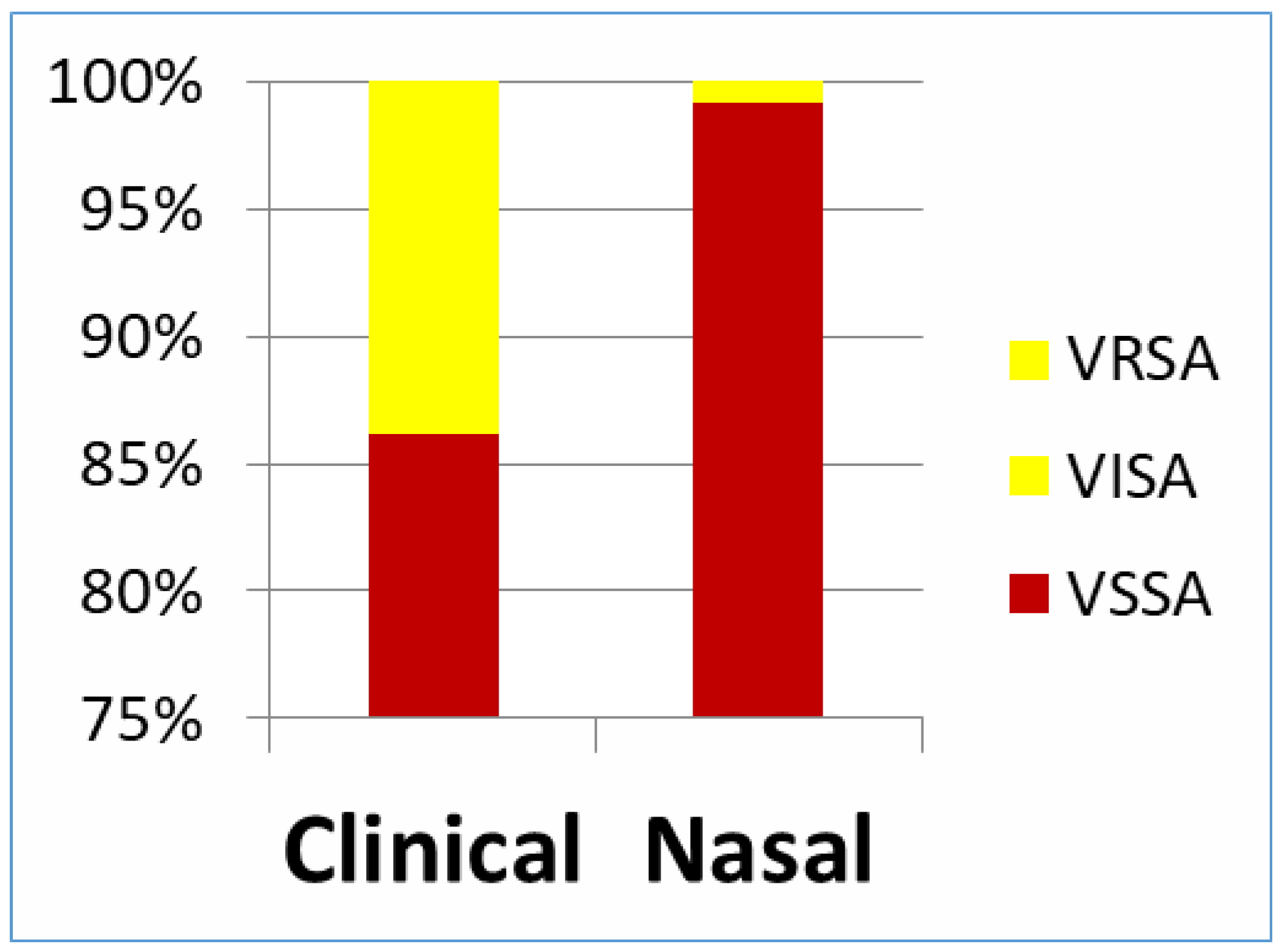
VRSA was only detected in clinical samples. Eleven VRSA isolates were detected, constituting 13.8% of the total S. aureus clinical isolates. Two of the 11 isolates had an MIC of ≥16 µg/mL, 6 had an MIC of ≥32 µg/mL and 3 showed a high MIC of ≥64 µg/mL. However, one strain of VISA was detected among the strains collected from nasal carriage from a healthcare worker (0.5%)—Figure 2. VRSA isolates were most resistant to ciprofloxacin (90.9%) followed by erythromycin (81.8%), sulfamethoxazole-trimethoprim, clindamycin and linezolid (63.6% each). Five isolates were resistant to all tested antibiotics.

Discussion

In the present study, even though we collected clinical samples from inpatients and outpatients, S. aureus was only detected in outpatients’ samples which means they were all community-acquired isolates. S. aureus isolates were most susceptible to linezolid and clindamycin. Higher levels of resistance were reported in India, where MRSA was found to constitute 45% of S. aureus isolates. Similar prevalence rates of MRSA were reported in Egypt among skin and soft tissue infections, where 47.4% of the isolated S. aureus strains were reported as MRSA [4,12]. Unfortunately, until the end of the study, there was no working microbiological laboratory in the hospital, so antibiotics were prescribed without performing a susceptibility test and were given empirically. This empiric use of antibiotics may not only lead to sub-optimal and ineffective treatment, but it may also select for antibiotic-resistant bacteria [13].
In the present study, 51.4% of patients and 42.9% of healthcare workers were colonized with MRSA. A similar MRSA colonization rate was reported by Moniri et al. (52.6%) [14]. The problem with this high carriage rate among both groups is the high risk of the development of subsequent MRSA infections either through transmission from healthcare workers to patients or through auto-infection [15]. The prevalence of MRSA in clinical and nasal carriage samples was comparable, probably because the studied infections were mostly from the outpatient clinic (i.e., mostly community-acquired MRSA). However, oxacillin disks were used in this study instead of cefoxitin so this can be considered as a limitation in the study.
Detection of vancomycin resistance is essential not only for an optimal therapy, but also for infection control measures and epidemiological purposes [1]. As recommended by the Clinical & Laboratory Standards Institute (CLSI), the agar dilution method was used for MIC determination for the detection of VISA and VRSA strains in this study. Most microbiology laboratories in Egypt depend only on the disk diffusion method to determine S. aureus susceptibility to vancomycin, which does not give reliable results. It can leave many VISA/VRSA isolates undetected and they will give inhibition zones with sizes similar to those of the vancomycin-susceptible ones [16]. In the present study, MIC determination detected 5.5% of the S. aureus isolates as VRSA and 0.5% of them as VISA. The prevalence of VRSA among clinical cases was 13.8% while none of the nasal carriers had VRSA. A lower prevalence rate was reported in other developing countries but they used the disk diffusion method, which means that the actual prevalence of VRSA might be different [17,18]. Gohniem et al. reported an even higher resistance rate (20.68% VISA and 20.68% VRSA) indicating that resistance rates are on the rise [19]. On the other contrary, Amr et al. reported a lower incidence (8.8%) of VRSA among clinical isolates. This difference is probably due to different geographical locations of both studies in Egypt [20].
Differences in the antibiotic resistance patterns vary widely between different geographic regions. Many factors contribute to those patterns including local infection control programs implemented, antibiotic prescribing policies and epidemiology of the studied strains themselves [1]. In developing countries the problem is more challenging due to frequent dispensing of antibiotics at drug stores without prescription [21]. Also, use of antibiotics without performing antibiotic susceptibility testing and the thus inappropriate prescribing of antibiotics that was observed in this study. Other factors include the uncontrolled use in agriculture and livestock [22].
Fortunately, none of the patients or community individuals were colonized with VISA or VRSA. Only one healthcare worker was colonized with VISA (0.5%). A comparable finding was reported by Ploy et al. in France, where 3.4% of healthcare workers were VISA nasal carriers [23].
Available data regarding the antibiotic susceptibility pattern of VRSA is sparse. All the detected VRSA and VISA strains were also methicillin-resistant [24]. An alarming observation was that five out of the eleven VRSA isolates in the present study showed resistance to all the tested antibiotics, including linezolid. Higher linezolid resistance (40%) in VRSA strains was also reported in Egypt [20]. Also, in another study linezolid was less effective against VRSA than other antibiotics [25]. Sanchez Garcia et al. reported that linezolid resistance probably develops on exposure [26].
Conclusions
The prevalence of VRSA was high in clinical samples (13.8%) collected from community-acquired infections suggesting that there is a high level of VRSA strains in Egypt that goes undetected since most labs only use disk diffusion for the detection of vancomycin resistance. Infection control measures and antimicrobial stewardship efforts should focus both on community and hospital resistant strains.

Author Contributions

NE was responsible for collection of data, samples and the laboratory work included in the study; she was also involved in literature search and writing of the article. MA was responsible for the analysis and interpretation of the results; AEKA was responsible for the conception and design of the research plan; she was also responsible for writing and revising the article. All authors read and approved the final version of the manuscript.

Funding

None to declare.

Conflicts of interest

All authors—none to disclose.

References

  1. Dhanalakshmi, T.A.; Umapathy, B.L.; Mohan, D.R. Prevalence of methicillin, vancomycin and multidrug resistance among Staphylococcus aureus. J Clin Diagn Res 2012, 6, 974–977. [Google Scholar]
  2. Hiramatsu, K.; Aritaka, N.; Hanaki, H.; et al. Dissemination in Japanese hospitals of strains of Staphylococcus aureus heterogeneously resistant to vancomycin. Lancet 1997, 350, 1670–1673. [Google Scholar] [CrossRef] [PubMed]
  3. Bierbaum, G.; Fuchs, K.; Lenz, W.; Szekat, C.; Sahl, H. Presence of Staphylococcus aureus with reduced susceptibility to vancomycin in Germany. Eur J Clin Microbiol Infect Dis 1999, 18, 691–696. [Google Scholar] [CrossRef] [PubMed]
  4. Goswami, N.N.; Trivedi, H.R.; Goswami, A.P.; Patel, T.K.; Tripathi, C.B. Antibiotic sensitivity profile of bacterial pathogens in postoperative wound infections at a tertiary care hospital in Gujarat, India. J Pharmacol Pharmacother 2011, 2, 158–164. [Google Scholar] [CrossRef] [PubMed]
  5. Palazzo, I.C.V.; Araujo, M.L.C.; Darini, A.L.C. First report of vancomycin-resistant staphylococci isolated from healthy carriers in Brazil. J Clin Microbiol 2005, 43, 179–184. [Google Scholar] [CrossRef]
  6. World Health Organization. WHO Publishes List of Bacteria for Which New Antibiotics Are Urgently Needed. 2017. Available online: http://www.who.int/mediacentre/news/releases/2017/bacteria-antibiotics-needed/en/ (accessed on 15 April 2017).
  7. NHS Trust. Procedure for Taking a Wound Swab. 2008. Available online: http://www.wirralct.nhs.uk/attachments/article/19/CP23Procedurefortakingawoundswab19Feb13.doc.pdf (accessed on 8 November 2013).
  8. National Health and Nutrition Examination Survey. Specimen Collection Procedures Manual. 2000. Available online: http://www.cdc.gov/nchs/data/nhanes/nhanes_01_02/specimen_collection_year_3.pdf (accessed on 5 May 2013).
  9. Forbes, B.; Sahm, D.; Weissfeld, A. Organisms. In Bailey and Scott’s Diagnostic Microbiology, 12th ed.; Forbes, B., Sahm, D., Weissfeld, A., Eds.; Mosby: St. Louis, MO, USA, 2007; pp. 254–264. [Google Scholar]
  10. Clinical and Laboratory Standards Institute. Performance Standards for antimicrobial disk susceptibility Tests; approved standard. 11th ed. M02-A11 ed.; Clinical and Laboratory Standards Institute: Wayne PA, USA, 2012. [Google Scholar]
  11. Wiegand, I.; Hilpert, K.; Hancock, R.E. Agar and broth dilution methods to determine the minimal inhibitory concentration (MIC) of antimicrobial substances. Nat Protoc 2008, 3, 163–175. [Google Scholar] [CrossRef]
  12. Sobhy, N.; Aly, F.; Abd El Kader, O.; Ghazal, A.; Elbaradei, A. Community-acquired methicillin-resistant Staphylococcus aureus from skin and soft tissue infections (in a sample of Egyptian population): Analysis of mec gene and staphylococcal cassette chromosome. Braz J Infect Dis 2012, 16, 426–431. [Google Scholar] [CrossRef] [PubMed]
  13. Paterson, D.L. “Collateral damage” from cephalosporin or quinolone antibiotic therapy. Clin Infect Dis 2004, 38, S341–S345. [Google Scholar] [CrossRef] [PubMed]
  14. Moniri, R.; Musav, G.A.; Fadavi, N. The prevalence of nasal carriage methicillin-resistant Staphylococcus aureus in hospitalized patients. Pak J Med Sci 2009, 25, 656–659. [Google Scholar]
  15. Safdar, N.; Bradley, E. The risk of infection after nasal colonization with Staphylococcus aureus. Am J Med 2008, 121, 310–315. [Google Scholar] [CrossRef] [PubMed]
  16. Clinical and Laboratory Standards Institute. Performance Standards for Antimicrobial Susceptibility Testing; Twenty-fourth informational supplement. M100-S24 ed.; Clinical and Laboratory Standards Institute: Wayne PA, USA, 2014. [Google Scholar]
  17. Kheder, S.I.; Ali, N.A.; Fathelrahman, A.I. Prevalence and antimicrobial susceptibility pattern of methicillin resistant staphylococci in a Sudanese surgical ward. Pharmacol Pharm 2012, 3, 103–108. [Google Scholar] [CrossRef]
  18. El-Jakee, J.K.; Atta, N.S.; Samy, A.A.; et al. Antimicrobial resistance in clinical isolates of Staphylococcus aureus from bovine and human sources in Egypt. Glob Vet 2011, 7, 581–586. [Google Scholar]
  19. Ghoniem, E.M.; El Hendawy, G.R.; Abdel Moteleb, T.M.; Hassan, H.A.; El Refai Khalil, H. Characterization of vancomycin-resistant Staphylococcus aureus in the National Liver Institute. Menoufia Med J 2014, 27, 825–832. [Google Scholar]
  20. Amr, G.E.; Gammal, S.A. Emergence of vancomycin resistant Staphylococcus aureus isolated from patients in ICUs of Zagazig University Hospitals. Egypt J Med Microbiol 2017, 26, 53–59. [Google Scholar] [CrossRef]
  21. Amin, M.E.; Amine, A.; Newegy, M.S. Injudicious provision of subtherapeutic doses of antibiotics in community pharmacies. Innov Pharm 2017, 8, 18. [Google Scholar] [CrossRef]
  22. Okeke, I.N.; Lamikanra, A.; Edelman, R. Socioeconomic and behavioral factors leading to acquired bacterial resistance to antibiotics in developing countries. Emerg Infect Dis 1999, 5, 18–27. [Google Scholar] [CrossRef] [PubMed]
  23. Ploy, M.C.; Francois, B.; Mounier, M.; Vignon, P.; Denis, F. Nasal carriage of vancomycin-intermediate Staphylococcus aureus among intensive care unit staff. Clin Infect Dis 2001, 33, 1951. [Google Scholar] [CrossRef] [PubMed]
  24. El-Baky, R.M.A.; Ahmed, H.R.; Gad, G.F.M. Prevalence and conjugal transfer of vancomycin resistance among clinical isolates of Staphylococcus aureus. Adv Res 2014, 2, 12–23. [Google Scholar] [CrossRef]
  25. El-Daker, M.A.; Mesbah, M.R.; El-Naggar, M.M.; Khalil, E.A.; El-Kenawy, M.F. The first two vancomycin resistant Staphylococcus aureus isolates in Mansoura University Hospital; epidemiology and antimicrobial study. Egypt J Med Microbiol 2008, 17, 31–42. [Google Scholar]
  26. Sánchez García, M.; De la Torre, M.A.; Morales, G.; et al. Clinical outbreak of linezolid-resistant Staphylococcus aureus in an intensive care unit. JAMA 2010, 303, 2260–2264. [Google Scholar] [CrossRef] [PubMed]
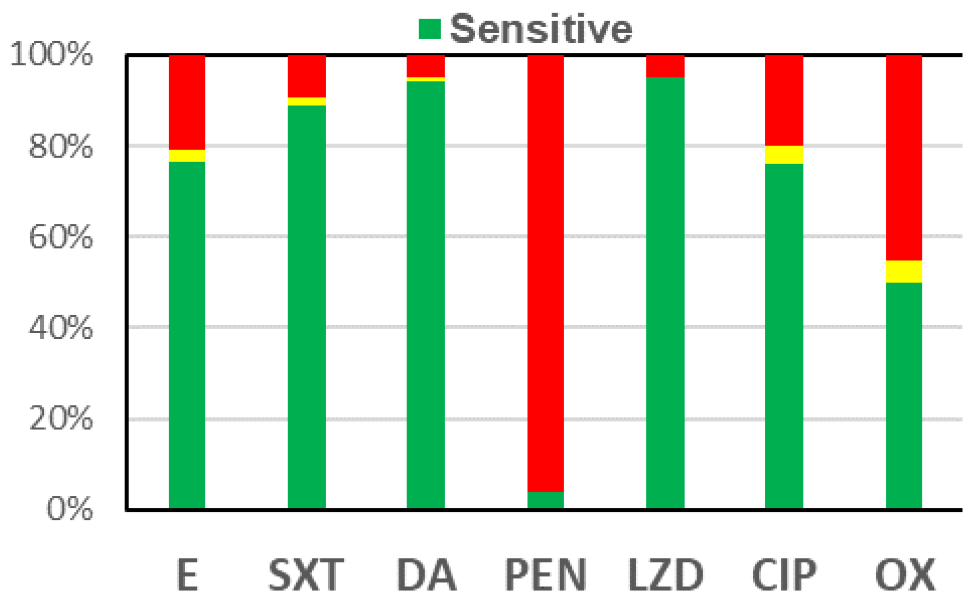
Figure 1. Antibiotic resistance among 200 S. aureus isolates. 
Germs 08 00134 g001
Figure 2. Susceptibility of 200 S. aureus isolates to vancomycin according to the type of sample. 
Germs 08 00134 g002
Table 1. S. aureus isolated from outpatients at Kafr El-Dawar General Hospital, 2013-2014. 
Abscess No (%) Diabetic foot
infection
No (%)
Postoperative wound
infection
No (%)
Skin infection
No (%)
Total
No
(%)
S. aureus42 (52.5) 16 (20.0) 5 (6.2) 17 (21.3) 80
(100)
Table 2. Distribution of isolation of S. aureus among nasal carriage groups, Kafr El-Dawar General Hospital, 2013-2014. 
Nasal carriage groupS. aureus nasal carriageTotal
PositiveNegativeNo.%
No.%No.%
Community55 12.4 387 87.6 442 46.0
Healthcare workers28 15.6 152 84.4 180 18.8
Patients37 10.9 301 89.1 338 35.2
Total120 12.5 840 87.5 960 100.0