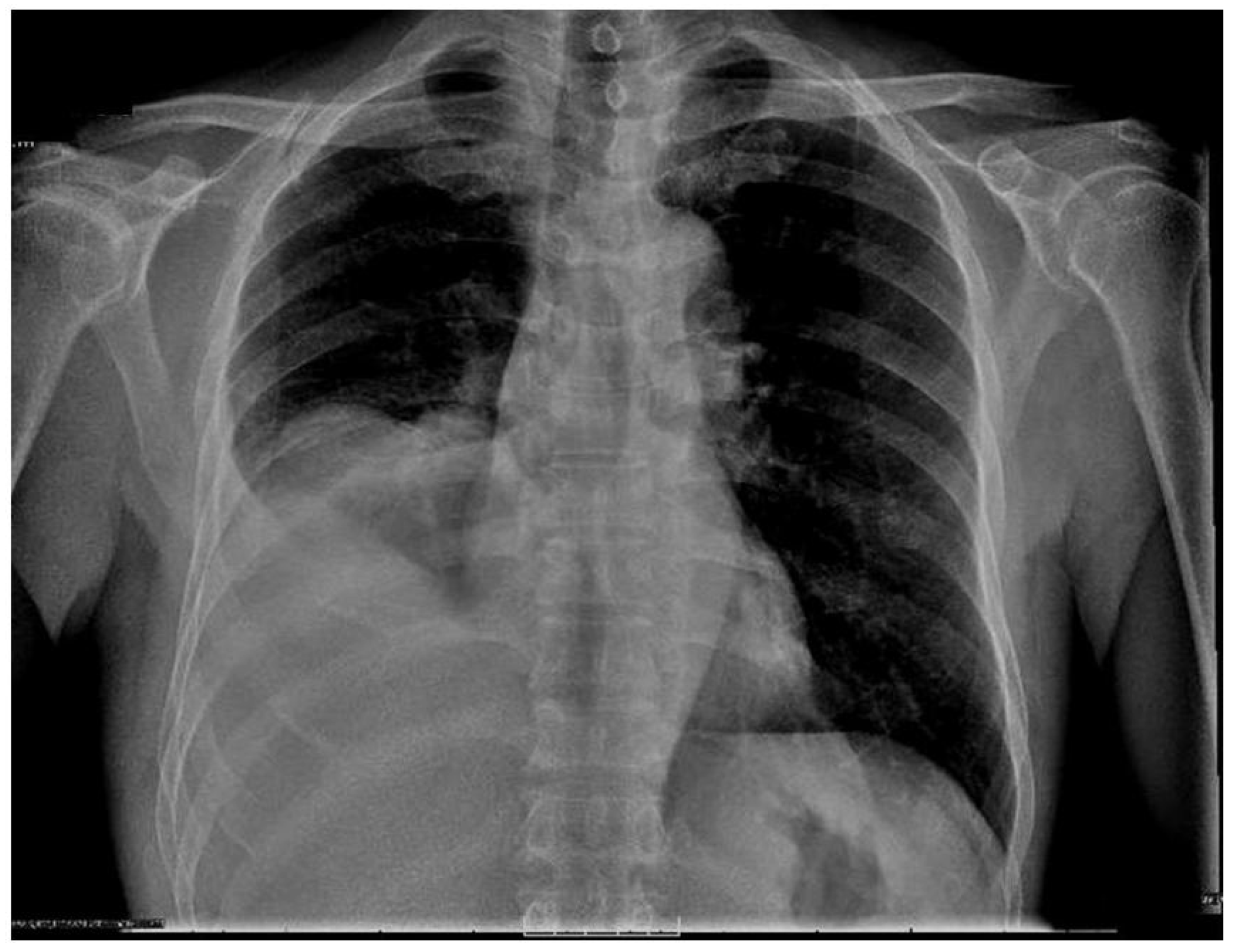
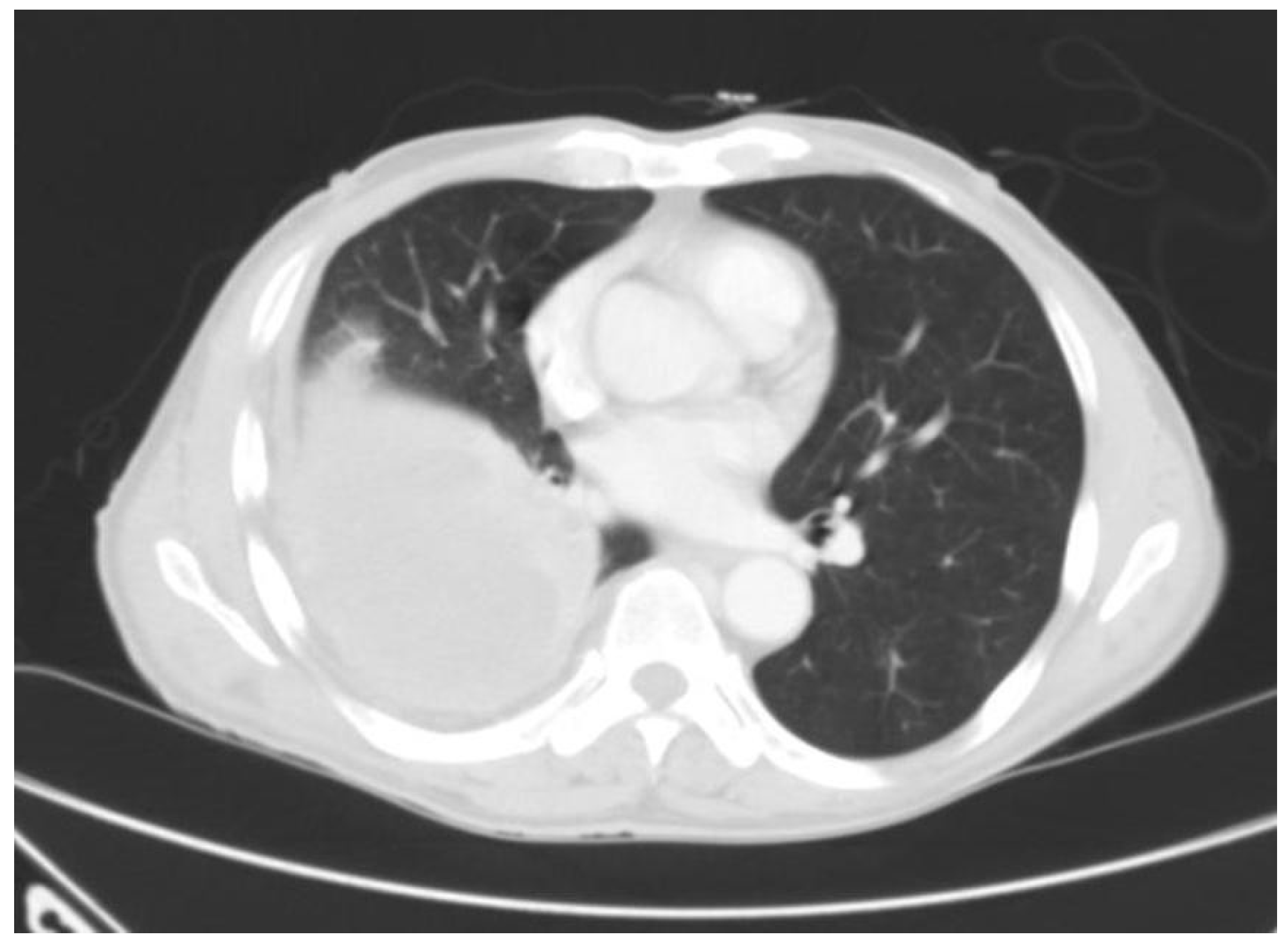
Tuberculous Empyema Presenting as a Persistent Chest Wall Mass: Case Report
Abstract
Introduction
Discussion
Conclusion
Conflicts of Interest
References
- Adamsson, K.; Bruchfeld, J.; Palme, I.B.; Julander, I. Extrapulmonary tuberculosis--an infection of concern in most clinical settings. Lakartidningen 2000, 97, 5622–5626. [Google Scholar] [PubMed]
- Kim, Y.T.; Han, K.N.; Kang, C.H.; Sung, S.W.; Kim, J.H. Complete resection is mandatory for tubercular cold abscess of the chest wall. Ann Thorac Surg 2008, 85, 273–277. [Google Scholar] [CrossRef] [PubMed]
- Aylk, S.; Qakan, A.; Aslankara, N.; Ozsöz, A. Tuberculous abscess on the chest wall. Monaldi Arch Chest Dis 2009, 71, 39–42. [Google Scholar][Green Version]
- Sahn, S.A.; Iseman, M.D. Tuberculous empyema. Semin Respir Infect 1999, 14, 82–87. [Google Scholar]
- Barrett, N.R. Tuberculosis of the Chest Wall. Tubercle 1939, 20, 445–59. [Google Scholar] [CrossRef]
- Tanaka, S.; Aoki, M.; Nakanishi, T.; et al. Retrospective case series analysing the clinical data and treatment options of patients with a tubercular abscess of the chest wall. Interact Cardiovasc Thorac Surg 2012, 14, 249–252. [Google Scholar] [CrossRef]
- Chaiyasate, K.; Hramiec, J. Images in clinical medicine. Tuberculosis empyema necessitatis. N Engl J Med 2005, 352, e8. [Google Scholar] [CrossRef]
- Senent, C.; Betlloch, I.; Chiner, E.; Llombart, M.; Moragón, M. Tuberculous empyema necessitatis. A rare cause of cutaneous abscess in the XXI century. Dermatol Online J 2008, 14, 11. [Google Scholar] [CrossRef] [PubMed]
- Ohta, K.; Yamamoto, Y.; Arai, J.; Komurasaki, Y.; Yoshimura, N. Solitary choroidal tuberculoma in a patient with chest wall tuberculosis. Br J Ophthalmol 2003, 87, 795. [Google Scholar] [CrossRef][Green Version]
- Huang, C.Y.; Su, W.J.; Perng, R.P. Childhood tuberculosis presenting as an anterior chest wall abscess. J Formos Med Assoc 2001, 100, 829–831. [Google Scholar]
- Gözübüyük, A.; Ozpolat, B.; Gürkök, S.; et al. Surgical management of chest wall tuberculosis. J Cutan Med Surg 2009, 13, 33–39. [Google Scholar] [CrossRef]
- Hossain, M.; Azzad, A.K.; Islam, S.; Aziz, M. Multiple chest wall tuberculous abscesses. J Pak Med Assoc 2010, 60, 589–591. [Google Scholar]
- Kim, Y.J.; Jeon, H.J.; Kim, C.H. Chest wall tuberculosis: Clinical features and treatment outcomes. Tuberc Respir Dis 2009, 67, 318–324. [Google Scholar] [CrossRef][Green Version]
- Abid, M.; Ben Amar, M.; Abdenadher, M.; Kacem, A.H.; Mzali, R.; Mohamed, I.B. Isolated abscess of the thoracic and abdominal wall: An exceptional form of tuberculosis. Rev Mal Respir 2010, 27, 72–75. [Google Scholar] [CrossRef] [PubMed]
- Iwata, Y.; Ishimatsu, A.; Hamada, M.; et al. A case of cervical and mediastinal lymph nodes tuberculosis, tuberculous pleurisy, spinal caries and cold abscess in the anterior chest wall. Kekkaku 2004, 79, 453–457. [Google Scholar]
- Gibbens, D.T.; Argy, N. Chest case of the day. Tuberculous empyema necessitatis. AJR Am J Roentgenol 1991, 156, 1295–1296. [Google Scholar] [CrossRef][Green Version]
- Yaita, K.; Suzuki, T.; Yoshimura, Y.; Tachikawa, N. Chest wall tuberculosis. Intern Med 2012, 51, 3231–3232. [Google Scholar] [CrossRef]
- Ansari, A.; Smith, M.A. Chest wall tuberculosis involving the second rib in a young Ethiopian woman. Hosp Med 2004, 65, 434–435. [Google Scholar] [CrossRef]
- Grover, S.B.; Jain, M.; Dumeer, S.; Sirari, N.; Bansal, M.; Badgujar, D. Chest wall tuberculosis—A clinical and imaging experience. Indian J Radiol Imaging 2011, 21, 28–33. [Google Scholar] [CrossRef] [PubMed]
- Gori, K.; Knudsen, P.B.; Nielsen, K.F.; Arneborg, N.; Jespersen, L. Alcohol-based quorum sensing plays a role in adhesion and sliding motility of the yeast Debaryomyces hansenii. FEMS Yeast Res 2011, 11, 643–652. [Google Scholar] [CrossRef]
- Buonsenso, D.; Focarelli, B.; Scalzone, M.; et al. Chest wall TB and low 25-hidroxy-vitamin D levels in a 15-month-old girl. Ital J Pediatr 2012, 38, 12. [Google Scholar] [CrossRef] [PubMed]
- Octavian, L.; Walton, J. Tuberculosis. In: Clinic C, ed. Current Clinical Medicine 2010: Expert Consult Premium Edition: Saunders, An Imprint of Elsevier; 2010: 789.
- Porcel, J.M.; Madroñero, A.B.; Bielsa, S. Tuberculous empyema necessitatis. Respiration 2004, 71, 191. [Google Scholar] [CrossRef]
- Brown, T.S. Tuberculosis of the ribs. Clin Radiol 1980, 31, 681–684. [Google Scholar] [CrossRef] [PubMed]
- Loureiro, A.I.; Pinto, C.S.; Oliveira, A.I.; Calvário, F.; Carvalho, A.; Duarte, R. Ulcerated lesion of the cecum as a form of presentation of gastrointestinal tuberculosis. Acta Med Port 2011, 24, 371–374. [Google Scholar]
- Tripathi, P.B.; Amarapurkar, A.D. Morphological spectrum of gastrointestinal tuberculosis. Trop Gastroenterol 2009, 30, 35–39. [Google Scholar]
- Mizell, K.N.; Patterson, K.V.; Carter, J.E. Empyema necessitatis due to methicillin-resistant Staphylococcus aureus: Case report and review of the literature. J Clin Microbiol 2008, 46, 3534–3536. [Google Scholar] [CrossRef]
- van Wiltenburg, R.T.; Gray, R.R. Residents’ corner. Answer to case of the month #28. Empyema necessitatis secondary to pulmonary actinomycosis. Can Assoc Radiol J 1994, 45, 482–484. [Google Scholar]
- Tonna, I.; Conlon, C.P.; Davies, R.J. A case of empyema necessitatis. Eur J Intern Med 2007, 18, 441–442. [Google Scholar] [CrossRef]
- Kim, Y.; Lee, S.W.; Choi, H.Y.; Im, S.A.; Won, T.; Han, W.S. A case of pyothorax-associated lymphoma simulating empyema necessitatis. Clin Imaging 2003, 27, 162–165. [Google Scholar] [CrossRef]
- Gokce, M.; Okur, E.; Baysungur, V.; Ergene, G.; Sevilgen, G.; Halezeroglu, S. Lung decortication for chronic empyaema: Effects on pulmonary function and thoracic asymmetry in the late period. Eur J Cardiothorac Surg 2009, 36, 754–758. [Google Scholar] [CrossRef] [PubMed]
- Kuzucu, A.; Soysal, O.; Günen, H. The role of surgery in chest wall tuberculosis. Interact Cardiovasc Thorac Surg 2004, 3, 99–103. [Google Scholar] [CrossRef] [PubMed]
- Keum, D.Y.; Kim, J.B.; Park, C.K. Surgical treatment of a tuberculous abscess of the chest wall. Korean J Thorac Cardiovasc Surg 2012, 45, 177–182. [Google Scholar] [CrossRef]
- Cho, S.; Lee, E.B. Surgical resection of chest wall tuberculosis. Thorac Cardiovasc Surg 2009, 57, 480–483. [Google Scholar] [CrossRef]
- Paik, H.C.; Chung, K.Y.; Kang, J.H.; Maeng, D.H. Surgical treatment of tuberculous cold abscess of the chest wall. Yonsei Med J 2002, 43, 309–314. [Google Scholar] [CrossRef] [PubMed]
- Akgül, A.G.; Örki, A.; Örki, T.; Yüksel, M.; Arman, B. Approach to empyema necessitatis. World J Surg 2011, 35, 981–984. [Google Scholar] [CrossRef]
- Lim, S.Y.; Pyon, J.K.; Mun, G.H.; Bang, S.I.; Oh, K.S. Reconstructive surgical treatment of tuberculosis abscess in the chest wall. Ann Plast Surg 2010, 64, 302–306. [Google Scholar] [CrossRef]
- Cho, K.D.; Cho, D.G.; Jo, M.S.; Ahn, M.I.; Park, C.B. Current surgical therapy for patients with tuberculous abscess of the chest wall. Ann Thorac Surg 2006, 81, 1220–1226. [Google Scholar] [CrossRef]
- Agrawal, V.; Joshi, M.K.; Jain, B.K.; Mohanty, D.; Gupta, A. Tuberculotic osteomyelitis of rib—A surgical entity. Interact Cardiovasc Thorac Surg 2008, 7, 1028–1030. [Google Scholar] [CrossRef] [PubMed]


© GERMS 2013.
Share and Cite
Madeo, J.; Patel, R.; Gebre, W.; Ahmed, S. Tuberculous Empyema Presenting as a Persistent Chest Wall Mass: Case Report. Germs 2013, 3, 21-25. https://doi.org/10.11599/germs.2013.1033
Madeo J, Patel R, Gebre W, Ahmed S. Tuberculous Empyema Presenting as a Persistent Chest Wall Mass: Case Report. Germs. 2013; 3(1):21-25. https://doi.org/10.11599/germs.2013.1033
Chicago/Turabian StyleMadeo, Jennifer, Rinal Patel, Wondwossen Gebre, and Shadab Ahmed. 2013. "Tuberculous Empyema Presenting as a Persistent Chest Wall Mass: Case Report" Germs 3, no. 1: 21-25. https://doi.org/10.11599/germs.2013.1033
APA StyleMadeo, J., Patel, R., Gebre, W., & Ahmed, S. (2013). Tuberculous Empyema Presenting as a Persistent Chest Wall Mass: Case Report. Germs, 3(1), 21-25. https://doi.org/10.11599/germs.2013.1033


