Next Article in Journal
Post-Procedural Infection Risk Following Laser Vaginal Rejuvenation: Clinical Experience from a Single Center
Previous Article in Journal
Extending Mental Health Support to the Survivors of Ebola Virus Disease by Overcoming Challenges
 
 
GERMS is published by MDPI from Volume 15 Issue 4 (2025). Previous articles were published by another publisher in Open Access under a CC-BY (or CC-BY-NC-ND) licence, and they are hosted by MDPI on mdpi.com as a courtesy and upon agreement with the former publisher Infection Science Forum.
Article

The Heart Rate Variability Changes in Measles, HIV, and Sars-Cov-2 Infections; A Subtle Autonomic Involvement—A Pilot Study

by
Ilinca Savulescu-Fiedler
1,2,
Teodora Deaconu
3,*,
Roxana Siliste
1,2,
Corina Homentcovschi
1,2,
Adriana Gurghean
1,2,
Ioana Toader
2 and
Serban Benea
1,3
1
Department of Internal Medicine, Faculty of Medicine, Carol Davila University of Medicine and Pharmacy, 050474 Bucharest, Romania
2
Department of Internal Medicine, Coltea Clinical Hospital, 030167 Bucharest, Romania
3
National Institute for Infectious Diseases “Prof. Dr. Matei Bals”, 021105 Bucharest, Romania
*
Author to whom correspondence should be addressed.
GERMS 2025, 15(2), 104-115; https://doi.org/10.18683/germs.2025.1460
Submission received: 5 April 2025 / Revised: 15 April 2025 / Accepted: 23 April 2025 / Published: 30 June 2025

Abstract

Introduction: Inflammation may induce dysautonomia, which is revealed by the decrease in heart rate variability (HRV) parameters. Our pilot study compares changes in HRV and the association between HRV and inflammatory markers in three RNA viral infections: acute (SARS-CoV-2, measles) and chronic (HIV). Methods: We evaluated 25 patients with viral infections and 8 control patients without viral disease, with similar mean age and comorbidities. Patients with measles and COVID-19 were hospitalized for mild to moderate respiratory symptoms, while those with HIV were assessed during follow-up visits. HRV parameters were assessed in time and spectral domains, under standardized conditions. Results: Significant differences were identified between measles and COVID-19 patients regarding SDNN (p = 0.016), rMSSD (p = 0.002), and between COVID-19 and HIV patients, both for SDNN (p = 0.029) and rMSSD (p = 0.017). SDNN and rMSSD had the highest value in COVID-19 group and the lowest value (lower than in the control group) in HIV and measles patients. All frequency-domain parameters reached their highest values in COVID-19 patients, whereas they were consistently lower in those with HIV and measles. A significant correlation of LF/HF ratio with serum fibrinogen was observed only in patients with measles and HIV infection (r = 0.793, p = 0.011, respectively r = 0.955, p < 0.001). Conclusions: Our study showed a significant decrease in HRV parameters in measles and HIV patients, with a more stable autonomic response in mild-moderate COVID-19 forms. A correlation between inflammation and markers of sympathetic dominance was found in patients with measles and HIV but not in COVID-19. Further studies may assess the relation between inflammation in viral infections and subtle changes in HRV parameters.

Introduction

The heart rate variability (HRV) represents the fluctuation in cardiac electrical signals and reflects the dynamic interplay between the sympathetic and parasympathetic branches of the autonomic nervous system (ANS) [1]. As such, HRV is widely recognized as a non-invasive marker of cardiac autonomic regulation. Evaluating HRV is valuable for estimating cardiovascular risk in specific situations, as reduced HRV has been consistently linked to an increased risk of cardiovascular mortality, including sudden cardiac death, and arrhythmic risk [2,3].
A growing body of literature demonstrates the relationship between both acute and chronic inflammation and the tone of the sympathetic nervous system. Bhati et al. revealed a connection between HRV and inflammatory biomarkers [4]. The parasympathetic and sympathetic systems have opposing effects on inflammation. The vagus nerve, which inhibits cortisol release and cytokine production by splenic macrophages, plays a crucial anti-inflammatory role [5]. Adrenergic signaling regulates various functions in immune cells, including cellular migration and cytokine secretion [6].
The present pilot study compares changes in parameters that assess HRV in short-term recordings across three types of RNA virus infections. It also investigates the potential link between HRV parameters and serum inflammatory markers commonly used in clinical practice, including C-reactive protein (CRP) and fibrinogen. The findings of our study will lay the groundwork for a larger trial. We selected three RNA viruses, measles and SARS-CoV-2, to analyze their impact on HRV during acute inflammation, as well as HIV to evaluate its influence on HRV in the context of chronic inflammation.
These viruses are selected due to the complex relationship between RNA viruses and cardiac disease. The SARS-CoV-2 virus has a high tropism for the angiotensin-converting enzyme 2 receptor, as well as for hypothalamic and brainstem structures, which may lead to dysautonomia [7]. The cytokine storm, a widespread inflammatory response, may contribute to ANS dysfunction observed in COVID-19 patients [7]. Additionally, low HRV parameters are directly associated with mortality in COVID-19 [8]. In the context of measles infection, the ANS can be significantly affected through several mechanisms, including central and peripheral neurotropism, the systemic inflammatory response, and baroreflex dysfunction [9,10,11]. Furthermore, abnormal autonomic function tests are commonly reported in people living with HIV, with many studies showing both sympathetic and parasympathetic dysfunctions [12].

Methods

The study is a pilot study that included 25 patients diagnosed with RNA viral infections, 9 with measles, 8 with HIV, and 8 with SARS-CoV2, recruited consecutively during various epidemiological periods between 2022 and 2024. Patients with measles and SARS-CoV-2 infection were hospitalized for mild to moderate respiratory symptoms, while those with HIV were assessed during scheduled follow-up visits. All participants were selected from two healthcare institutions: the National Institute for Infectious Diseases “Prof. Dr. Matei Balș” and the Colțea Clinical Hospital, both from Bucharest, Romania. An additional control group consisted of 8 individuals with an average age similar to that of the entire group of patients with viral infection, as well as a similar distribution of comorbidities compared to the entire group of studied patients. Patients in the control group did not have a viral infection with any of the studied viruses.
Diagnosis in each group was established based on clinical and laboratory criteria. Measles cases presented with a typical maculopapular rash, fever, and/or conjunctivitis, and were confirmed serologically through the detection of measles-specific IgM antibodies. HIV-positive patients had a confirmed diagnosis, were receiving stable antiretroviral therapy, and showed no signs of opportunistic infections at the time of evaluation. SARS-CoV-2 infection were confirmed through RT-PCR testing.
Exclusion criteria encompass severe respiratory symptoms, confirmed or suspected myocarditis, prior significant pulmonary diseases, decompensated cardiac conditions, cancer, and other associated infections (bacterial, fungal, or viral). The small sample size was determined by the number of measles patients and the constraints of the aforementioned exclusion criteria.
All enrolled patients underwent evaluation of inflammatory status, including measurement of serum fibrinogen and CRP. We sought to investigate the correlation between common inflammation assessment parameters and HRV. Short-term HRV assessments were conducted using the Inner Balance app–emWavePro device, a validated non-invasive recording system. The emWave Pro by HeartMath is a heart rhythm monitoring system that provides insights into the autonomic nervous system. It uses a pulse sensor to measure HRV, including advanced HRV analysis, which measures time-domain and frequency-domain variables.
Recordings were performed during the morning (between 8:30 and 12), under standardized conditions: patients remained seated in a quiet environment, following a 10minute rest period. The HRV data were collected over 5 to 10 minutes, with the sensor positioned at the ear. The shortest recording time required for all time-domain (rMSSD and SDNN) and spectral-domain (TP, VLF, ULF, HF, LF) recordings is 5 minutes [13,14,15]. Patients with measles and COVID-19 infection were evaluated during the acute infection period (maximum 2 weeks from diagnosis).
HRV parameters were analyzed to reflect various components of autonomic function. Parasympathetic activity was evaluated using SDNN (standard deviation of the NN intervals), rMSSD (root mean square of successive RR interval differences), HF (high frequency band), nHF, and nhf (normalized power in the High Frequencies band); sympathetic activity was assessed via VLF (power in the Very Low Frequency band), LF (power in the Low Frequency band), nLF, and nlf (normalized power in the Low Frequency band); and the sympathovagal balance was interpreted through the LF/HF ratio. Additional composite parameters included HF+LF/TP (total power band), HF+LF/VLF, HF+LF/VLF+ULF (power in the Ultralow Frequency band), VLF/TP, and VLF+ULF/TP. The complete set of HRV indices comprised heart rate (HR), time domain metrics (SDNN, rMSSD), absolute frequency domain parameters (TP, HF, LF, VLF, ULF, ULF+VLF), and relative frequency values (nLF, nlf, nHF, nhf, LF/HF). HRV analysis is divided into time-based and frequency-based categories, each providing unique insights into heart rhythm and autonomic nervous system activity. The main parameters followed in our study and their significance are described in Table 1.
All statistical analyses were performed using SPSS software (version 26.0, IBM Corp., Armonk, NY, USA). Group comparisons were conducted using the independent samples t-test for two-group comparisons and ANOVA for comparisons among multiple groups. Pearson correlation was used to assess relationships between HRV parameters and inflammatory markers. Linear regression models were applied to determine the impact of inflammatory markers on HRV parameters, including R-squared values and beta coefficients. A significance level of p<0.05 was considered statistically significant for all analyses.
This study was conducted under the principles of the Declaration of Helsinki. Ethical approval for the study was granted by the Coltea Clinical Hospital Ethical Committee (approval number: 6006/10.04.2024) and The National Institute for Infectious Diseases "Prof. Dr. Matei Balș" Ethical Committee (approval number: C04530/26.04.2024). Informed consent was obtained from all individual participants included in the study. Patient confidentiality and data protection were ensured throughout the study, and all collected data were anonymized before analysis.

Results

We included 33 patients in our study group, 9 with measles, 8 with COVID-19, 8 with HIV infection, and 8 in the control group. The median age of the study population was 48 years (IQR: 41.5, 65.5). The patients in the COVID-19 group had the highest median age, 67 years (IQR: 63.5, 73.0), while the patients in the measles and HIV groups had the lowest median age with 46 years (IQR: 38.0, 47.5) and 46.5 years (IQR: 35.3, 54.5), respectively. The male-to-female ratio varied among the groups, with a higher proportion of females in the measles group (7:2) and a higher proportion of males in the control group (5:3). The HIV and COVID-19 groups exhibited the following gender distribution, between males and females (3:5).
The highest prevalence of comorbidities was noted in the COVID-19 group, where high blood pressure and diabetes were the most common conditions. Four out of eight patients had a diagnosis of heart failure. In the HIV group, the prevalent comorbidities included high blood pressure (n=3) and diabetes (n=2). The demographic data, comorbidities, and inflammatory markers of the patients are summarized in Table 2.
The median values for HRV parameters for each group and the statistical comparisons with the control group are summarized in Table 3. The HRV parameters showed differences only for the COVID-19 group compared with the control group, except for HF+LF/VLF variation for HIV patients. In contrast, the COVID-19 group exhibited significantly higher median values for TP (3203.05 ms², IQR:1210.5 - 6503.4, p=0.042), and HF (1470.6 ms², IQR: 688.4 - 2923.2, p=0.036), compared to the control group. In COVID-19 patients, we noticed a significantly higher median value for VLF (588.6 ms², IQR:154.4 - 1185.6, p=0.046), a higher median value for LF (315.3-2190.7, p=0.067) and significantly higher value for VLF+LF (162.51502.3, p=0.043) than the control group. SDNN (72.6-166.9, p=0.075) and rMSSD (109.5-218.7, p=0.063) values were higher in COVID-19 patients than in the control group. For HIV patients, we mentioned a significantly lower value for HF+LF/VLF (0.45-0.72, p=0.006) than the control group. Statistical comparisons between infectious diseases are presented in Table 4.
Significant differences were identified between measles and COVID-19 patients in terms of SDNN (p=0.016), rMSSD (p=0.002), and between COVID-19 and HIV patients, both for SDNN (p=0.029) and rMSSD (p=0.017). Both parameters, SDNN and rMSSD, had the highest value in COVID-19 patients and the lowest value (lower than in the control group) in HIV and measles patients.
Our analysis revealed that frequency-domain parameters, including TP, LF, HF, VLF, and the combined VLF+ULF, reached their highest values in patients with COVID-19, whereas they were consistently lower in those with HIV and measles, often falling below the control group values. Specifically, TP was significantly higher in the COVID-19 group compared to both measles (p=0.021) and HIV patients (p=0.002). A similar pattern was observed for HF (p= 0.023 vs measles; p=0.001 vs HIV) and LF (p=0.056 vs measles; p=0.023 vs HIV). LF and VLF values followed the same trend, with higher levels in COVID-19 compared to measles, for LF (p=0.056) and VLF (p=0.052) and compared with HIV, for LF (p=0.023) and VLF (p=0.035), as did the combined VLF+ULF component (p=0.056 vs measles; p=0.044 vs HIV). The LF/HF ratio was lowest in COVID-19 patients, with a significant difference only when compared to measles (p=0.026).
Regarding derived ratios, HIV patients exhibited the lowest HF+LF/VLF values, significantly different from both measles (p=0.010) and COVID-19 patients (p=0.002). Similarly, HF+LF/TP was lowest in HIV patients and significantly lower than in the COVID-19 group (p=0.021). Conversely, HIV patients showed the highest VLF+ULF/TP and VLF/TP ratios, with significant differences compared to COVID-19 patients (p=0.021 and p=0.039, respectively).
Normalized frequency parameters (nLF, nlf, nHF, nhf) displayed significant differences among the viral infection groups but not relative to the control group. COVID-19 patients had the lowest nLF and nlf values, while exhibiting the highest nHF and nhf values. These differences were statistically significant when compared to measles (nLF: p=0.014; nlf: p=0.013) and HIV (nlf: p=0.042; nHF: p=0.030; nhf: p=0.042).
The correlation between the inflammatory markers, CRP and fibrinogen, and HRV parameters for each infectious disease are presented in Table 5. In the measles group, a significant correlation was observed between fibrinogen and the LF/HF ratio (r=0.793, p=0.011), with the LF/HF ratio increasing in correlation with an increase of fibrinogen levels. We also found in measles patients a significant direct correlation between fibrinogen and nlf (r=0.797, p=0.010) and, an inverse correlation with nhf (r=-0.797, p=0.010) and nHF (r=-0.722, p=0.028). The only inverse, significantly, correlation for CRP values and HRV parameters in measles patients was between CRP and nHF (r=-0.669, p=0.049). A significant correlation was observed in the HIV group, between both fibrinogen and CRP and the LF/HF ratio (r=0,767, p=0,026 and r=0,955, p<0,001, respectively). Additionally, a significant decrease in rMSSD (r=-0.732, p=0.039) with fibrinogen was reported in the HIV group. HF showed a negative correlation with inflammatory markers, though not statistically significant (r = -0.628, p = 0.096 for fibrinogen). Fibrinogen and CRP levels are directly and significantly correlated with nlf (r=0.785, p=0.021, respectively r=0.992, p=0.002), and inversely correlated with nhf (r=0.786, p=0.021, respectively r=-0.902, p=0.002). In the COVID-19 group, no significant correlations were identified between CRP, Fbg, and HRV parameters.

Discussion

We analyzed the parameters that quantify the parasympathetic function (HF power, rMSSD), sympathetic function (VLF and LF power), and sympathetic-parasympathetic balance (SDNN, LF/HF ratio) for each group in the time and spectral domains. HF power and rMSSD are widely accepted as reliable indicators of parasympathetic nervous system activity [19]. While influenced by both branches of the autonomic nervous system, the LF component is primarily associated with sympathetic activity [14,20]. The LF/HF ratio reflects the overall sympathetic/parasympathetic balance [21]. Elevated VLF values are also linked to increased sympathetic output, whereas the origins of ULF power remain unclear, with suggestions of contributions from both autonomic branches. SDNN is affected by both sympathetic and parasympathetic modulation, demonstrating strong correlations with TP and its spectral components, except for HF power [18].
We also expressed HF and LF in relative values, as the relative proportion of HF and LF bands in the TP of the power spectral domain, referred to as nHF and nLF, respectively. Some authors calculate the relative values of LF and HF using TP-VLF as the denominator, while others use HF+LF as the denominator [14,15]. Thus, we obtain two results for both LF and HF, represented by the two formulas: nLF = LF/TPVLF and nlf = LF/HF+LF, as well as nHF = HF/TP-VLF and nhf = HF/HF+LF.
The differences of HRV parameters between the viral groups. SDNN and rMSSD exhibited the highest values in COVID-19 patients, while showing the lowest values (lower than in the control group) in those with HIV and measles. TP, LF, HF, VLF, and the combined VLF+ULF reached their peak values among COVID-19 patients, whereas these metrics were consistently lower in individuals with HIV and measles, often falling below the control group values.
The LF/HF ratio was lowest in COVID-19 patients, with a significant difference only when compared to those with measles. COVID-19 patients presented the lowest nLF and nlf values, while demonstrating the highest nHF and nhf values. These differences were statistically significant compared to measles and HIV. All these findings suggest a higher adaptability and greater parasympathetic tone in patients with mild to moderate COVID-19, alongside a higher sympathetic tone and/or reduced parasympathetic tone in patients with HIV or mild to moderate measles infection.
Our results align with other studies, indicating a dual effect on HRV based on the severity of respiratory manifestations in COVID19 patients. Some authors observed an increase in VLF power in severely ill COVID-19 patients and a decrease in the VLF power band in cases of mild disease [7]. Additionally, some researchers found that SDNN and rMSSD were higher in COVID-19 patients compared to those without the virus, with all results suggesting an elevated parasympathetic tone in COVID-19 patients [22]. Notably, one study found that an SDNN of less than 70 ms was associated with a higher mortality rate in patients with severe respiratory forms of COVID-19 [23].
In HIV patients, we observed the lowest levels of SDNN and rMSDD, indicating a higher sympathetic tone and an increased risk of cardiac events and mortality in this group. Some studies have found reductions in SDNN, HF, and LF in HIV patients, suggesting dysfunction in the parasympathetic and sympathetic nervous systems among people living with HIV [12]. Mittal et al. suggest that HRV is diminished in individuals living with HIV, even in the absence of clinical evidence of autonomic dysfunction [24].
Regarding derived ratios, HIV patients exhibited the lowest HF+LF/VLF values, significantly differing from both measles and COVID-19 patients. Similarly, HF+LF/TP was lowest in HIV patients and significantly lower than in the COVID-19 cohort. In contrast, HIV patients showed the highest VLF+ULF/TP and VLF/TP ratios, with significant differences compared to COVID-19 patients. In HIV patients, the LF/HF ratio, which reflects sympatho-vagal balance, was significantly elevated, suggesting increased sympathetic activity and/or reduced parasympathetic function modulation.
HRV parameters in measles infection showed no variation compared to controls; however, these findings should be interpreted considering the higher mean age and greater number of comorbidities in the control group compared to those with measles. Riabokon et al. demonstrated that adult patients with measles exhibit a functional state of the autonomic nervous system (ANS) characterized by a decrease in HRV parameters, signifying a shift towards increased sympathetic tone compared to healthy individuals (P < 0.01). Furthermore, in patients with a complicated course of measles, the parameters SDNN, TP, and VLF were lower (P < 0.05), while nLF was higher (P < 0.05) than in patients without complications from this infection [25].
HRV parameters and inflammatory markers in the analyzed subgroups. In our study group of COVID-19 patients, we found no significant correlations between the inflammatory markers (CRP and fibrinogen) and HRV parameters, suggesting a more stable autonomic system response, at least in mild to moderate cases of the disease. In the research by Komaenthammasophon et al., HRV parameters were lower in patients who died from COVID-19 pneumonia compared to survivors. Consistent with our findings, the authors noted that the group with lower SDNN had higher levels of inflammatory markers than the group with higher SDNN [23]. The linear regression analysis revealed significant associations between inflammatory markers and HRV parameters in HIV patients, highlighting the impact of systemic inflammation on autonomic regulation. This suggests that increased fibrinogen levels are linked to a reduction in parasympathetic activity, reflected by lower rMSSD, indicating impaired vagal modulation of heart rate. Similarly, the LF/HF ratio, an indicator of sympathovagal balance, was significantly correlated with higher values of CRP but not with the serum fibrinogen. The positive association between CRP and LF/HF suggests a shift towards sympathetic dominance, likely driven by systemic inflammation. Fibrinogen showed a significant positive relationship with the VLF/TP ratio, indicating an association between systemic inflammation and alterations in VLF autonomic rhythms, a marker of sympathetic activity. CRP also exhibited a significant negative association with nHF, further supporting the hypothesis that increased inflammation contributes to a suppression of parasympathetic activity/increase in sympathetic tone. Moreover, nHF was significantly reduced, while nLF was increased in patients with higher fibrinogen and CRP levels, further supporting a shift toward increased sympathetic dominance. These findings indicate that systemic inflammation in HIV patients is associated with a dysregulated autonomic response, characterized by increased sympathetic activity and reduced parasympathetic modulation.
In the measles group, we observed a significant correlation between fibrinogen levels and the LF/HF ratio, indicating a higher sympathetic tone and an inflammation-related autonomic imbalance. Moreover, the nHF parameter demonstrated a significant inverse association with CRP and fibrinogen levels, suggesting an inverse relationship between inflammation and parasympathetic activity. Meanwhile, nLF displayed a direct correlation with fibrinogen levels, pointing to a trend toward sympathetic predominance.
The small sample size due to logistical challenges, such as the difficult recording of HRV in isolated COVID-19 patients and the limited number of hospitalized measles patients, affects the statistical significance of the analyzed results. The main limitations of our pilot study are as follows: first, the small sample size and the heterogeneity of the studied groups, which were not matched by sex or age; second, we only analyzed three ARN viral infections; third, we did not include data related to associated pathologies that may influence the evolution of HRV parameters and their analysis. Additionally, the absence of follow-up on the recovery of HRV after the acute episode is another limiting factor.

Conclusions

We observed a concordance across all the groups studied between the time domain parameters (SDNN, rMSSD) and the spectral parameter (HF) that assess the parasympathetic nervous system. Our study showed a significant decrease in HRV parameters in measles and HIV patients indicating a reduced autonomic flexibility and a shift toward sympathetic dominance. There was a more stable autonomic response in mild-moderate COVID-19 forms. SDNN and rMSSD values were more significant in COVID-19 patients with mild to moderate respiratory symptoms, indicating that these patients had enhanced vagal modulation. A correlation between inflammation and markers of sympathetic dominance was found in patients with measles and HIV but not in COVID-19. The linear regression analysis revealed significant associations between inflammatory markers and HRV parameters in HIV patients, where increased fibrinogen levels correlate with a reduction in parasympathetic activity, and CRP levels correlate with an increase in sympathetic dominance and a decrease in parasympathetic tone. In measles, fibrinogen levels are correlated with an increase in sympathetic tone. New studies could provide valuable insight regarding the subtle relation between inflammation and the autonomous nervous system, including the effect of various viral infections on HRV parameters.

Author Contributions

Conceptualization: ISF and SNB; methodology: ISF, SB, RS, CH, AG; formal analysis: TD, IT; investigation: TD, IT; data curation: ISF, RS, TD, IT, SB; writing original draft: ISF, SB, TD, CH, AG, RS; writing review and editing: ISF and SB. All authors have read and agreed to the published version of the manuscript.

Funding

None to declare.

Data Availability Statement

The data supporting the findings of this study are available from the corresponding author upon reasonable request.

Conflicts of Interest

All authors – none to declare.

References

  1. Malik, M.; Hnatkova, K.; Huikuri, H.V.; Lombardi, F.; Schmidt, G.; Zabel, M. CrossTalk proposal: heart rate variability is a valid measure of cardiac autonomic responsiveness. J Physiol. 2019, 597, 2595–2598. [Google Scholar] [CrossRef]
  2. Buccelletti, E.; Gilardi, E.; Scaini, E.; et al. Heart rate variability and myocardial infarction: systematic literature review and metanalysis. Eur Rev Med Pharmacol Sci. 2009, 13, 299–307. [Google Scholar]
  3. Ling, N.; Li, C.L.; Wang, Z.Z.; Zhang, H.N.; Xu, H.; An, X.J. Heart rate variability in children with myocarditis presenting with ventricular arrhythmias. Eur Rev Med Pharmacol Sci. 2018, 22, 1102–1105. [Google Scholar] [PubMed]
  4. Bhati, P.; Alam, R.; Moiz, J.A.; Hussain, M.E. Subclinical inflammation and endothelial dysfunction are linked to cardiac autonomic neuropathy in type 2 diabetes. J Diabetes Metab Disord. 2019, 18, 419–428. [Google Scholar] [CrossRef]
  5. Rosas-Ballina, M.; Olofsson, P.S.; Ochani, M.; et al. Acetylcholine-synthesizing T cells relay neural signals in a vagus nerve circuit. Science. 2011, 334, 98–101. [Google Scholar] [CrossRef] [PubMed]
  6. Sharma, D.; Farrar, J.D. Adrenergic regulation of immune cell function and inflammation. Semin Immunopathol. 2020, 42, 709–717. [Google Scholar] [CrossRef]
  7. Milovanovic, B.; Djajic, V.; Bajic, D.; et al. Assessment of autonomic nervous system dysfunction in the early phase of infection with SARS-CoV-2 virus. Front Neurosci. 2021, 15, 640835. [Google Scholar] [CrossRef]
  8. Tsuji, H.; Venditti, F.J.; Manders, E.S.; et al. Reduced heart rate variability and mortality risk in an elderly cohort. The Framingham Heart Study. Circulation. 1994, 90, 878–883. [Google Scholar] [CrossRef]
  9. Tatsuo, H.; Ono, N.; Tanaka, K.; Yanagi, Y. SLAM (CDw150) is a cellular receptor for measles virus. Nature. 2000, 406, 893–897. [Google Scholar] [CrossRef]
  10. Blecharz-Lang, K.G.; Wagner, J.; Fries, A.; et al. Interleukin 6-mediated endothelial barrier disturbances can be attenuated by blockade of the IL6 receptor expressed in brain microvascular endothelial cells. Transl Stroke Res. 2018, 9, 631–642. [Google Scholar] [CrossRef] [PubMed]
  11. Nance, D.M.; Sanders, V.M. Autonomic innervation and regulation of the immune system (1987-2007). Brain Behav Immun. 2007, 21, 736–745. [Google Scholar] [CrossRef]
  12. Mueller, B.R.; Mehta, M.; Campbell, M.; et al. The autonomic nervous system (ANS)-immune network in people living with HIV. Res Sq [Preprint]. 2024, rs.3.rs5504909. [Google Scholar] [CrossRef]
  13. Kleiger, R.E.; Stein, P.K.; Bigger, J.T. Heart rate variability: measurement and clinical utility. Ann Noninvasive Electrocardiol Off J Int Soc Holter Noninvasive Electrocardiol Inc. 2005, 10, 88–101. [Google Scholar] [CrossRef]
  14. Koenig, J.; Thayer, J.F. Sex differences in healthy human heart rate variability: a meta-analysis. Neurosci Biobehav Rev. 2016, 64, 288–310. [Google Scholar] [CrossRef] [PubMed]
  15. Burr, R.L. Interpretation of normalized spectral heart rate variability indices in sleep research: a critical review. Sleep. 2007, 30, 913–919. [Google Scholar] [CrossRef]
  16. Rajendra Acharya, U.; Paul Joseph, K.; Kannathal, N.; Lim, C.M.; Suri, J.S. Heart rate variability: a review. Med Biol Eng Comput. 2006, 44, 1031–1051. [Google Scholar] [CrossRef] [PubMed]
  17. Shaffer, F.; McCraty, R.; Zerr, C.L. A healthy heart is not a metronome: an integrative review of the heart's anatomy and heart rate variability. Front Psychol. 2014, 5, 1040. [Google Scholar] [CrossRef] [PubMed]
  18. Umetani, K.; Singer, D.H.; McCraty, R.; Atkinson, M. Twenty-four hour time domain heart rate variability and heart rate: relations to age and gender over nine decades. J Am Coll Cardiol. 1998, 31, 593–601. [Google Scholar] [CrossRef]
  19. Penttilä, J.; Helminen, A.; Jartti, T.; et al. Time domain, geometrical and frequency domain analysis of cardiac vagal outflow: effects of various respiratory patterns. Clin Physiol Oxf Engl. 2001, 21, 365–376. [Google Scholar] [CrossRef]
  20. Ergun, U.; Demirci, M.; Nurlu, G.; Komürcü, F. Power spectral analysis of heart rate variability: normal values of subjects over 60 years old. Int J Neurosci. 2008, 118, 1165–1173. [Google Scholar] [CrossRef]
  21. Shaffer, F.; Ginsberg, J.P. An overview of heart rate variability metrics and norms. Front Public Health. 2017, 5, 258. [Google Scholar] [CrossRef] [PubMed]
  22. Kaliyaperumal, D.; Rk, K.; Alagesan, M.; Ramalingam, S. Characterization of cardiac autonomic function in COVID-19 using heart rate variability: a hospital based preliminary observational study. J Basic Clin Physiol Pharmacol. 2021, 32, 247–253. [Google Scholar] [CrossRef] [PubMed]
  23. Komaenthammasophon, C.; Pachinburavan, M.; Chokesuwattanaskul, R. Heart rate variability and mortality in critically ill COVID-19 pneumonia patients. Heliyon. 2024, 10, e34842. [Google Scholar] [CrossRef] [PubMed]
  24. Mittal, C.M.; Wig, N.; Mishra, S.; Deepak, K.K. Heart rate variability in human immunodeficiency virus-positive individuals. Int J Cardiol. 2004, 94, 1–6. [Google Scholar] [CrossRef]
  25. Riabokon, Y.Y.; Bilokobyla, S.O.; Riabokon, O.V. Clinical description of measles complications in adults in connection with changes in the functional state of the autonomic nervous system. Pathologia. 2019, 16, 339–344. [Google Scholar] [CrossRef]
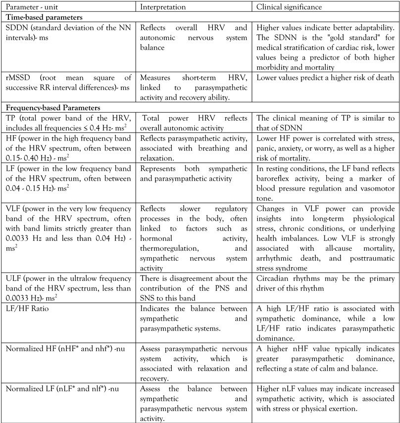
Table 1. HRV parameters: description, interpretation, and clinical relevance [13,16,17,18].
Germs 15 00104 i001
*nLF= LF/TP-VLF (14), nlf= LF/HF+LF (15); nHF= HF/TP-VLF (14), nhf= HF/HF+LF [15].
Table 2. Clinical and laboratory characteristics of the study and control groups.
Measles SARS-CoV-2 HIV Control
Number of patients 9 8 8 8
Age in years, median (IQR) 46 (38.0-47.5) 67 (63.5-73.0) 46.50 (35.3-54.5) 54.5
(41.0-70.8)
Male:female 2:7 3:5 3:5 5:3
High blood pressure, n (%) 2, (22.2) 6 (75.0) 3 (37.5) 2 (25.0)
Diabetes, n (%) 0 (0.0) 6 (62.5) 2 (25.0) 2 (25.0)
Heart failure, n (%) 0 (0.0) 4 (50.0) 0 (0.0) 3 (37.5)
Atrial fibrillation, n (%) 0 (0.0) 3 (37.5) 0 (0.0) 1 (12.5)
Ischemic heart disease, n (%) 1 (12.5) 3 (37.5) 1 (12.5) 2 (25.0)
CRP (mg/dL), median (IQR) 21.6
(17.8-61.7)
2.1
(1.4-11.6)
2.1
(0.4-8.0)
1.5
(0.6-3.9)
Fibrinogen (mg/dL), median (IQR) 428.0
(376.5-509.0)
463.0
(365.8-599.3)
284.5
(237.0-381.8)
319.0
(267.8-474.8)
Table 3. Median values of heart rate variability parameters and inflammatory markers across study groups.
Variables
(median) (IQR)
MeaslespSARS-CoV-2pHIVpControl group
SDNN 62.70
(33.10-65.55)
0.217 114.00
(72.57-166.90)
0.075 40.40
(28.80-102.90)
0.486 64.80
(51.62-90.67)
rMSSD 68.60
(31.55-81.50)
0.161 157.65
(109.50-218.72)
0,063 42.00
(23.40-134.60)
0.441 76.60
(56.42-129.45)
TP 318.70
(214.10-1493.75)
0.817 3203,05
(1210.50-6503.37)
0.042 493,85
(106.55-970.25)
0.271 988.50
(283.05-1381.52)
HF 98.00
(52.30-604.85)
0.682 1470.60
(688.40-2923.15)
0.036 45.70
(26.87-481.95)
0.203 304.90
(78.52-703.80)
LF 135.70
(51.65-488.05)
0.824 1044.65
(315.25-2190.65)
0.067 132.75
(25.20-305.47)
0.234 344.45
(60,60-638,65)
VLF 134.10
(82.10-346.70)
0.835 588.60
(154.40-1185.6)
0.046 109.90
(49.25-280.95)
0.613 190.35
(57.57-259.70)
LF/HF 0.90
(0.85-1.85)
0.719 0.61
(0.49-0.85)
0.172 0.90
(0.62-2.07)
0.611 0.71
(0.39-2.12)
ULF 11.80
(5.40-44.85)
0.488 21.45
(8.15-272.12)
0.206 9.40
(1.02-132.85)
0.243 17.90
(3.35-26.50)
VLF+ULF 134.20
(91.85-478.95)
0.677 695.50
(162.55-1502.30)
0.043 113.45
(53.20-412.12)
0.994 204.45
(72.87-268.57)
HF+LF/TP 0.70
(0.44-0.73)
0.264 0.78
(0.75-0.84)
0.636 0.53
(0.45-0.72)
0.098 0.79
(0.54-0.86)
VLF/TP 0.25
(0.23-0.45)
0.232 0.17
(0.14-0.24)
0.790 0.39
(0.18-5.35)
0.114 0.19
(0.12-0.23)
VLF+ULF/TP 0.27
(0.26-0.56)
0.264 0.21
(0.15-0.25)
0.637 0.47
(0.28-0.55)
0.098 0.20
(0.14-0.46)
HF+LF/VLF+ULF 2.38
(0.79-2.73)
0.157 3.67
(3.07-5.57)
0.985 1.12
(0.83-2.74)
0.112 3.92
(1.45-6.07)
HF+LF/VLF 2.75
(1.26-3.00)
0.109 4.44
(3.12-5.95)
0.895 0.53
(0.45-0.72)
0.006 4.14
(2.37-7.16)
Variables
(median) (IQR)
MeaslespSARS-CoV-2pHIVpControl group
nHF= HF/TP-VLF 0.52
(0.32-0.54)
0.353 0.56
(0.52-0.66)
0.323 0.45
(0.32-0.59)
0.294 0.57
(0.34-5.94)
nLF= LF/TP-VLF 0.45
(0.43-0.51)
0.442 0.36
(0.32-0.43)
0.571 0.43
(0.38-0.48)
0.711 0.40
(0,24-0,62)
nhf=HF/HF+LF 0.53
(0.35-0.55)
0.258 0.62
(0.54-0.67)
0.531 0.53
(0.34-0.60)
0.329 0.59
(0.37-0.73)
nlf=LF/HF+LF 0.46
(0.44-0.64)
0.255 0.38
(0.33-0.46)
0.534 0.47
(0.39-0.66)
0.326 0.41
(0.27-0.63)
Table 4. p-value between groups according median values of heart rate variability parameters and inflammatory markers.
Measles vs. HIV Measles vs.
SARS-CoV-2
HIV vs. SARS-CoV-2
SDNN 0.794 0.016 0.029
rMSSD 0.699 0.002 0.017
TP 0.445 0.021 0.018
HF 0.437 0.023 0.011
LF 0.424 0.056 0.023
VLF 0.502 0.052 0.035
LF/HF 0.775 0.026 0.120
Ncoh 0.120 0.140 0.022
ULF 0.773 0.369 0.471
VLF+ULF 0.703 0.056 0.044
HF+LF/TP 0.466 0.075 0.021
VLF/TP 0.601 0.097 0.039
VLF+ULF/TP 0.466 0.075 0.021
HF+LF/VLF+ULF 0.711 0.111 0.075
HF+LF/VLF 0.010 0.890 0.002
nHF= HF/TP-VLF 0.799 0.027 0.030
nLF= LF/TP-VLF 0.594 0.014 0.138
nhf=HF/HF+LF 0.980 0.013 0.042
nlf=LF/HF+LF 0.980 0.013 0.042
Table 5. Correlation between inflammatory markers and HRV parameters in HIV, measles, and SARSCoV-2 patients.
HIV Measles SARS-CoV-2
Fibrinogen CRP Fibrinogen CRP Fibrinogen CRP
SDNN r -0.598 -0.266 0.147 -0.156 -0.358 0.300
p 0.118 0.524 0.706 0.688 0.383 0.470
rMSSD r -0.732 -0.463 -0.085 -0.343 -0.374 0.149
p 0.039 0.248 0.827 0.366 0.362 0.724
TP r -0.335 -0.002 -0.160 -0.122 -0.148 0.278
p 0.418 0.996 0.681 0.755 0.726 0.504
HF r -0.628 -0.342 -0.221 -0.195 -0.177 0.193
p 0.096 0.407 0.567 0.614 0.674 0.646
LF r -0.339 -0.099 -0.137 -0.151 -0.146 0.422
p 0.412 0.815 0.726 0.698 0.731 0.298
VLF r 0.253 0.649 -0.121 0.069 -0.098 0.049
p 0.546 0.081 0.757 0.860 0.817 0.909
LF/HF r 0.767 0.955 0.793 0.576 0.163 0.497
p 0.026 <0.001 0.011 0.104 0.701 0.210
ULF r 0.085 0.286 0.278 0.387 -0.003 0.682
p 0.841 0.492 0.469 0.304 0.994 0.062
VLF+ULF r 0.207 0.558 0.015 0.215 -0.087 0.213
p 0.622 0.151 0.969 0.578 0.838 0.613
HF+LF/TP r -0.702 -0.601 -0.314 -0.607 -0.654 -0.237
p 0.052 0.115 0.410 0.083 0.079 0.573
VLF/TP r 0.718 0.502 0.163 0.406 0.605 0.053
p 0.045 0.205 0.675 0.278 0.112 0.901
VLF+ULF/TP r 0.702 0.601 0.314 0.607 0.654 0.237
p 0.052 0.115 0.410 0.083 0.079 0.573
HF+LF/VLF+ULF r -0.689 -0.371 -0.303 -0.448 -0.417 -0.248
p 0.059 0.366 0.428 0.227 0.304 0.554
HF+LF/VLF r -0.702 -0.601 -0.273 -0.414 -0.445 -0.045
p 0.052 0.115 0.478 0.268 0.269 0.915
nHF= HF/TP-VLF r -0.682 -0.834 -0.722 -0.669 -0.453 -0.642
p 0.062 0.010 0.028 0.049 0.260 0.086
nLF= LF/TP-VLF r 0.609 0.440 0.781 0.329 -0.007 0.292
p 0.109 0.276 0.013 0.388 0.986 0.482
nhf=HF/HF+LF r -0.786 -0.902 -0.797 -0.624 -0.186 -0.493
p 0.021 0.002 0.010 0.073 0.658 0.215
nlf=LF/HF+LF r 0.786 0.992 0.797 0.626 0.186 0.493
p 0.021 0.002 0.010 0.073 0.658 0.215