Next Article in Journal
Varicella Zoster Virus Central Nervous System Infection—A Retrospective Study from a Tertiary Center in Greece
Previous Article in Journal
Vitamin D and COVID-19: Comparative Analysis with Other Respiratory Infections and impact of Comorbidities
 
 
GERMS is published by MDPI from Volume 15 Issue 4 (2025). Previous articles were published by another publisher in Open Access under a CC-BY (or CC-BY-NC-ND) licence, and they are hosted by MDPI on mdpi.com as a courtesy and upon agreement with the former publisher Infection Science Forum.
Article

Comparative Phenotypic and Proteomic Analysis of Colistin-Exposed Pseudomonas aeruginosa

by
Nguyen Bao Vy Tran
1,
Thuc Quyen Huynh
1,2,
Hong Loan Ngo
1,
Ngoc Hoa Binh Nguyen
1,
Thi Hiep Nguyen
3,
Thi Hang Tong
1,
Thi Truc Ly Trinh
1,
Van Dung Nguyen
1,
Le Nhat Minh Pham
1,4,
Prem Prakash Das
5,
Teck Kwang Lim
5,
Qingsong Lin
5 and
Thi Thu Hoai Nguyen
1,2,*
1
School of Biotechnology, International University, Vietnam National University, Ho Chi Minh City 700000, Vietnam
2
Research Center for Infectious Diseases, International University, Vietnam National University, Ho Chi Minh City 700000, Vietnam
3
School of Biomedical Engineering, International University, Vietnam National University, Ho Chi Minh City 700000, Vietnam
4
Cho Ray Hospital, Ho Chi Minh City 700000, Vietnam
5
Protein and Proteomics Centre, Department of Biological Sciences, National University of Singapore, Singapore 119260, Singapore
*
Author to whom correspondence should be addressed.
GERMS 2024, 14(3), 246-266; https://doi.org/10.18683/germs.2024.1436 (registering DOI)
Submission received: 13 March 2024 / Revised: 26 June 2024 / Accepted: 11 September 2024 / Published: 30 September 2024

Abstract

Introduction: The emergence of colistin resistance threatens the treatment of Pseudomonas aeruginosa infections. Methods: In this study, in vitro development of colistin resistance was investigated using comparative phenotypic and proteomic analysis of P. aeruginosa ATCC 9027, its 14-day colistin sub-MIC exposed strain (Col-E1), and 10-day antibiotic-free cultured Col-E1 strain (Col-E2). Antibiotic susceptibility, morphology, virulence factors, and proteomic changes were assessed using disc-diffusion, agar-based, spectrophotometry, SEM, and iTRAQ-LC-MS/MS methods. Results: Colistin-exposed strains decreased susceptibility to colistin while remaining susceptible to other antibiotics. Col-E1 reduced the cell lengths by 17.67% and the colony size by 36.16% compared to the initial strain. The reduction remained in Col-E2. The pyocyanin production was reduced in Col-E1 (p = 0.025, Tukey HSD) and increased again in Col-E2 (p = 0.005, Tukey HSD). In contrast, no significant changes in elastase, protease, rhamnolipid, pyoverdine, and biofilm production were observed (p > 0.05, Tukey HSD). In Col-E1, the proteome analysis showed 135 differentially expressed proteins (DEPs) of which 94 DEPs (69.23%) maintained their expression change in Col-E2. Among DEPs, 82 were involved in metabolism and protein synthesis. Some DEPs (6/135) played a role in stress response such as GrpE (fold change: 14.93) and Hmp (fold change: 12.08). In particular, membrane proteins like OprD, DdlB, and OprI showed significant colistin response with fold change of -8.47, 6.43 and 6.19, respectively. Conclusions: In summary, colistin response in P. aeruginosa seemed to affect morphology, production of pyocyanin, and proteins of metabolism, protein synthesis, stress response and membrane.

Introduction

Pseudomonas aeruginosa is an important nosocomial pathogen causing a wide range of infections, particularly for immunocompromised patients. For the last decade, it emerges as a big threat to public health due to the ability to resist to multiple antibiotics [1]. Under exposure to sub- minimum inhibitory concentration (MIC) values of antibiotics, the bacterium could significantly increase its MIC values over time [2]. Several studies showed that sub-MIC values induced the alteration of bacterial characteristics and virulence expression but detailed mechanisms are not known [3].
Colistin (polymyxin E) is commonly used in human and animal health care [4]. By interacting with lipid A of lipopolysaccharide (LPS), colistin causes damage to the outer membrane of Gram- negative bacteria resulting in cell death [5]. Resistance to polymyxins involves outer membrane change, upregulation of efflux systems, and acquisition of some resistance determinants. Mutations in lpxA, lpxC, and lpxD lead to dysfunction of lipid A biosynthesis, resulting in loss of LPS; overexpression of outer membrane protein OprH prevents the binding of colistin to the LPS, and can also result in colistin resistance [6].
Proteomics is a powerful tool for studying molecular responses to antibiotics [7]. However, limited proteomic studies have been performed to understand the colistin resistance mechanisms [8]. In this work, the colistin adaptive resistance of P. aeruginosa was investigated under the perspectives of morphology, virulence factors, and proteome.

Methods

Bacterial strains

P. aeruginosa ATCC 9027 (American Type Culture Collection, USA) was the original strain in this study. The microdilution assay in Mueller Hinton (MH) broth following a previous study[9] was used to assess colistin MIC values of P. aeruginosa ATCC 9027 and generate Col-E1 strain. We collected the bacteria at the well next to the MIC well, where the growth of P. aeruginosa was still evident for the subsequent exposure. Antibiotic concentrations used were adjusted based on the obtained MIC values. The exposure process was performed for 14 days to achieve the 14-day colistin sub-MIC exposed strain (Col-E1). Col-E1 was then continuously exposed to colistin-free medium for 10 days. MIC values of colistin were checked daily using the microdilution method mentioned previously [9].

Antibiotic susceptibility testing

P. aeruginosa ATCC 9027, Col-E1, and Col- E2 strains were cultured in MH broth (HiMedia, India) for 24 h at 37°C. Then the diluted cultures (OD600nm = 0.08) were spread on MH agar. The commercial antibiotic paper discs of different antibiotics (meropenem (Me) (10 µg), tobramycin (Tb) (10 µg), ceftazidime (Cz) (30 µg), gentamicin (Ge) (10 µg), ciprofloxacin (Ci) (5 µg), cefepime (Cm) (30 µg), piperacillin/ tazobactam (Pt) (100/10 µg), imipenem (Im) (10 µg)) (NAM KHOA Biotek Company, Vietnam) were placed on the surface of the agar and incubated overnight at 37°C. The inhibition zones were measured and evaluated following Clinical and Laboratory Standards Institute (CLSI) 2022 and CLSI 2023 (USA).

Morphology and virulence factor assessment

Colony morphology and cell length observation

P. aeruginosa ATCC 9027, Col-E1, and Col- E2 strains were cultured in tryptic soy broth (TSB) for 24 h at 37°C. The cultures were adjusted to OD600nm = 0.08, then 10-6 dilution cultures were prepared to ensure that the colony count on each Petri dish remained below 15. This practice aimed to facilitate optimal morphology observation when spreading the cultures on tryptic soy agar as the recommendation from a previous study [10]. Following the spreading, the dishes were incubated overnight at 37°C. Colony morphology was observed using a stereo microscope. The experiment was performed in triplicate.
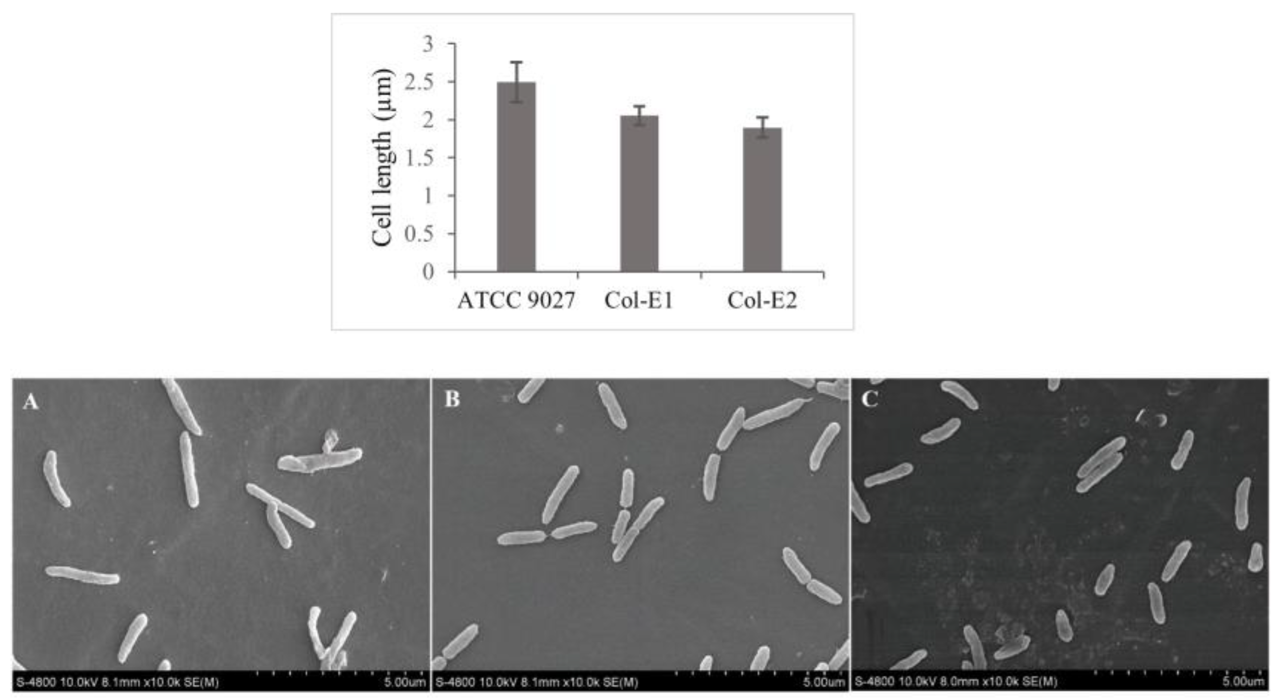
Bacterial cell length was measured by using scanning electron microscopy (SEM). The sample preparation for SEM was performed following our previous study [11]. Each strain was embedded on plastic discs by culturing overnight in LB broth. The plastic discs were subsequently washed with 1X PBS buffer and fixed by 10% formalin for 24 hours. Following fixation, specimens were rinsed with 1X PBS and dehydrated by series of graded ethanol (25%, 50%, 70%, 80%, 90%, 100%). For air drying, the specimens were treated with t-butyl alcohol for 24 hours at 4°C. Finally, the individual cell length was analyzed by SEM COXEM machine (South Korea) and ImageJ software (NIH, USA). Ten cells from each sample were measured.

Virulence factor assessment

Elastase, protease, rhamnolipid, pyocyanin, pyoverdine, and biofilm production of P. aeruginosa ATCC 9027, Col-E1, and Col-E2 were analyzed. P. aeruginosa ATCC 9027 was used as a positive control for the tests because it could produce tested virulence factors in this study. For negative control, Escherichia coli ATCC 4157 was used for protease, pyocyanin and pyoverdine tests, while Staphylococcus aureus ATCC 29213 was used for elastase test [12]. Each virulence test was performed in triplicate.
Elastase, protease, and rhamnolipid testing: The elastase, protease, and rhamnolipid production of P. aeruginosa strains were observed by spotting method on elastin–nutrient plate agar (0.3% elastin), skim milk agar, methylene blue (MB) and cetyltrimethylammonium bromide (CTAB) containing agar (HiMedia Laboratories, USA and Sigma-Aldrich, USA), respectively. Then the plates were incubated at 37°C for 24 h and at 30°C 48 h for elastase test. The enzymatic activity was visualized by the halos around the spot cultures.
For pyocyanin testing, the strains were cultured in glycerol-alanine (Gly-Ala) broth medium to maximize the yield of pyocyanin production [13]. After culturing overnight at 37°C with shaking, the culture was centrifuged at 6000 rpm for 20 min to collect the supernatant. After filtering, 3 mL of the supernatant were mixed well with 1.8 mL of chloroform (VN-CHEMSOL Co., Ltd., Vietnam). The raw pyocyanin (red layer) was extracted by mixing the blue layer with 0.2 N HCl (2:1). The pyocyanin absorbance was measured at 520 nm using BioTek Synergy HTX Multimode Reader (Agilent, USA). The pyocyanin concentration (µg/mL) was estimated by multiplying OD520nm by 17.072 which is the molar extinction coefficient of pyocyanin at 520 nm [14].
For pyoverdine testing, King B broth (HiMedia Laboratories,USA) was used to maximize the yield of pyoverdine production. After culturing overnight at 37°C with shaking, the culture was centrifuged at 6000 rpm for 20 min to collect the supernatant. After filtering, the fluorescence intensity of the supernatant was measured by BioTek Synergy HTX Multimode Reader at an excitation 405 nm and emission 450 nm [15].
Biofilm testing: The crystal violet (HiMedia Laboratories, USA) staining method was used to test for biofilm production [16]. The OD550nm was measured using BioTek Synergy HTX Multimode Reader.

iTRAQ LC-MS/MS proteomic analysis

Quantitative iTRAQ- LC-MS/MS analysis

Total protein extraction was performed following the procedure described in a previous study [17]. Proteins were trypsinized and labeled with iTRAQ tags from the Reagent 8-plex kit (Sigma-Aldrich, USA). iTRAQ-labeled peptide mixtures were analyzed using strong-cation- exchange (SCX) coupled with liquid chromatography-tandem mass spectrometry (LC- MS/MS). Ultimate 3000 LC system and 5800 MALDI-TOF/TOF analyzer were used to run the samples. Protein identification and quantification using the MS/MS spectra was performed using Protein Pilot Software 4.5 (Applied Biosystems) according to the Swiss-Prot/UniProt database [18].

Quantitative reverse transcription PCR (RT- qPCR) analysis

Total RNA was extracted using Monarch® Total RNA Miniprep Kit (New England Biolabs, USA). The quantity and quality of the extracted RNA were assessed using BioTek Synergy HTX Multimode Reader. Then, it was transcribed into complementary DNA (cDNA) using the SensiFASTTM cDNA Synthesis Kit and qPCR was performed using SensiFASTTM SYBR No-ROX Kit (Meridian Bioscience, USA). The thermal cycle setting included 2 min at 95°C, followed by 40 cycles of 5 s at 95°C and 30 s at 60°C. The rpoD and rplU were used as internal controls to normalize fold changes [19,20]. Primers in this study were designed using NCBI Primer-BLAST (https://www.ncbi.nlm.nih.gov/tools/primer-blast/) – Table S1. The fold change was evaluated against P. aeruginosa ATCC 9027 according to the 2-ΔΔCt method from a study of Kumar and Lorand 2021 [21]. Values greater than 1 were up-regulated genes, while values smaller than 1 were down- regulated genes. The experiment was triplicated.

Data analysis

IBM® SPSS® Statistics 20.0 (IBM Corp., USA) was used to analyze the data. The Tukey HSD test was used to determine the difference in morphology and virulence factor production of P. aeruginosa strains. The p-value was set to be <0.05. Raw MS/MS data were analyzed by using ProteinPilot Software 4.5 (AB SCIEX). The proteins were considered to be differentially expressed if their iTRAQ ratios were ≥2 (up- regulation) and ≤0.5 (or ≤-2) (down-regulation). For protein identification, a threshold applied was >0.05 (CI, 10%) with setting ProtScore at 2.0 and the false discovery rate (FDR) <1%. The differentially expressed proteins (DEPs) were analyzed by the Gene Ontology (GO) annotation from the UniProt database. A protein-protein interaction network was constructed using the web resource STRING 11.5. To classify the protein functions, UniProt, STRING 11.5, and PANTHER 17.0 were applied.

Results

Antibiotic susceptibility profile of P. aeruginosa under colistin exposure

After continuously being exposed to sub-MIC of colistin for 14 days, the colistin MIC value of P. aeruginosa ATCC 9027 increased from 4 µg/mL to 16 µg/mL (Figure S1). And after 10 days of being cultured back in antibiotic-free medium, the colistin MIC dropped to 8 µg/mL. Furthermore, Col-E1 and Col-E2 did not develop cross resistance to other antibiotics, including meropenem, tobramycin, ceftazidime, gentamicin, ciprofloxacin, cefepime, piperacillin/tazobactam and imipenem (Table S2).

Morphology of P. aeruginosa under colistin exposure

The colony sizes of Col-E1 and Col-E2 were significantly different from the P. aeruginosa ATCC 9027. The average colony sizes of Col-E1 and Col-E2 were smaller than the original strain, which were 3.92±0.51 mm, 3.10±0.25 mm, and 6.14±0.19 mm, respectively (p<0.05, Tukey HSD) – Figure 1.
The cell length of P. aeruginosa ATCC 9027 (2.49±0.45 µm) decreased significantly after exposure to colistin in which Col-E1 was 2.05±0.21 µm and Col-E2 was 1.90±0.23 µm (p<0.05, Tukey HSD) – Figure 2.

Elastase, protease, rhamnolipid, pyocyanin, pyoverdine, and biofilm production of P. aeruginosa under colistin exposure

The obtained data indicated that P. aeruginosa ATCC 9027 reduced the production of pyocyanin under the exposure to colistin (p=0.025, Tukey HSD test) and increased again after colistin was removed from the culture medium (p=0.005, Tukey HSD test). However, the production of elastase, protease, rhamnolipid, pyoverdine, and biofilm remained in both Col-E1 and Col-E2 (p>0.05, Tukey HSD test) – Table 1.

Proteome response of P. aeruginosa to colistin

The same set of 947 different proteins were identified in P. aeruginosa ATCC 9027, Col-E1 and Col-E2 strain using iTRAQ LC-MS/MS. DEPs were determined when quantitatively comparing Col-E1 and Col-E2 strain to the initial strain P. aeruginosa ATCC 9027. In total, for both Col-E1 and Col-E2, more up-regulated proteins (Col-E1: 108, Col-E2: 101) than down-regulated proteins (Col-E1: 27, Col-E2: 41) were found (cut-off: ≥2: up-regulation or ≤-2: down- regulation). The fold change ranged from -8.47 to 17.95. Interestingly, 94 DEPs in Col-E1 kept their expression change even after 10 days being cultured in antibiotic-free medium (Col-E2), including 82 up- and 12 down-regulated proteins (Figure S2, Table S3). On the other hand, 40 DEPs reverted their expression to the initial state before the colistin exposure, and only 1 protein turned from up to down regulation after colistin was withdrawn (PA0038; fold change: Col-E1: 2.74, Col-E2: -3.09) – Table S4.

The DEPs related to metabolism and protein synthesis

To classify the function of DEPs of P. aeruginosa in responding to colistin, PANTHER 17.0 was used. Two big classified functional groups were molecular function and biological process (Figure 3). Most DEPs belonged to structural molecule activity, catalytic activity in molecular function group (Figure 3A) and metabolic process, and cellular process in biological process group (Figure 3B). From STRING analysis, there were 22 clusters detected from DEPs of Col-E1. The biggest cluster included 25 ribosomal proteins (Figure S3). They were all up-regulated and the fold change ranged from 2.63 to 10.05.

The DEPs related to stress response

There were 7 up-regulated proteins (GrpE, Hmp, MsrA, LptF, PhoP, PA2575, and PA4061) and 1 down-regulated protein (TrxB1) related to stress response detected in P. aeruginosa under colistin exposure as classified by protein function from UniProt. The fold change ranged from -2.68 to 14.93 (Figure 4, Tables S3 and S4).

The DEPs related to membrane modification

Following UniProt, 16 DEPs with membrane-related functions were detected, including 8 up-regulated and 8 down-regulated proteins (fold change: -8.47 to 2.10). The up- regulated membrane proteins were DdlB (6.43), OprI (6.19), SltB1 (3.68), Ffh (3.63), BamD (2.91), PagL (2.64), WapR (2.37), and PA5378 (2.17). The down-regulated proteins were OprD (- 8.47), PA2760 (-2.47), PilP (-2.36), LptE (-2.34), BamE (-2.33), Alr (-2.16), PA1288 (-2.15), and PA0833 (-2.05) – Figure 4, Tables S3 and S4.

Quantitative reverse transcription PCR (RT- qPCR) analysis

Selected DEPs found via proteomic analysis also presented the same trend of change at gene expression level. However, the fold change from the RT-qPCR analysis was overall lower than that from the iTRAQ LC-MS/MS analysis. It was 1.26, 1.30, 2.00, 1.05 and 0.57 for grpE, hmp, fur, mvaT and oprD, respectively (Figure S4, Table S5).

Discussion

After exposure to the sub-MIC of colistin for a while, P. aeruginosa ATCC 9027 increased MIC value of colistin, which was consistent with previous observations that the pathogen could become less susceptible to an antibiotic to which it was exposed[2] and changed its properties [3]. Interestingly, the increasing fold in MIC for colistin is relatively low, only 2-4 folds compared to the increasing fold induced by other antibiotics [22]. Furthermore, long-term exposure to colistin luckily did not trigger cross resistance to other antibiotics.
As colistin inhibited cell growth by disrupting cell membrane, it is under expectation that colony size was reduced under colistin exposure [23]. Besides, shorter cell length observed after colistin exposure was also consistent with other studies about P. aeruginosa morphological changes in responding to colistin exposure in vitro or during treatment [24,25]. It has been known that cell length or cell size was dependent to the growth rate which positively links to DNA content and protein synthesis [26,27], thus it is understandable that antibiotics which interfere with cellular process have impact on cell size.
Prolonged exposure to sub-MIC levels of colistin in P. aeruginosa did not significantly impact the production of most virulence factors, except for pyocyanin. Pyocyanin production was observed to decrease in the presence of colistin, returning to normal upon colistin removal. It has been known that the sub-MIC of antibiotics, including colistin, could inhibit the production of pyocyanin [28]. In a previous study, elastase exceptionally increased activity when being incubated with colistin [29]. Unlike colistin, ciprofloxacin exposure significantly reduced elastase, protease, and biofilm production in P. aeruginosa [22]. In colistin-resistant derivatives of Acinetobacter baumannii ATCC 19606, reduction of biofilm production and loss of LPS were also noted [30].
At protein level, virulence-related proteins did not show much change in responding to colistin. For instance, Fur (3.53) and BfrB (2.08), which were responsible for iron uptake of P. aeruginosa [31], were slightly up-regulated in our study. Regarding biofilm production, MvaT (2.37), which took part in biofilm formation [32], was also only slightly up-regulated. LpxA (1.15), LpxC (1.01), and LpxD (-1.53) which were shown to be involved in the virulence of colistin- resistant pathogens, especially in reducing biofilm production [30], also did not change expression under colistin exposure. PtsP (-1.20), which contributed to the pyocyanin production [33], showed a minor decrease in expression in the presence of colistin.
Most found DEPs were related to metabolism and protein synthesis. Among ribosomal proteins, RplJ had the highest fold change (10.05). The highest upregulated protein involved in metabolism was MetF (14.32). This protein indirectly influenced protein synthesis via the methionine biosynthesis process [34]. The lowest down protein of the metabolism group was PyrC (-3.15), which is involved in pyrimidine biosynthesis. The pyrimidine biosynthesis process is known to be crucial for bacterial proliferation [35], thus downregulation of PyrC is an appropriate adaptation to antibiotics stress including colistin.
For proteins involved in stress response, GrpE (Col-E1: 14.93, Col-E2: 11.22) and Hmp (Col-E1: 12.08, Col-E2: 19.31) had the highest fold change. GrpE prevented the aggregation of stress-denatured proteins when P. aeruginosa faced to hyperosmotic or heat shock [36], while Hmp was essential in response to nitrosative stress or NO2 pollutant condition [37]. LptF (Col—E1: 3.39, Col- E2: 3.21) was an OmpA-like outer membrane protein, reported to be involved in resistance to oxidative stress response of P. aeruginosa [38]. PhoP (Col-E1: 2.56 and Col-E2: 2.41) was a member of the two-component regulatory system PhoP/PhoQ which played a role in the resistant regulation to cationic antibiotics [39].
Among membrane protein, OprI was highly up-regulated (Col-E1: 6.19 and Col-E2: 6.82). It may be because OprI compensated the decline of OprD for the integrity of the outer membrane [40]. Other membrane DEPs including LptE and BamD, BamD and BamE, Alr and DdlB, particularly PagL and PhoP, were shown to be involved in disrupting polymyxin penetration [41]. Some DEPs such as PA2760, PA0833, PA1288, and PA5378, could be potential factors in the colistin adaption of P. aeruginosa which would need further investigation. The summary of DEPs and their cellular locations indicated clearly the involvement of membrane proteins and protein- synthesis related proteins in the colistin response of the bacterium (Figure 4).
In general, even when colistin was removed from the culture for many generations during 10 days of continuous culture, a great number of DEPs maintained their expression trend while only a few, such as PA4061 (2.17 to 1.54), TrB1 (- 2.68 to -1.50) and OprD (-8.47 to -1.94), returned to their normal expression. OprD was thought to be the most prevalent mechanism for carbapenem resistance[42] and colistin response[43] in P. aeruginosa. Previous studies showed that the resistance phenotype could be reverted, a characteristic feature of adaptive resistance, when the antibiotic stress is removed [44]. However, in our study, most of the changed phenotypic characteristics and many DEPs remained after the exposure to colistin was stopped. We assumed that colistin-exposed P. aeruginosa may have epigenetic memory[45] or changes have been made in its genome. It would be more likely the epigenetic changes as the expected resistance- related mutation rates were lower than the observed survival rates in sub-MIC of antibiotics [46].
Sub-MIC exposure can clearly cause long- term effect on the protein expression changes of colistin-exposed P. aeruginosa, thus the use of antibiotics must be done with even greater caution. It is obvious that surviving P. aeruginosa under sub-MIC colistin exposure develop sustainable colistin resistance without acquiring any external resistant genes, thus antibiotic dosage and duration must be reasonable to eradicate the pathogen completely. In addition, combination therapy could be one option. An in vitro study showed that combining colistin with aminoglycoside, carbapenems, fluoroquinolones, and others could significantly improve outcomes for infections caused by multidrug-resistant Klebsiella pneumoniae, a common antibiotic- resistant Gram-negative bacterium like P. aeruginosa [47]. However, some in vivo studies on combination of colistin and carbapenem, although displaying a positive effect, did not provide significant improvement [48,49,50]. One good point from the studies is that the toxicity or adverse effect of colistin combination was similar to monotherapy [48,49,50].
Moreover, DEPs identified in our study could potentially serve as biomarkers for colistin- resistant isolates. Rapid detection of resistance is essential for clinicians to administer more effective treatment strategies as well as to prevent the spread of resistant isolates.
This study provides valuable insights into the response of P. aeruginosa ATCC 9027 to colistin exposure and identifies potential biomarkers for colistin resistance. However, it has some significant limitations. First, it did not provide direct evidences showing the role of each protein to the colistin resistance development or phenotypic changes. Second, the changes in genome, transcriptome or metabolome of the bacterium in responding to colistin were not analyzed. Third, whether this response is the same for different strains and different antibiotics is still not investigated. Therefore, further investigations should be performed to unlock this complicated problem.

Conclusions

After colistin exposure, surviving P. aeruginosa decreased colony size, cell length, and the production of pyocyanin, but did not dramatically change production of elastase, protease, rhamnolipid, pyoverdine, and biofilm. Luckily, surviving P. aeruginosa did not develop high-fold increased resistance to colistin and cross resistance to other antibiotic classes. A substantial proportion of DEPs maintained the expression changes even though P. aeruginosa was no longer in colistin containing environment, suggesting an epigenetic regulation memory in responding to antibiotics.

Supplementary Materials

The following are available online at www.mdpi.com/germs-14-00246/s1.

Author Contributions

NBVT contributed to investigation (lead); data curation (lead); formal analysis (lead); validation (lead); writing – original draft preparation (lead). TQH contributed to investigation (supporting); data curation (supporting); formal analysis (supporting); validation (supporting). HLN contributed to investigation (supporting); data curation (supporting). NHBN contributed to investigation (supporting). THN contributed to methodology (supporting). THT contributed to methodology (supporting). TTLT contributed to methodology (supporting). VDN contributed to methodology (supporting). LNMP contributed to methodology (supporting). PPD contributed to methodology (supporting); data curation (supporting). TKL contributed to data curation (supporting), writing-review (supporting). QSL contributed to supervision (supporting), writing-review (supporting). TTHN contributed to conceptualization (lead); resources (lead); formal analysis (lead); funding acquisition (lead); project administration (lead), supervision (lead); writing-review and editing (lead). All authors read and approved the final version of the manuscript.

Funding

This research was supported by Vietnam National Foundation for Science and Technology Development (NAFOSTED) under grant number 108.04-2018.08.

Data Availability Statement

All data generated or analyzed during this study are included in this published article.

Acknowledgments

The authors would like to thank their colleagues at the International University and Protein and Proteomics Center of National University of Singapore for their support.

Conflicts of Interest

All authors – none to declare.

References

  1. Yero, D.; Díaz-Lobo, M.; Costenaro, L.; et al. The Pseudomonas aeruginosa substrate-binding protein Ttg2D functions as a general glycerophospholipid transporter across the periplasm. Commun Biol. 2021, 4, 448. [Google Scholar] [CrossRef]
  2. Sandoval-Motta, S.; Aldana, M. Adaptive resistance to antibiotics in bacteria: a systems biology perspective. WIREs Syst Biol Med. 2016, 8, 253–267. [Google Scholar] [CrossRef]
  3. de Andrade, J.P.L.; de Macêdo Farias, L.; Ferreira, J.F.G.; et al. Sub-inhibitory concentration of piperacillin-tazobactam may be related to virulence properties of filamentous Escherichia coli. Curr Microbiol. 2016, 72, 19–28. [Google Scholar] [CrossRef] [PubMed]
  4. Gogry, F.A.; Siddiqui, M.T.; Sultan, I.; Haq, Q.M.o.h.d.R. Current update on intrinsic and acquired colistin resistance mechanisms in bacteria. Front Med (Lausanne). 2021, 8, 677720. [Google Scholar] [CrossRef] [PubMed]
  5. Dößelmann, B.; Willmann, M.; Steglich, M.; et al. Rapid and consistent evolution of colistin resistance in extensively drug-resistant Pseudomonas aeruginosa during morbidostat culture. Antimicrob Agents Chemother. 2017, 61, e00043–17. [Google Scholar] [CrossRef] [PubMed]
  6. Saleh, N.M.; Hesham, M.S.; Amin, M.A.; Samir Mohamed, R. Acquisition of colistin resistance links cell membrane thickness alteration with a point mutation in the lpxD gene in Acinetobacter baumannii. Antibiotics (Basel). 2020, 9, 164. [Google Scholar] [CrossRef]
  7. Tsakou, F.; Jersie-Christensen, R.; Jenssen, H.; Mojsoska, B. The role of proteomics in bacterial response to antibiotics. Pharmaceuticals (Basel). 2020, 13, 214. [Google Scholar] [CrossRef]
  8. Hashemi, M.M.; Holden, B.S.; Coburn, J.; et al. Proteomic analysis of resistance of Gram-negative bacteria to chlorhexidine and impacts on susceptibility to colistin, antimicrobial peptides, and ceragenins. Front Microbiol. 2019, 10, 210. [Google Scholar] [CrossRef]
  9. Kowalska-Krochmal, B.; Dudek-Wicher, R. The minimum inhibitory concentration of antibiotics: methods, interpretation, clinical relevance. Pathogens. 2021, 10, 165. [Google Scholar] [CrossRef]
  10. Sousa, A.M.; Machado, I.; Nicolau, A.; Pereira, M.O. Improvements on colony morphology identification towards bacterial profiling. J Microbiol Methods. 2013, 95, 327–335. [Google Scholar] [CrossRef]
  11. Nguyen, N.H.B.; Pham, T.T.V.; Huynh, T.Q.; Nguyen, T.H.; Nguyen, T.T.H. Sample preparative procedure for Pseudomonas aeruginosa observation under scanning electron microscope. Vietnam J Biotechnol. 2022, 20, 717–726. [Google Scholar] [CrossRef]
  12. Garcia Dde, O.; Timenetsky, J.; Martinez, M.B.; Francisco, W.; Sinto, S.I.; Yanaguita, R.M. Proteases (caseinase and elastase), hemolysins, adhesion and susceptibility to antimicrobials of Stenotrophomonas maltophilia isolates obtained from clinical specimens. Braz J Microbiol. 2002, 33, 157–162. [Google Scholar] [CrossRef]
  13. Ahamed, F.; Devnath, P. Extraction, purification and characterization of pyocyanin produced by Pseudomonas aeruginosa and evaluation for its antimicrobial activity. Int Res J Biol Sci. 2017, 6, 1–7. [Google Scholar]
  14. Essar, D.W.; Eberly, L.; Hadero, A.; Crawford, I.P. Identification and characterization of genes for a second anthranilate synthase in Pseudomonas aeruginosa: interchangeability of the two anthranilate synthases and evolutionary implications. J Bacteriol. 1990, 172, 884–900. [Google Scholar] [CrossRef]
  15. Dao, K.H.T.; Hamer, K.E.; Clark, C.L.; Harshman, L.G. Pyoverdine production by Pseudomonas aeruginosa exposed to metals or an oxidative stress agent. Ecol Appl. 1999, 9, 441–448. [Google Scholar] [CrossRef]
  16. Banin, E.; Vasil, M.L.; Greenberg, E.P. Iron and Pseudomonas aeruginosa biofilm formation. Proc Natl Acad Sci U S A. 2005, 102, 11076–11081. [Google Scholar] [CrossRef] [PubMed]
  17. Thai, V.C.; Lim, T.K.; Le, K.P.U.; Lin, Q.; Nguyen, T.T.H. iTRAQ-based proteome analysis of fluoroquinolone- resistant Staphylococcus aureus. J Glob Antimicrob Resist. 2017, 8, 82–89. [Google Scholar] [CrossRef] [PubMed]
  18. Zhou, Y.; Chen, W.N. iTRAQ-Coupled 2-D LC-MS/MS analysis of membrane protein profile in Escherichia coli incubated with apidaecin IB. PLoS One. 2011, 6, e20442. [Google Scholar] [CrossRef] [PubMed]
  19. Sanz-García, F.; Hernando-Amado, S.; Martínez, J.L. Mutational evolution of Pseudomonas aeruginosa resistance to ribosome-targeting antibiotics. Front Genet. 2018, 9, 451. [Google Scholar] [CrossRef]
  20. Savli, H.; Karadenizli, A.; Kolayli, F.; Gundes, S.; Ozbek, U.; Vahaboglu, H. Expression stability of six housekeeping genes: a proposal for resistance gene quantification studies of Pseudomonas aeruginosa by real-time quantitative RT-PCR. J Med Microl. 2003, 52, 403–408. [Google Scholar] [CrossRef]
  21. Kumar, A.; Lorand, D. Robust ΔΔct estimate. Genomics. 2021, 113, 420–427. [Google Scholar] [CrossRef]
  22. Huynh, T.Q.; Nguyen, T.T.H. The effects of sub-MIC ciprofloxacin exposure on antibiotic susceptibility and virulence factors in Pseudomonas aeruginosa ATCC 9027. Int J Life Sci Res Arch. 2022, 3, 70–77. [Google Scholar] [CrossRef]
  23. Sabnis, A.; Hagart, K.L.; Klöckner, A.; et al. Colistin kills bacteria by targeting lipopolysaccharide in the cytoplasmic membrane. eLife. 2021, 10, e65836. [Google Scholar] [CrossRef] [PubMed]
  24. Mortensen, N.P.; Fowlkes, J.D.; Sullivan, C.J.; et al. Effects of colistin on surface ultrastructure and nanomechanics of Pseudomonas aeruginosa cells. Langmuir. 2009, 25, 3728–3733. [Google Scholar] [CrossRef]
  25. O'Driscoll, N.H.; Cushnie, T.P.T.; Matthews, K.H.; Lamb, A.J. Colistin causes profound morphological alteration but minimal cytoplasmic membrane perforation in populations of Escherichia coli and Pseudomonas aeruginosa. Arch Microbiol. 2018, 200, 793–802. [Google Scholar] [CrossRef]
  26. Basan, M.; Zhu, M.; Dai, X.; et al. Inflating bacterial cells by increased protein synthesis. Mol Syst Biol. 2015, 11, 836. [Google Scholar] [CrossRef]
  27. Schaechter, M.; MaalØe, O.; Kjeldgaard, N.O. Dependency on medium and temperature of cell size and chemical composition during balanced growth of Salmonella typhimurium. Microbiology. 1958, 19, 592–606. [Google Scholar] [CrossRef]
  28. Seiffein, N.L.; Ali, G.H. Effect of subinhibitory concentrations of selected antibiotics and propolis on pyocyanin and biofilm production among Pseudomonas aeruginosa isolates in Alexandria, Egypt. Egypt J Med Microbiol. 2021, 30, 129–137. [Google Scholar] [CrossRef]
  29. Jones, A.; Elphick, H.; Pettitt, E.; Everard, M.L.; Evans, G.S. Colistin stimulates the activity of neutrophil elastase and Pseudomonas aeruginosa elastase. Eur Respir J. 2002, 19, 1136–1141. [Google Scholar] [CrossRef] [PubMed]
  30. Carretero-Ledesma, M.; García-Quintanilla, M.; Martín- Peña, R.; Pulido, M.R.; Pachón, J.; McConnell, M.J. Phenotypic changes associated with colistin resistance due to lipopolysaccharide loss in Acinetobacter baumannii. Virulence. 2018, 9, 930–942. [Google Scholar] [CrossRef] [PubMed]
  31. Cianciulli Sesso, A.; Lilić, B.; Amman, F.; Wolfinger, M.T.; Sonnleitner, E.; Bläsi, U. Gene expression profiling of Pseudomonas aeruginosa upon exposure to colistin and tobramycin. Front Microbiol. 2021, 12, 937. [Google Scholar] [CrossRef] [PubMed]
  32. Vallet, I.; Diggle, S.P.; Stacey, R.E.; et al. Biofilm formation in Pseudomonas aeruginosa: fimbrial cup gene clusters are controlled by the transcriptional regulator MvaT. J Bacteriol. 2004, 186, 2880–2890. [Google Scholar] [CrossRef]
  33. Xu, H.; Lin, W.; Xia, H.; et al. Influence of ptsP gene on pyocyanin production in Pseudomonas aeruginosa. FEMS Microbiol Lett. 2005, 253, 103–109. [Google Scholar] [CrossRef]
  34. Kozak, M. Comparison of initiation of protein synthesis in procaryotes, eucaryotes, and organelles. Microbiol Rev. 1983, 47, 1–45. [Google Scholar] [CrossRef]
  35. Lipowska, J.; Miks, C.D.; Kwon, K.; et al. Pyrimidine biosynthesis in pathogens - structures and analysis of dihydroorotases from Yersinia pestis and Vibrio cholerae. Int J Biol Macromol. 2019, 136, 1176–1187. [Google Scholar] [CrossRef]
  36. UniProt. 2023. grpE - Protein GrpE - Pseudomonas aeruginosa. UniProtKB. Available online: https://www.uniprot.org/uniprotkb/A0A069Q2K4/entry (accessed on 18 July 2023).
  37. Poole, R.K. Flavohaemoglobin: the pre-eminent nitric oxide-detoxifying machine of microorganisms. F1000Res. 2020, 9, 7. [Google Scholar] [CrossRef]
  38. Damron, F.H.; Napper, J.; Teter, M.A.; Yu, H.D. Lipotoxin F of Pseudomonas aeruginosa is an AlgU-dependent and alginate-independent outer membrane protein involved in resistance to oxidative stress and adhesion to A549 human lung epithelia. Microbiology (Reading). 2009, 155, 1028–1038. [Google Scholar] [CrossRef]
  39. Macfarlane, E.L.; Kwasnicka, A.; Ochs, M.M.; Hancock, R.E. PhoP-PhoQ homologues in Pseudomonas aeruginosa regulate expression of the outer-membrane protein OprH and polymyxin B resistance. Mol Microbiol. 1999, 34, 305–316. [Google Scholar] [CrossRef]
  40. Lin, Y.M.; Wu, S.J.; Chang, T.W.; et al. Outer membrane protein I of Pseudomonas aeruginosa is a target of cationic antimicrobial peptide/protein. J Biol Chem. 2010, 285, 8985–8994. [Google Scholar] [CrossRef] [PubMed]
  41. Han, M.L.; Velkov, T.; Zhu, Y.; et al. Polymyxin-induced lipid A deacylation in Pseudomonas aeruginosa perturbs polymyxin penetration and confers high-level resistance. ACS Chem Biol. 2018, 13, 121–130. [Google Scholar] [CrossRef] [PubMed]
  42. Li, H.; Luo, Y.F.; Williams, B.J.; Blackwell, T.S.; Xie, C.M. Structure and function of OprD protein in Pseudomonas aeruginosa: from antibiotic resistance to novel therapies. Int J Med Microbiol. 2012, 302, 63–68. [Google Scholar] [CrossRef]
  43. Muller, C.; Plésiat, P.; Jeannot, K. A two-component regulatory system interconnects resistance to polymyxins, aminoglycosides, fluoroquinolones, and β-lactams in Pseudomonas aeruginosa. Antimicrob Agents Chemother. 2011, 55, 1211–1221. [Google Scholar] [CrossRef]
  44. Ghosh, D.; Veeraraghavan, B.; Elangovan, R.; Vivekanandan, P. Antibiotic resistance and epigenetics: more to it than meets the eye. Antimicrob Agents Chemother. 2020, 64, e02225–19. [Google Scholar] [CrossRef]
  45. Riber, L.; Hansen, L.H. Epigenetic memories: the hidden drivers of bacterial persistence? Trends Microbiol. 2021, 29, 190–194. [Google Scholar] [CrossRef]
  46. Adam, M.; Murali, B.; Glenn, N.O.; Potter, S.S. Epigenetic inheritance based evolution of antibiotic resistance in bacteria. BMC Evol Biol. 2008, 8, 52. [Google Scholar] [CrossRef] [PubMed]
  47. Ontong, J.C.; Ozioma, N.F.; Voravuthikunchai, S.P.; Chusri, S. Synergistic antibacterial effects of colistin in combination with aminoglycoside, carbapenems, cephalosporins, fluoroquinolones, tetracyclines, fosfomycin, and piperacillin on multidrug resistant Klebsiella pneumoniae isolates. PLoS One. 2021, 16, e0244673. [Google Scholar] [CrossRef] [PubMed]
  48. Paul, M.; Daikos, G.L.; Durante-Mangoni, E.; et al. Colistin alone versus colistin plus meropenem for treatment of severe infections caused by carbapenem-resistant Gram- negative bacteria: an open-label, randomised controlled trial. Lancet Infect Dis. 2018, 18, 391–400. [Google Scholar] [CrossRef] [PubMed]
  49. Kaye, K.S.; Marchaim, D.; Thamlikitkul, V.; et al. Colistin monotherapy versus combination therapy for carbapenem-resistant organisms. NEJM Evid. 2022, 2, EVIDoa2200131. [Google Scholar] [CrossRef] [PubMed]
  50. Huang, C.; Chen, I.; Tang, T. Colistin Monotherapy versus colistin plus meropenem combination therapy for the treatment of multidrug-resistant Acinetobacter baumannii infection: a meta-analysis. J Clin Med. 2022, 11, 3239. [Google Scholar] [CrossRef] [PubMed]
  51. Sanz-García, F.; Hernando-Amado, S.; Martínez, J.L. Mutational evolution of Pseudomonas aeruginosa resistance to ribosome-targeting antibiotics. Front Genet. 2018, 9, 451. [Google Scholar] [CrossRef]
  52. Savli, H.; Karadenizli, A.; Kolayli, F.; et al. Expression stability of six housekeeping genes: a proposal for resistance gene quantification studies of Pseudomonas aeruginosa by real-time quantitative RT-PCR. J Med Microbiol. 2003, 52, 403–408. [Google Scholar] [CrossRef] [PubMed]
  53. Tomás, M.; Doumith, M.; Warner, M.; et al. Efflux pumps, OprD porin, AmpC beta-lactamase, and multiresistance in Pseudomonas aeruginosa isolates from cystic fibrosis patients. Antimicrob Agents Chemother. 2010, 54, 2219–2224. [Google Scholar] [CrossRef] [PubMed]
  54. Kumar, A.; Lorand, D. Robust ΔΔct estimate. Genomics 2021, 113, 420–427. [Google Scholar] [CrossRef] [PubMed]
Figure 1. Colony morphology of P. aeruginosa in response to colistin exposure. Upper image: average colony size from three replications of each strain: P. aeruginosa ATCC 9027, Col-E1, and Col-E2. Lower image: representative colony image of (A) P. aeruginosa ATCC 9027, (B) Col-E1, and (C) Col-E2. The bacterium was cultured on TSA at 37°C for 24h.
Germs 14 00246 g001
Figure 2. Cell morphology of P. aeruginosa in response to colistin exposure. Upper image: Average cell size of 10 cells of each strain: P. aeruginosa ATCC 9027, Col-E1, and Col-E2. Lower image: representative scanning electron microscopy (SEM) images of (A) P. aeruginosa ATCC 9027, (B) Col-E1, and (C) Col-E2.
Germs 14 00246 g002
Figure 3. The function classification of DEPs of P. aeruginosa after exposure to colistin (Col-E1). (A) Molecular function and (B) Biological process were grouped by PANTHER 17.0 (http://www.pantherdb.org/).
Germs 14 00246 g003
Figure 4. Proteomic response of Pseudomonas aeruginosa to colistin exposure. The colistin responsive proteins primarily involved membrane, stress response and protein synthesis. OprI and OprD alternative expression was shown to be particularly important in colistin response. The protein function, location, and interactions were classified by STRING 11.5 (https://string-db.org/) and UniProt (https://www.uniprot.org/).
Germs 14 00246 g004
Table 1. Elastase, protease, rhamnolipid, pyocyanin, pyoverdine, and biofilm production of P. aeruginosa ATCC 9027, Col-E1, and Col-E2.
Virulence factorsP. aeruginosa
ATCC 9027
Col-E1Col-E2
Elastase (mm)3.58 ± 0.633.50 ± 0.503.67 ± 1.61
Protease (mm)3.17 ± 0.142.75 ± 0.252.66 ± 0.38
Rhamnolipid (mm)2.92 ± 0.762.44 ± 0.642.58 ± 0.40
Pyocyanin (µg/mL)*1.26 ± 0.130.40 ± 0.241.60 ± 0.42
Pyoverdine (excitation: 405
nm, emission: 450 nm)
175.46 ± 19.97169.92 ± 6.37210.39 ± 3.71
Biofilm (OD550nm)5.95 ± 1.342.42 ± 2.007.96 ± 3.85
There were no significant differences in the production of elastase, protease, rhamnolipid, pyoverdine, and biofilm between these strains (p>0.05, Tukey HSD). The pyocyanin production of Col-E1 was reduced significantly (*p=0.025, Tukey HSD), while Col-E2 increased the pyocyanin production again (*p=0.005, Tukey HSD).