Next Article in Journal
Infective Endocarditis by Carbapenem-Resistant Gram-Negative Bacteria—A Systematic Review
Previous Article in Journal
Clinical, Epidemiological and Molecular Aspects of Patients with Mpox in Romania
 
 
GERMS is published by MDPI from Volume 15 Issue 4 (2025). Previous articles were published by another publisher in Open Access under a CC-BY (or CC-BY-NC-ND) licence, and they are hosted by MDPI on mdpi.com as a courtesy and upon agreement with the former publisher Infection Science Forum.
Article

Linoleic Acid Acts as a Potential Anti-Virulence Agent in Klebsiella pneumoniae

by
Jayalaxmi Wangkheimayum
1,
Tuhina Banerjee
2,
Somorita Baishya
3,
Swati Sharma
2,
Manabendra Dutta Choudhury
3,
Monjur Ahmed Laskar
3 and
Amitabha Bhattacharjee
1,*
1
Department of Microbiology, Assam University, Silchar, India
2
Department of Microbiology, Institute of Medical Sciences, Banaras Hindu University, Varanasi, India
3
Department of Life Science and Bioinformatics, Assam University, Silchar, India
*
Author to whom correspondence should be addressed.
GERMS 2024, 14(2), 136-148; https://doi.org/10.18683/germs.2024.1426 (registering DOI)
Submission received: 9 February 2024 / Revised: 26 May 2024 / Accepted: 9 June 2024 / Published: 30 June 2024

Abstract

Introduction: The rise in antimicrobial resistance among bacterial pathogens is a global concern, and anti-virulence therapy may be an alternative strategy to address the issue. Multidrug resistant (MDR) hypervirulent Klebsiella pneumoniae (HvKp) is known to be associated with healthcare associated infections. These are often challenging to treat and here anti-virulence therapy may be a treatment option. The study of anti-virulence compounds against HvKp by in-silico prediction, in-vitro experiments and in-vivo assay enables to determine which anti-virulence compounds are suitable for an alternative approach MDR HvKp. Methods: Modeling of the proteins, ligand binding and molecular docking were performed targeting different hypervirulence genes viz., rmpA, rmpA2 and, iroC by in-silico analysis using different bioinformatics tool and software. Minimum inhibitory concentration (MIC) was determined for six antivirulence compounds; curcumin, eugenol, reserpine, linoleic acid, Ɛ-anethole, and α-thujone by standard protocol. Quantitative real-time PCR was performed selecting two isolates harboring rmpA, rmpA2 and iroC genes. Galleria mellonella larva killing assay was used for in-vivo experiment. Results: In-silico analysis observed that linoleic acid could be the best fit in comparison with the other compounds. None of the anti-virulence compounds showed any inhibitory activity and upon transcriptional expression analysis of the hypervirulence genes; rmpA was marginally increased for both the isolates when linoleic acid exposure was given. Conclusions: In-vivo study revealed that linoleic acid and reserpine showed anti-virulence activity.

Introduction

Antimicrobial resistance is a global threat and the situation has become increasingly critical with no new group of antimicrobials on the pipeline [1]. Anti-virulence seems to be the most promising alternate strategy to tackle this global threat. This also aims to block the virulence factors within a bacterium thereby limiting its 1pathogenic behavior [2]. This strategy is found to be advantageous as emergence of resistance against anti-virulence compounds is very low [3]. Many compounds are investigated for potential anti-virulence properties and appear to be successfull [4]. Hypervirulent Klebsiella pneumoniae is known to be linked with healthcare associated infections and the investigators have already reported multidrug resistant (MDR) hypervirulent Klebsiella pneumoniae associated with fatal bloodstream infections within neonatal intensive care unit of a tertiary referral hospital, where it was evident that strategy regarding management of such infections is lacking [5]. The World Health Organization has included MDR K. pneumoniae as global priority[1] and recently the department of Biotechnology, Government of India, published the Indian priority pathogen list[6] where MDR K. pneumoniae has been included in the critical priority list of pathogens [6].
With reference to hypervirulence in K. pneumoniae, several virulence genes have been implicated. In this regard, the iroC gene encodes an ABC (ATP-binding cassette) transporter required for transport of salmochelins[7] and rmpA and rmpA2 is a regulator of mucoid phenotype A, which contributes to the hypervirulent phenotype [8]. At present there is no consensus on management of hypervirulent K. pneumoniae and an associated high mortality has been reported in earlier studies [9,10]. With this background and with the endeavor to look for a solution for these hypervirulent strains causing infections, in the present study in-silico prediction, in-vitro expression and in-vivo assessment of antivirulence compounds were done against multidrug resistant hypervirulent Klebsiella pneumoniae.

Methods

Modeling of the proteins

For this study, hypervirulence and hypermucoviscous genes rmpA, rmpA2 and iron acquisition gene, iroC were considered. In silico studies were performed to determine the best fit for these hypervirulence genes. Structural profiles of these genes were studied to reveal their structural stability. However, owing to the unavailability of crystal structure of rmpA, rmpA2 and iroC proteins in NCBI-BLASTp search in Protein Data Bank, computational modeling was employed for prediction of the protein structures. Complete structure of rmpA, rmpA2 and iroC proteins were modeled using I-TASSER (https://zhanglab.ccmb.med.umich.edu/ITASSE R/). This 3D structure prediction server uses available amino acid sequences to deduce protein structures following their default algorithm. The best template having highest significance generated following the default algorithm was selected [11,12,13].

Ligands

Six natural compounds possessing antivirulence properties were selected as ligands for docking analysis based on literature survey (Supplementary Table S1). Imipenem was considered as the known inhibitor of bacterial growth. The SMILES of ligands were extracted from PubChem (https://pubchem.ncbi.nlm.nih.gov/) and converted to sdf format using Open Babel toolbox [14].

Molecular docking studies

For docking studies, Molegro Virtual Docker (MVD) was employed in order to find the best fit for the proteins under consideration. Individual receptors were imported into the software, followed by import of the ligands. The ligands were docked against the active site/cavities of the receptors. Docking radius of 20 Å and axis 50 × 50 × 50 was constructed around the cavities to focus on the specific area during docking. Overall, 10 independent docking runs, and 1500 iterations were set for each ligand which returned 1 pose in MVD. The details of the interacting residues and the chemical compounds have been provided as Supplementary Tables S2, S3 and Supplementary Figures S1-S3. MolDock score and Hydrogen Bond score generated after the run showed the efficacy of the target and ligand and were considered for further analysis. This score was derived from piecewise linear potential (PLP) based on cavity prediction and differential evolution [15,16,17,18].

Bacterial strains and identification

The study was conducted at the Institute of Medical Sciences, Banaras Hindu University, in Varanasi, and Assam University, Silchar, Assam, India. Previously characterized and published isolates of MDR K. pneumoniae from cases of neonatal sepsis were considered. A total of 9 isolates were revived by subculture on MacConkey agar and were re-identified by a VITEK 2 compact system (bioMérieux, USA).

Assessment of inhibitory activity of selected anti-virulence compounds

Minimum inhibitory concentration (MIC) of isolates against anti-virulence compounds namely: curcumin, eugenol, reserpine, linoleic acid, Ɛanethole and α-thujone (Sigma Aldrich, USA) were determined. Imipenem with a concentration range of 4-16 µg/mL was used as control. The MIC of the anti-virulence compounds was evaluated by Resazurin microtiter assay against all the bacterial isolates [19]. MICs were determined by the microbroth dilution method using a concentration range from 0.5 to 1024 μL/mL. Suspension of the bacteria at log growth phase was made in cation adjusted Mueller Hinton broth (CAMHB) and matched with 0.5 MacFarland turbidity standard. Bacterial suspensions were incubated overnight with serial dilution of all the six compounds in 96 well microplate at 37 °C. Following this, the plates were observed for visible turbidity prior to addition of resazurin. Standard antibiotic solutions of imipenem (Sigma-Aldrich Laboratories, India) were used as control at the same concentration range. Wells containing only sterile media were used as media control to validate the experiment. All experiments were performed in duplicates.

Transcriptional analysis of hypervirulence genes with and without anti-virulence compounds

Two isolates based on their imipenem MIC profile and carriage of rmpA, rmpA2 and iroC (BK1 and BK2) were selected for expressional analysis. Expression of rmpA, rmA2 and iroC genes was determined by quantitative real-time PCR assay (Applied Biosystem, USA). Oligonucleotides specific for the respective genes were used for the study (Table 1). The isolates were inoculated into a freshly prepared 5 mL Luria Bertani broth (Hi-Media Laboratories Pvt. Ltd., India) containing 2 µL volume of each extract (curcumin 0.1 g/mL, eugenol 1.067 g/mL, reserpine 0.2 g/mL, linoleic acid 0.5 g/mL, Ɛ-anethole 0.968 g/mL, α-thujone 0.914 g/mL) respectively. Isolates were grown without anti-virulence compounds and incubated in a shaker incubator (160 rpm) at 37 °C for 1216 h. Mid-log phase bacterial cultures at OD600 (OD value 0.4-0.5) were used for the experiment. Total mRNA was extracted by using RNeasy mini kit (Qiagen, Germany) following the manufacturer’s instructions and reversetranscribed into cDNA by using QuantiTect® Reverse Transcription kit (Qiagen, Germany). Quantification of rmpA, rmpA2 and iroC transcripts was performed by Picodrop (Pico 200, UK) and real-time PCR was performed using Power Sybr Green Master Mix (Applied Biosystem, UK) using Step One Plus real-time detection system (Applied Biosystem, USA), and expression was calculated based on their respective CT value.

In-vivo analysis of anti-virulence in Galleria mellonella infection model

The Galleria mellonella (wax moth) and larva were obtained from UDeS Honey Farms, Varanasi, India and reared in the parasitology section of the laboratory at 30 °C in dark with natural beeswax diet. The last instar larvae, weighing 250 mg–350 mg was used for the experiment. G. mellonella experiment was performed as described previously[17] with slight modification. Briefly, bacterial suspension of hypervirulent K. pneumoniae was prepared by mixing 2-3 pure isolated colonies into sterile PBS, from overnight growth on MacConkey agar plates. The bacterial cell count was adjusted to 1.5 × 10⁸ CFU/mL according to 0.5 McFarland. A total of 5 µL of the bacterial suspension were injected into the last proleg of the larva with a microliter™ syringe (10 µL glass syringe FN (701N) P/n 80300, Hamilton® Reno, USA). Thirty minutes post-injection, 2 µL volume of each extract (ɑ-thujone 0.914 g/mL, curcumin 0.1 g/mL, eugenol; 1.067 g/mL, anethol; 0.968 g/mL, linoleic acid; 0.5 g/mL, reserpine; 0.2 g/mL) was injected into the second-to-last proleg of the larvae. A set of control group i.e., larva injected with only bacterial suspension, injected with sterile PBS and non-injected larva were included with each experiment. The larva was kept at 37 °C in dark and observed every 24 h for consecutive 6 days. During this period, the survival of larva was assessed by any response to physical stimuli. The experiments were repeated two times with 4 larvae in each experimental group.

Statistical analysis

The Log-rank (Mantel-Cox) Test was used to determine the activity of extract and duration of death. Statistical analysis was done GraphPad Prism version 5.0 for Windows, GraphPad Software, USA.

Results

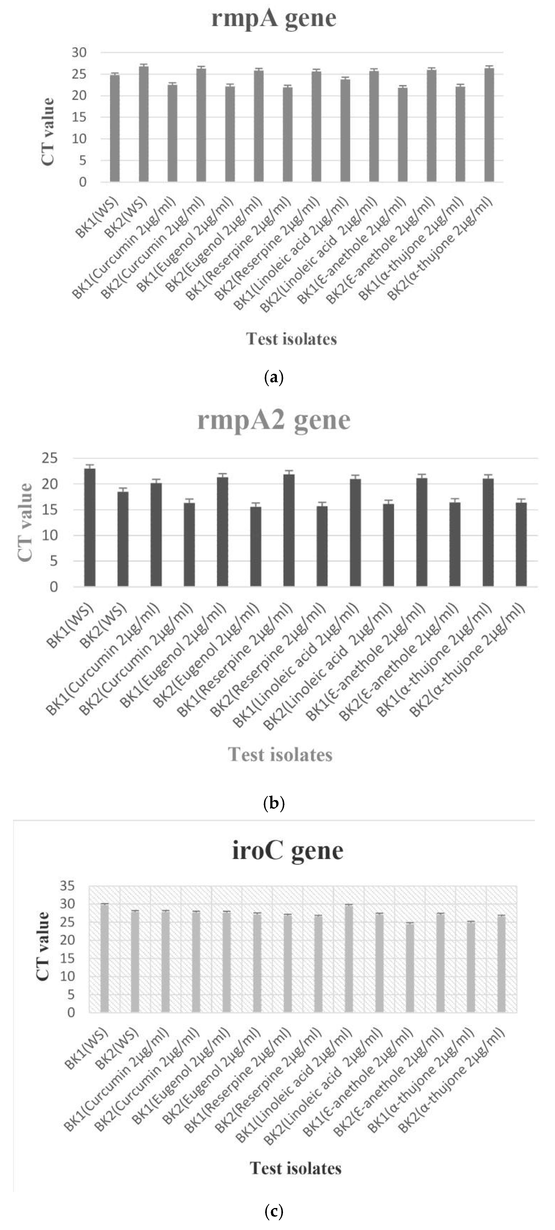
The final predicted structures of rmpA, rmpA2 and iroC are depicted in Figure 1 A, B, and C respectively (Figure 1). In case of all the 3 receptors, the computational model showed that except Reserpine and Curcumin all the ligands could effectively bind to the active sites, but Linoleic acid showed the best fit (Supplementary Table S4). For H-bonds, among the effectively bonded complexes, linoleic acid had the highest number of H-bonds for all the 3 receptors. The compounds did not show any inhibitory activity on the hypervirulent K. pneumoniae isolates, even at concentrations ranging from 512-1024 µg/mL, and isolates showed growth in all the concentrations of the said compounds whereas they were inhibited by imipenem within 4-8 µg/mL range. Transcriptional expression pattern of the isolates (BK1 and BK2) harboring rmpA was marginally increased when linoleic acid stress was applied. However, no significant change in expression of the above-mentioned gene was observed the with rest of the compounds (Figure 2A). Similarly, rmpA2 and iroC too did not show any significant expressional pattern with either of the anti-virulence compounds (Figure 2B,C).
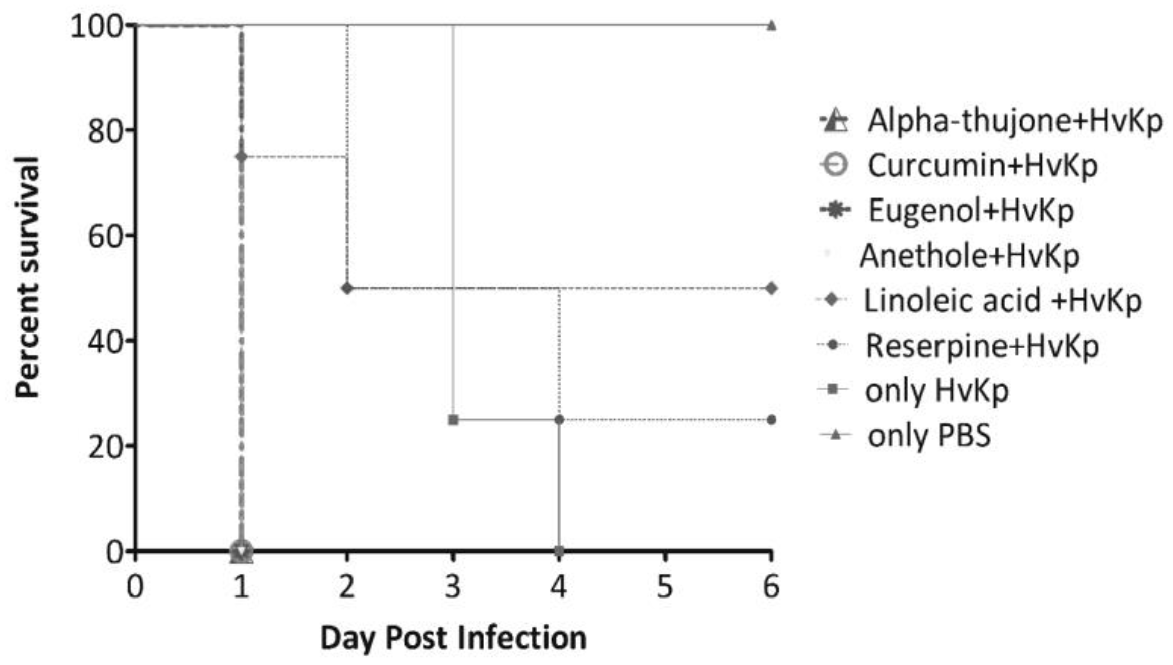
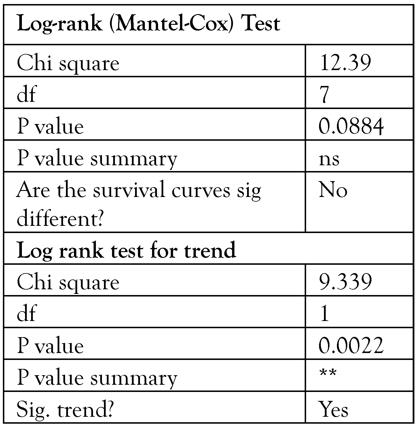
The in-vivo G. mellonella infection study showed that ɑ-thujone, curcumin, eugenol and anethol had no activity against hypervirulent K. pneumoniae (BK1 and BK2) as 100% of larvae died on day 1 to day 2 post-infection. However, linoleic acid and reserpine exhibited antivirulence activity as the larvae showed 50% and 25% survival rates respectively at day 6 postinfection (Table 2). All the larva injected only with sterile PBS showed 100% survival rate. By day 3, 25% and by day 4, 100% larva, injected only with hypervirulent K. pneumoniae strain died. The log-rank (Mantel-Cox) test defined that the time to death for hypervirulent Klebsiella pneumoniae infected larva was reduced compared to extracts treated larvae (p=0.08)—Figure 3.

Discussion

Anti-virulence compounds have an advantage of imposing little evolutionary and selection pressure in development of resistance compared to conventional antibiotics. As these compounds target virulence factors within a pathogen, it is less likely that they will have an impact on normal flora [19,20]. This study tried to search for compounds to combat hypervirulence in K. pneumoniae, which is one of the emerging issues and there have been few studies in this respect.
Molecular docking is a powerful drug designing tool that predicts binding pattern/orientation of ligand with protein at lowest potential energy. This released energy is the measure of stability of the ligand-protein complex i.e., the higher the docking score the higher is the ability of the ligand to inhibit the receptor’s activity [21,22,23]. In earlier studies it has been observed that in Staphylococcus mutans and K. pneumoniae, linoleic acid could reduce biofilm accumulation without affecting their viability [9,10,11]. Reports have suggested that anethole affects the virulence cascade in Vibrio cholerae [4]. Eugenol affects exotoxin production in Staphylococcus aureus, thus depicting an anti-virulence activity besides affecting biofilm formation in Escherichia coli, Pseudomonas aeruginosa, K. pneumoniae, S. aureus [12], while α-thujone interferes with quorum sensing system of P. aeruginosa [9,12]. In the expression analysis, transcriptional response of hypervirulence genes did not show any specific pattern in presence of the studied anti-virulence compounds. We may assume that this antivirulence compound has no interference with the virulence genes and the binding of this compound takes place with translational form of these virulence genes.
Hypervirulence in K. pneumoniae represents a serious public health threat globally. In this study, we have investigated hypervirulent isolates with multiple anti-virulence compounds. In a very recent study by Zarandi et al., it has been reported that curcumin acquire antibiofilm, antiefflux and anti-capsule activities, which may be used to boost anti-Klebsiella treatment [24,31]. Several virulence factors of K. pneumoniae enable its spread and emergence as a global threat. There are several genetic markers of hypervirulence in K. pneumoniae; among these, as previously described, the most important ones include regulators of the mucoid phenotype, the genes rmpA and rmpA2, siderophores such as aerobactin, enterobactin, yersiniabactin, and salmochelin, and genes that regulate iron uptake and allantoin metabolism [8,25]. Furthermore, rmpA/rmpA2 genes along with siderophores appear to be associated with invasive infections [26]. All the MDR hypervirulent K. pneumoniae isolates in a study were phenotypically and genotypically hypermucoviscous with several types of siderophore-encoding genes; their high invasivity reflected in their ability to cause 100% mortality in the neonatal unit, as previously reported [5].
The in-vivo experiment underscored antivirulence activity of linoleic acid and reserpine in G. mellonella model, which is also in agreement with the in-silico finding. The present study has included a small number of bacterial isolates and a small number of insect larva and this warrants further investigation with a larger number of isolates. This study was successful with its approach for searching an effective anti-virulence compound by in-silico, in-vitro, and in-vivo analysis.

Conclusions

This study documented that linoleic acid and reserpine have anti-virulence property without any antimicrobial activity against the hypervirulent isolates. This study also laid the grounds for further studies on this aspect of curbing or tackling virulence, to combat morbidity and mortality thus caused by hypervirulent K. pneumoniae strains.

Supplementary Materials

The following supporting information can be downloaded at: https://www.mdpi.com/article/10.18683/germs.2024.1426/s1.

Author Contributions

Data curation, TB; Investigation, JW, SB and SS; Methodology, TB, SB and AB; Software, SB and MAL; Supervision, AB; Validation, TB and MDC; Writing—original draft, JW and AB; Writing—review & editing, AB. All authors read and approved the final version of the manuscript.

Funding

None to declare.

Institutional Review Board Statement

The study was approved by the Ethics Committee of Assam University, Silchar, India.

Acknowledgments

The authors would like to acknowledge DBT (Govt. of India) sponsored Bioinformatics and Computational Biology Centre of Assam University, India and DelCON’s e-Journal Access Facility and DBT sponsored Advanced Level Institutional Biotech Hub (Phase II), Assam University, Silchar, India for providing infrastructural support.

Conflicts of Interest

All authors—none to declare.

References

  1. World Health Organization. Global priority list of antibiotic-resistant bacteria to guide research, discovery, and development of new antibiotics; WHO Press: Geneva, Switzerland, 2017. [Google Scholar]
  2. Choby, J.E.; Howard-Anderson, J.; Weiss, D.S. Hypervirulent Klebsiella pneumoniae clinical and molecular perspectives. J. Intern. Med. 2020, 287, 283–300. [Google Scholar] [CrossRef] [PubMed]
  3. Wyres, K.L.; Nguyen, T.N.T.; Lam, M.M.C.; et al. Genomic surveillance for hypervirulence and multi-drug resistance in invasive Klebsiella pneumoniae from South and Southeast Asia. Genome Med. 2020, 12, 11. [Google Scholar] [CrossRef]
  4. Buroni, S.; Chiarelli, L.R. Antivirulence compounds: A future direction to overcome antibiotic resistance? Future Microbiol. 2020, 155, 299–301. [Google Scholar] [CrossRef]
  5. Banerjee, T.; Wangkheimayum, J.; Sharma, S.; Kumar, A.; Bhattacharjee, A. Extensively drug resistant hypervirulent Klebsiella pneumoniae from a series of neonatal sepsis in a tertiary care hospital, India. Front. Med. 2021, 8, 645955. [Google Scholar] [CrossRef]
  6. WHO Country Office for India, Department of Biotechnology, Government of India. Indian Priority Pathogen List to Guide research, discovery, and development of new antibiotics in India. 2017. [Google Scholar]
  7. Crouch, M.L.; Castor, M.; Karlinsey, J.E.; Kalhorn, T.; Fang, F.C. Biosynthesis and IroC-dependent export of the siderophore salmochelin are essential for virulence of Salmonella enterica serovar Typhimurium. Mol. Microbiol. 2008, 67, 971–983. [Google Scholar] [CrossRef]
  8. Lai, Y.C.; Peng, H.L.; Chang, H.Y. RmpA2, an activator of capsule biosynthesis in Klebsiella pneumoniae CG43, regulates K2 cps gene expression at the transcriptional level. J. Bacteriol. 2003, 185, 788–800. [Google Scholar] [CrossRef]
  9. Shankar, C.; Basu, S.; Lal, B.; et al. Aerobactin seems to be a promising marker compared with unstable RmpA2 for the identification of hypervirulent carbapenem-resistant Klebsiella pneumoniae: In silico and in vitro evidence. Front. Cell Infect. Microbiol. 2021, 11, 709681. [Google Scholar] [CrossRef] [PubMed]
  10. Yu, V.L.; Hansen, D.S.; Ko, W.C.; et al. Virulence characteristics of Klebsiella and clinical manifestations of K. pneumoniae bloodstream infections. Emerg. Infect. Dis. 2007, 13, 986–993. [Google Scholar] [CrossRef]
  11. O’Boyle, N.M.; Banck, M.; James, C.A.; Morley, C.; Vandermeersch, T.; Hutchison, G.R. Open Babel: An open chemical toolbox. J. Cheminform. 2011, 3, 33. [Google Scholar] [CrossRef]
  12. Lin, J.C.; Chang, F.Y.; Fung, C.P.; et al. High prevalence of phagocytic-resistant capsular serotypes of Klebsiella pneumoniae in liver abscess. Microbes Infect. 2004, 6, 1191–1198. [Google Scholar] [CrossRef] [PubMed]
  13. Araújo, B.F.; Ferreira, M.L.; Campos, P.A.; et al. Hypervirulence and biofilm production in KPC-2producing Klebsiella pneumoniae CG258 isolated in Brazil. J. Med. Microbiol. 2018, 67, 523–528. [Google Scholar] [CrossRef]
  14. Wu, S.C.; Liu, F.; Zhu, K.; Shen, J.Z. Natural products that target virulence factors in antibiotic-resistant Staphylococcus aureus. J. Agric. Food Chem. 2019, 67, 13195–13211. [Google Scholar] [CrossRef] [PubMed]
  15. Bitencourt-Ferreira, G.; de Azevedo, W.F., Jr. Molegro Virtual Docker for docking. Methods Mol. Biol. 2019, 2053, 149–167. [Google Scholar] [CrossRef]
  16. Thomsen, R.; Christensen, M.S. MolDock: A new technique for high-accuracy molecular docking. J. Med. Chem. 2006, 49, 3315–3321. [Google Scholar] [CrossRef]
  17. Hong, S.S.; Kim, K.; Huh, J.Y.; Jung, B.; Kang, M.S.; Hong, S.G. Multiplex PCR for rapid detection of genes encoding class A carbapenemases. Ann. Lab. Med. 2012, 32, 359–361. [Google Scholar] [CrossRef]
  18. Poirel, L.; Walsh, T.R.; Cuvillier, V.; Nordmann, P. Multiplex PCR for detection of acquired carbapenemase genes. Diagn. Microbiol. Infect. Dis. 2011, 70, 119–123. [Google Scholar] [CrossRef] [PubMed]
  19. Sarker, S.D.; Nahar, L.; Kumarasamy, Y. Microtitre platebased antibacterial assay incorporating resazurin as an indicator of cell growth, and its application in the in vitro antibacterial screening of phytochemicals. Methods 2007, 42, 321–324. [Google Scholar] [CrossRef] [PubMed]
  20. Jacobs, A.C.; Thompson, M.G.; Black, C.C.; et al. AB5075, a highly virulent isolate of Acinetobacter baumannii, as a model strain for the evaluation of pathogenesis and antimicrobial treatments. mBio 2014, 5, e01076-14. [Google Scholar] [CrossRef]
  21. Nie, L.; Lv, Y.; Yuan, M.; et al. Genetic basis of high-level aminoglycoside resistance in Acinetobacter baumannii from Beijing, China. Acta Pharm. Sin. B. 2014, 4, 295–300. [Google Scholar] [CrossRef]
  22. Kusumaningrum, S.; Budianto, E.; Kosela, S.; Sumaryono, W.; Juniarti, F. The molecular docking of 1,4naphthoquinone derivatives as inhibitors of Polo-like kinase 1 using Molegro Virtual Docker. J. Appl. Pharm. Sci. 2014, 4, 47–53. [Google Scholar]
  23. Morris, G.M.; Lim-Wilby, M. Molecular docking. Methods Mol. Biol. 2008, 443, 365–382. [Google Scholar] [CrossRef]
  24. Karami-Zarandi, M.; Ghale, H.E.; Ranjbar, R. Characterization of virulence factors and antibacterial activity of curcumin in hypervirulent Klebsiella pneumoniae. Future Microbiol. 2022, 17, 529–540. [Google Scholar] [CrossRef]
  25. Effah, C.Y.; Sun, T.; Liu, S.; Wu, Y. Klebsiella pneumoniae: An increasing threat to public health. Ann. Clin. Microbiol. Antimicrob. 2020, 19, 1. [Google Scholar] [CrossRef]
  26. Holt, K.E.; Wertheim, H.; Zadoks, R.N.; et al. Genomic analysis of diversity, population structure, virulence, and antimicrobial resistance in Klebsiella pneumoniae, an urgent threat to public health. Proc Natl Acad Sci USA 2015, 112, E3574-81. [Google Scholar] [CrossRef]
  27. Silva, L.N.; Zimmer, K.R.; Macedo, A.J.; Trentin, D.S. Plant natural products targeting bacterial virulence factors. Chem. Rev. 2016, 116, 9162–9236. [Google Scholar] [CrossRef]
  28. Zahid, M.S.; Awasthi, S.P.; Asakura, M.; et al. Suppression of virulence of toxigenic Vibrio cholerae by anethole through the cyclic AMP (cAMP)-cAMP receptor protein signaling system. PLoS ONE 2015, 10, e0137529. [Google Scholar] [CrossRef] [PubMed]
  29. Wu, S.C.; Liu, F.; Zhu, K.; Shen, J.Z. Natural products that target virulence factors in antibiotic-resistant Staphylococcus aureus. J. Agric. Food Chem. 2019, 67, 13195–13211. [Google Scholar] [CrossRef] [PubMed]
  30. Choudhury, D.; Paul, D.; Ghosh, A.S.; et al. Effect of singledose carbapenem exposure on transcriptional expression of blaNDM-1 and mexA in Pseudomonas aeruginosa. J. Glob. Antimicrob. Resist. 2016, 7, 72–77. [Google Scholar] [CrossRef] [PubMed]
  31. Karami-Zarandi, M.; Rahdar, H.A.; Esmaeili, H.; Ranjbar, R. Klebsiella pneumoniae: An update on antibiotic resistance mechanisms. Future Microbiol. 2023, 18, 65–81. [Google Scholar] [CrossRef]
Figure 1. (A) represents predicted structures of rmpA, (B) represents predicted structures of rmpA2 and (C) showing predicted structures of iroC.
Germs 14 00136 g001
Figure 2. (A) Expressional analysis of rmpA gene harboring isolates against different anti-virulence compounds and without anti-virulence compound (Control).X-axis denotes test isolates whereas Yaxis shows CT value of the corresponding isolates. (B) Expressional analysis of rmpA2 gene harboring isolates against different anti-virulence compounds and without anti-virulence compound (Control) X-axis denotes test isolates whereas Y-axis shows CT value of the corresponding isolates. (C) Expressional analysis of iroC gene harboring isolates against different anti-virulence compounds and without anti-virulence compound (Control). X-axis denotes test isolates whereas Yaxis shows CT value of the corresponding isolates.
Germs 14 00136 g002
Figure 3. Kaplan-Meier survival curves showing survival of Galleria mellonella larvae infected with hypervirulent Klebsiella pneumoniae (HvKp).
Germs 14 00136 g003
Table 1. Oligonucleotide primers used for Real time PCR.
Germs 14 00136 i001
Table 2. Comparison of Survival Curves.
Germs 14 00136 i002