Next Article in Journal
Facial Nerve Palsy: An Early Sign of COVID-19
Previous Article in Journal
Adverse Events Following Immunization (AEFI): COVID-19 Vaccine Dose and Characteristics in Central Java, Indonesia
 
 
GERMS is published by MDPI from Volume 15 Issue 4 (2025). Previous articles were published by another publisher in Open Access under a CC-BY (or CC-BY-NC-ND) licence, and they are hosted by MDPI on mdpi.com as a courtesy and upon agreement with the former publisher Infection Science Forum.
Article

Enterobacteriaceae Isolates from Clinical and Household Tap Water Samples: Antibiotic Resistance, Screening for Extended-Spectrum, Metallo- and AmpC-Beta-Lactamases, and Detection of blaTEM, blaSHV and blaCTX-M in Uyo, Nigeria

by
Olajide J. Akinjogunla
1,*,
Adebowale T. Odeyemi
2,
Edinam-Abasi S. Udofia
1,
Oyetayo O. Adefiranye
3,
Clarence S. Yah
4,
Igbagbo Ehinmore
5 and
Idongesit U. Etukudo
6
1
Department of Microbiology, Faculty of Science, University of Uyo, P.M.B. 1017, Uyo, Akwa Ibom State, Nigeria
2
Department of Microbiology, College of Pure and Applied Science, Landmark University, Omu-Aran, Kwara State, Nigeria
3
Department of Microbiology, University of Lagos, Akoka, Lagos State, Nigeria
4
School of Health Systems and Public Health, Faculty of Health Sciences, University of Pretoria, South Africa
5
Department of Biological Sciences, Lagos State University of Science and Technology, Ikorodu, Lagos State, Nigeria
6
Department of Microbiology, Abia State University, Uturu, Abia State, Nigeria
*
Author to whom correspondence should be addressed.
GERMS 2023, 13(1), 50-59; https://doi.org/10.18683/germs.2023.1366
Submission received: 5 October 2022 / Revised: 18 February 2023 / Accepted: 1 March 2023 / Published: 31 March 2023

Abstract

Introduction: Infections caused by multidrug-resistant (MDR) bacteria, extended spectrum β-lactamase (ESBL), metallo-β-lactamase (MBL) and AmpC-β-lactamase (AmpC-βL)-producers are increasing globally. This study identified bacteria in clinical and tap water samples and determined the prevalence of MDR, and β-lactamase enzymes and genes. Methods: Isolates were identified by the Vitek 2 (bioMérieux, France) automated system. Antibiotic resistance and screening for β-lactamase enzymes and genes was done using disc diffusion method and Vitek 2 automated system, CHROMagar-ESBL, combined double disc, inhibition-based method and multiplex polymerase chain reaction, respectively. Results: The Enterobacteriaceae isolates obtained were Escherichia coli, Klebsiella pneumoniae, Citrobacter freundii, Salmonella spp., Proteus mirabilis, Enterobacter aerogenes, Shigella sonnei, Proteus vulgaris, Enterobacter sakazakii, Klebsiella oxytoca, Citrobacter diversus, and Serratia liquefaciens. Of the 674 isolates from clinical samples, 36.5%, 28.5%, and 19.9% were ESBL, MBL, and AmpC-βL producers, respectively. A low prevalence of AmpC-βL and MBL producers were obtained, with no significant difference (p<0.05) between the prevalence of ESBL and non-ESBL producers. Isolates exhibited varied levels of resistance to gentamicin, amoxicillin-clavulanic acid, ciprofloxacin, and tetracycline. The results showed that 54.6% of ESBL producers, 57.9% of MBL producers, and 62.8% of AmpC-βL producers were MDR strains. Of the 141 representative isolates tested, 36.9%, 15.6%, and 20.6% had only blaTEM, blaSHV, and blaCTX-M, respectively; 5.7% possessed both blaTEM and blaSHV; 7.1% possessed both blaTEM and blaCTX-M and 4.3% had both blaSHV and blaCTX-M. Conclusions: This study found a high prevalence of β-lactamase producers, indicating the need for further research on the molecular epidemiology of β-lactamase producers and their impacts in the region.

Introduction

The prevalence of multidrug-resistant (MDR) Enterobacteriaceae isolates is a serious public health threat due to its steadily increasing rate over the years, which has posed significant challenges in combating infections, particularly in developing countries. [1] Among the Enterobacteriaceae isolates that have been evidently identified as major cause of MDR bacterial infections were K. pneumoniae, Salmonella spp., and Enterobacter spp. [1]
Extended-spectrum β-lactamases (ESBL), AmpC β-lactamase (AmpC-βL) and metallo β-lactamase (MBL) are enzymes responsible for microbial resistance to β-lactam antibiotics. [2] ESBL, AmpC-βL, and MBL producing Enterobacteriaceae (PE) isolates from humans, animals, and environmental sources have been reported globally. [3] AmpC-βLs and ESBLs are clinically significant cephalosporinases, mutant forms of β-lactamase enzymes produced by Gram-negative bacteria, that are encoded on plasmid and/or chromosome, and mediate resistance to penicillin and third generation cephalosporins, [4,5] but do not affect cephamycins or carbapenems. AmpC-βL-PE reported included Serratia spp., Providencia spp., and C. freundii. [4] ESBLs producing strains usually manifest resistance to multiple antibiotic classes, thus, the emergence of ESBL-PE has critically compromised the efficacy of antibiotics. Infections caused by ESBL-PE are a major public health threat worldwide and are usually associated with high morbidity and mortality. [4]
Acquired MBLs have emerged as one of the main worrisome resistance mechanisms by Enterobacteriaceae isolates due to their ability to hydrolyze β-lactams and carbapenems. MBLs are zinc ion-dependent with broad substrate specificity that are not inhibited by clavulanic acid, sulbactam, or tazobactam. [6] Hence, this study determined the occurrence of MDR, β-lactamase enzymes and genes in isolates from clinical and tap water samples in Uyo, Nigeria.

Methods

Collection of tap water and clinical samples

Tap water samples (n=106) were collected in Uyo, Akwa Ibom State. The tap water was allowed to run for a few minutes before 200 mL were collected using sterile screw-cap containers. A total of 414 clinical samples, comprising mid-stream urine (n=148), stool (n=70), wound swabs (n=64), ear discharge (n=38), and blood (n=94), were collected using sterile leak-proof vials and swab sticks from patients in Uyo, Akwa Ibom State. All the samples were transported in an icebox to the Microbiology Laboratory for bacteriological analyses.

Inclusion criteria

Patients who agreed and gave verbal informed consent to participate in the study.

Exclusion criteria

Patients who were on antibiotics within one week of enrollment and/or declined to participate in the study.

Bacteriological analysis of clinical samples

One milliliter (1 mL) of each well-mixed mid-stream urine (MSU) sample was inoculated on plates of Cysteine Lactose Electrolyte Deficient Agar (CLED), MacConkey Agar (MCA) without salt, and Violet Red Bile Glucose Agar (VRBGA). Each stool sample was serially diluted, and 1 mL of each aliquot was inoculated onto plates of MCA, VRBGA, and Eosine Methylene Blue (EMB) agar. Each wound swab/ear discharge was separately dipped into a test tube containing sterile dH2O (9 mL), and 0.1 mL was pipetted and surface-inoculated on plates of MCA, VRBGA, and blood agar. All the plates were aerobically incubated for 24 h at 37 °C. After incubation, colonies on plates with bacterial growths were subcultured on Nutrient agar plates and aerobically incubated for 24 h at 37 °C. Each blood sample (1 mL) was inoculated into 5 mL of Brain Heart Infusion (BHI) Broth, VRBGA and incubated. After incubation, 1 mL of the inoculated broth medium was subcultured on a plate of blood agar. Pure isolates were streaked on nutrient agar slants, incubated for 24 h at 37 °C and stored in the refrigerator at 4 °C.

Detection of Enterobacteriaceae isolates in tap water

Each water sample (100 mL) was aseptically filtered through a MF-milliporeTM membrane filter with a 0.45 μm pore size. The membranes were aseptically removed and placed on plates of Membrane Faecal Coliform Agar and mEndo agar for faecal coliforms (FC) and total coliforms (TC), respectively. All the plates were incubated at 37 °C except the plates of mFC agar, which were incubated at 45 °C for 24 h. After incubation, colonies on the filter were enumerated and recorded; the results were expressed as the number of FC and TC in 100 mL of water, and colonies were subcultured onto nutrient agar plates and incubated at 37 °C for 24 h. Pure cultures of isolates were streaked onto nutrient agar slants, incubated at 37 °C for 24 h, and stored in the refrigerator at 4 °C.

Identification of Enterobacteriaceae isolates

The identification of Enterobacteriaceae isolates was carried out by Vitek 2 automated system (Vitek card GN (Ref.) 21341, BioMérieux Inc, France).

Antibiotic susceptibility testing

Antibiotic susceptibility was performed by disc diffusion method and results were interpreted according to the Clinical and Laboratory Standards Institute guidelines. A loopful of bacterial colony was taken, transferred to a tube containing 5 mL sterile dH2O, mixed gently until it formed a homogenous suspension. The Muller Hinton Agar plate was uniformly flooded with bacterial suspension using sterile swab sticks. The antibiotic discs of gentamycin (10 μg); cefoxitin (30 μg); ceftazidime (30 μg); imipenem (10 μg); cefotaxime (30 μg); amoxicillin-clavulanic acid (30 μg); ciprofloxacin (5 μg); meropenem (10 μg) and tetracycline (30 μg) were aseptically placed on the surfaces of the inoculated plates. the plates were inverted, incubated at 37 °C for 16h and the diameters of inhibition zones were measured. The antibiotic susceptibilities of isolates were also determined by Vitek 2 automated systems. Isolates that were resistant to ≥3 antibiotic classes were considered as MDR strains. [7]

Screening of extended spectrum β-lactamase (ESBL) producers

The CHROMagar ESBL, a chromogenic agar, was used for the phenotypic screening of ESBL-PE isolates following the manufacturer’s instructions.

Screening of metallo β-lactamase(MBL) and AmpC-βL producers

Screening of MBL-PE was determined using Combined Double Disc Test. A disc of imipenem (10 μg, IMP) and imipenem-EDTA disc were placed 20 mm apart on an MHA plate inoculated with 10µL bacterial suspension and incubated for 16-18h at 37 °C. An increase of ≥7 mm in inhibitory zone around IMP-EDTA disc compared to IMP disc alone indicated MBL production. Screening of AmpC-βL-PE was determined by Inhibition-Based method. A disc of cefoxitin (30μg, CEF) and cefoxitin-boronic acid disc were placed 30 mm apart on an MHA plate inoculated with 10µL bacterial suspension and incubated for 16 h at 37 °C. An increase of ≥5 mm in inhibitory zone around the CEF-BA disc compared to CEF disc alone indicated AmpC-βL production. [3]

Extraction of genomic DNA of isolates

The genomic DNA (gDNA) of 141 representative β-lactamases resistant isolates was extracted by boiling method. Briefly, 100 µL of an overnight freshly grown culture was centrifuged at 2,300 x g for 5 min. The supernatant was discarded, and the pellet was re-suspended into 100 µL of phosphate buffer solution, boiled in a water bath at 100 °C for 10 min, cooled on ice for 10 min in each step, and centrifuged at 2,300 x g for 10 min. The supernatant containing gDNA was collected, stored at -20 °C in an Eppendorf tube, and later used as a DNA template.

PCR amplification of β-lactamase genes in Enterobacteriaceae isolates

The multiplex polymerase chain reaction (PCR) amplification assay for detection of the blaTEM, blaSHV, and blaCTX-M genes in isolates was carried out. A final PCR reaction volume (25 μL) contained 12.5 µL of PCR-master mix, 1 μL of each primer, 3 μL of template DNA and 6.5 µL of RNase-free water. The PCR amplification conditions were as follows: initial denaturation at 95 °C for 5 min; followed by 30 cycles of denaturation at 95 °C for 1 min; annealing temperature at 55 °C for 1 min; extension at 72 °C for 1 min; and a final extension at 72 °C for 10 min. The amplified PCR products were electrophoresed on 1.5% agarose gel containing 0.5 μg/mL of ethidium bromide and visualized under a UV transilluminator.

Statistical analysis

The Statistical Package for Social Sciences (SPSS) version 22 (IBM Corp, USA) was used for all statistical analyses. The significant difference between β-lactamase and non β-lactamase producing isolates at ap-value <0.05 were determined using a Chi-square statistical test.

Results

A total of 796 isolates were recovered from the clinical and tap water samples (Table 1). Based on different clinical samples, E. coli, K. pneumoniae, and C. freundii were the most common isolates from urine, stool, wounds, and ear discharge. A total of 122 isolates were obtained from tap water, of which 38 were K. pneumoniae, 22 were Salmonella spp., 14 were E. coli, 10 were P. mirabilis, 10 were S. sonnei, 6 were E. aerogenes, 6 were C. freundii, 2 were E. sakazakii, and 2 were P. vulgaris, while S. liquefaciens, K. oxytoca, and C. diversus had 4 each (Table 1).
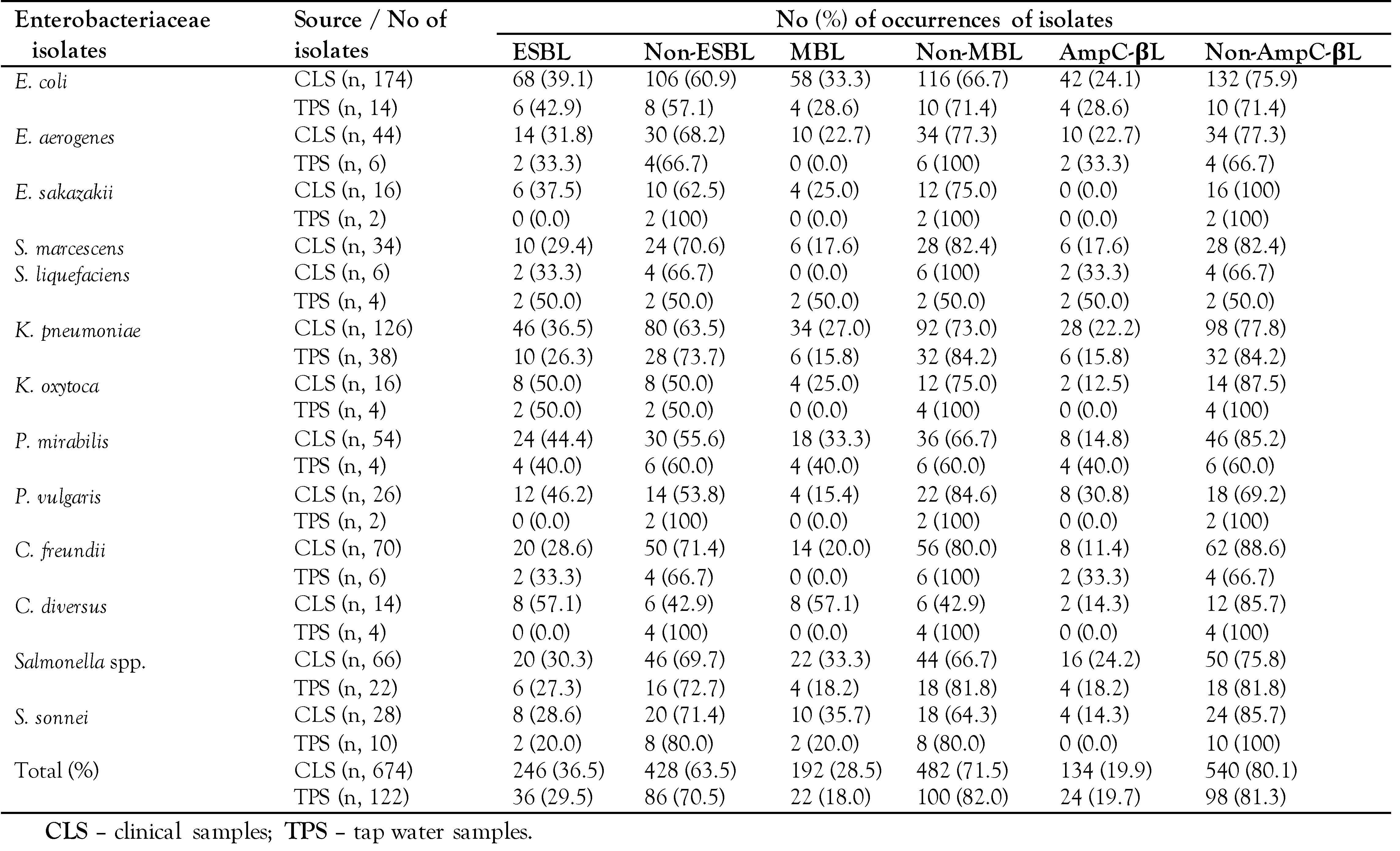
Among the ESBL producers from clinical samples, C. diversus had the highest prevalence, of 57.1% (8/14), while the least frequency was observed for C. freundii (28.6%, 20/70) and S. sonnei (28.6%, 8/28) – Table 2. The highest and lowest AmpC-βL producers were S. liquefaciens (33.3%) and C. freundii (11.4%), respectively. The isolates (n=192) were positive for MBL production, and the most prevalent phenotype was C. diversus (57.1%), while the least prevalent phenotype was P. vulgaris (15.4%) – Table 2. The prevalence of the ESBL producers from tap water were: E. coli (42.9%), E. aerogenes (33.3%), S. liquefaciens (50%), K. pneumoniae (26.3%), K. oxytoca (50%), P. mirabilis (40%), C. freundii (33.3%), Salmonella spp. (27.3%), and S. sonnei (20%). Only 22 MBL-producers, comprising E. coli (n=4), S. liquefaciens (n=2), K. pneumoniae (n=6), P. mirabilis (n=4), Salmonella spp. (n=4), and S. sonnei (n=2), were obtained from tap water samples. Table 2 shows the overall prevalence of AmpC-βL producers in tap water as 19.7% (24/122). S. liquefaciens had the highest prevalence (50.0%), while isolates with the least prevalence were K. pneumoniae (15.8%).
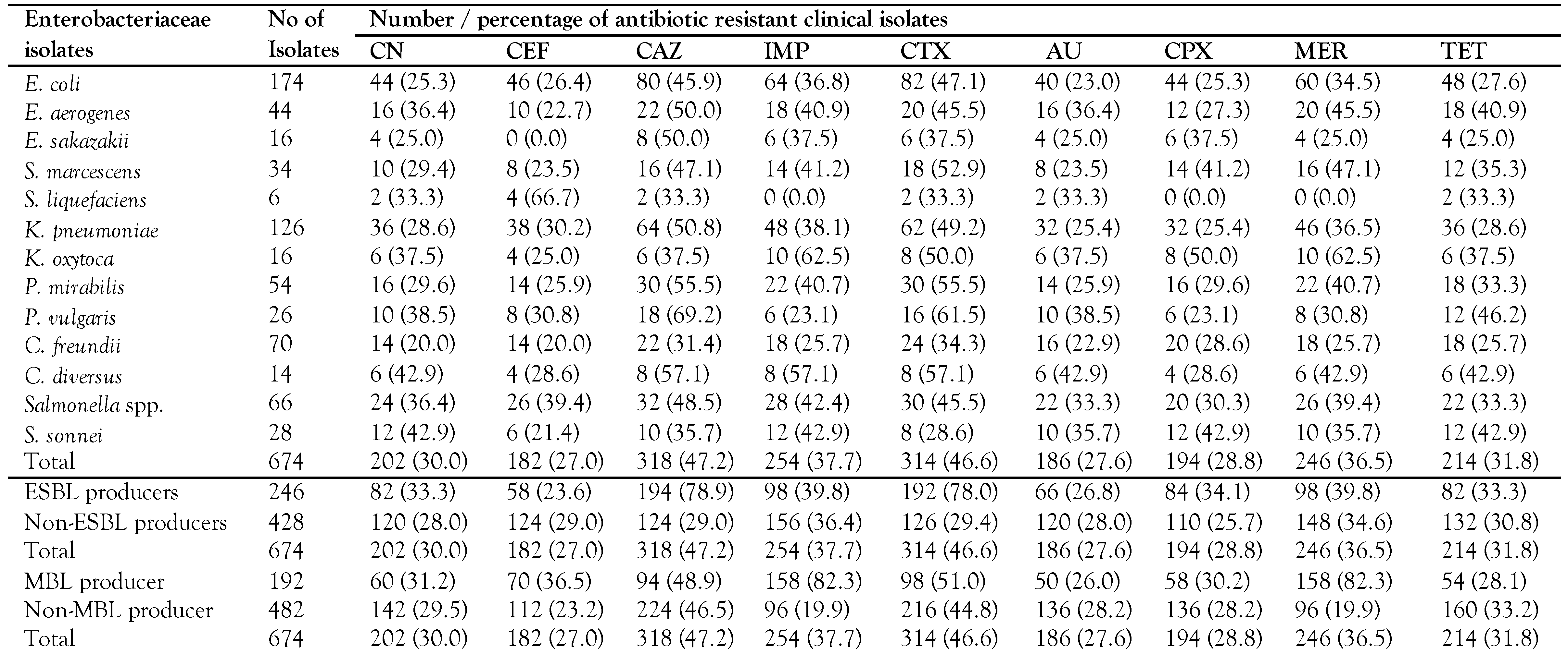
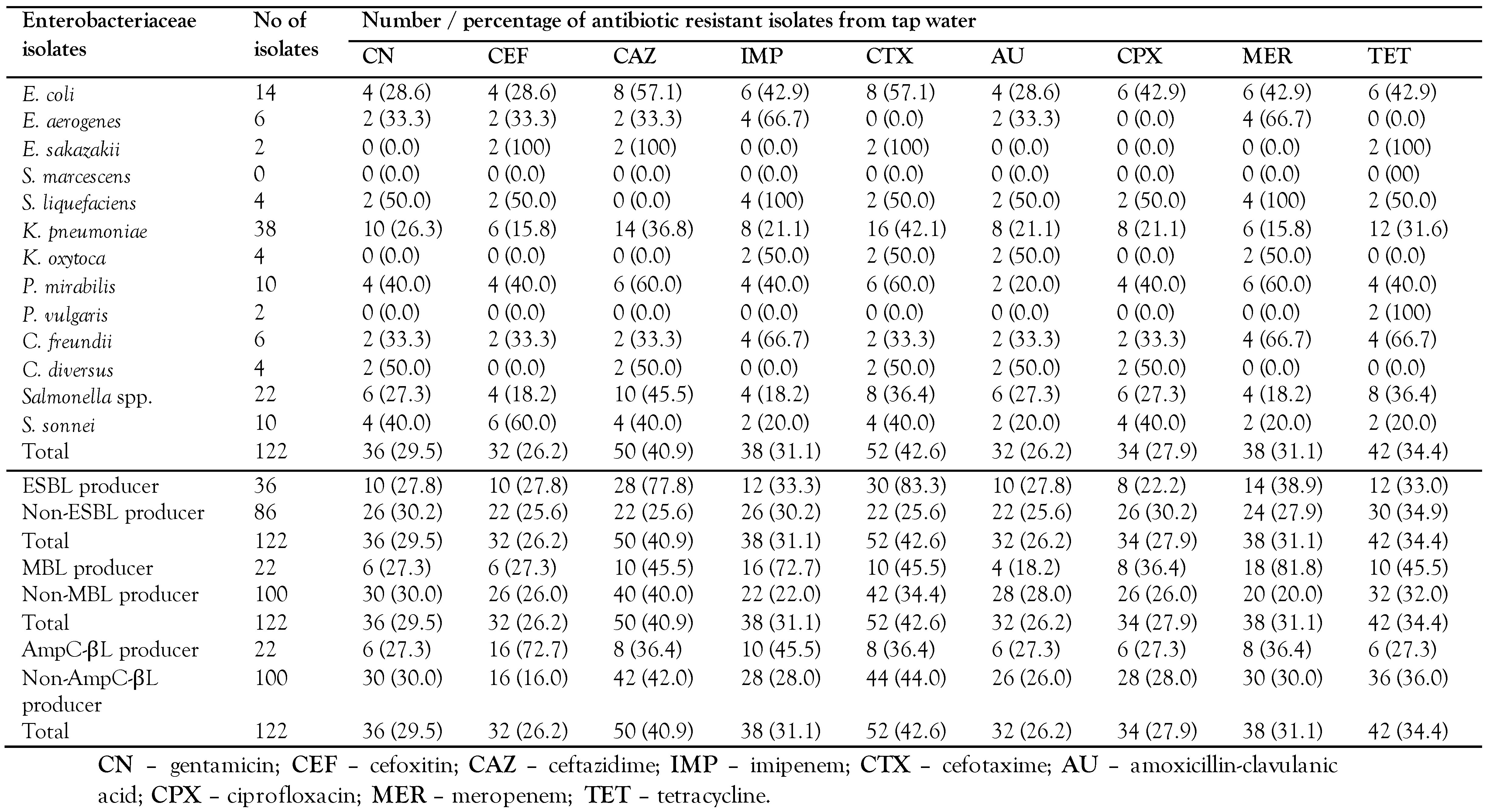
Overall, the isolates exhibited a high level of resistance to ceftazidime (47.2%) and cefotaxime (46.6%). The resistance of isolates to gentamicin, cefoxitin and amoxicillin-clavulanic acid was 30%, 27% and 27.6%, respectively. A high cefoxitin resistance was exhibited by S. liquefaciens (66.7%), while imipenem resistant K. oxytoca was 62.5%. ESBL producers were more resistant to antibiotics than non-ESBL producers: cefotaxime (78% vs. 29.4%) and gentamicin (33.3% vs 28%). The non-MBL showed a low resistance (19.9%) to meropenem, while ≤51% MBL- and AmpC-βL-producers were resistant to ceftazidime (Table 3a). Of the 122 isolates obtained from tap water samples, 42.6% were resistant to cefotaxime, 40.9% were resistant to ceftazidime, and 34.4% were resistant to tetracycline, while 27.9% exhibited resistance to ciprofloxacin. S. sonnei, S. liquefaciens, E. sakazakii and P. mirabilis were highly resistant (>40%) to cefotaxime, whereas a low rate of resistance to cefoxitin and meropenem was seen in Salmonella spp. and K. pneumoniae. The non-ESBL producing isolates were more resistant to gentamicin and tetracycline than ESBL producers. The MBL and AmpC-βL producers were highly resistant (>36.4%) to ceftazidime, imipenem, cefotaxime and meropenem (Table 3b).
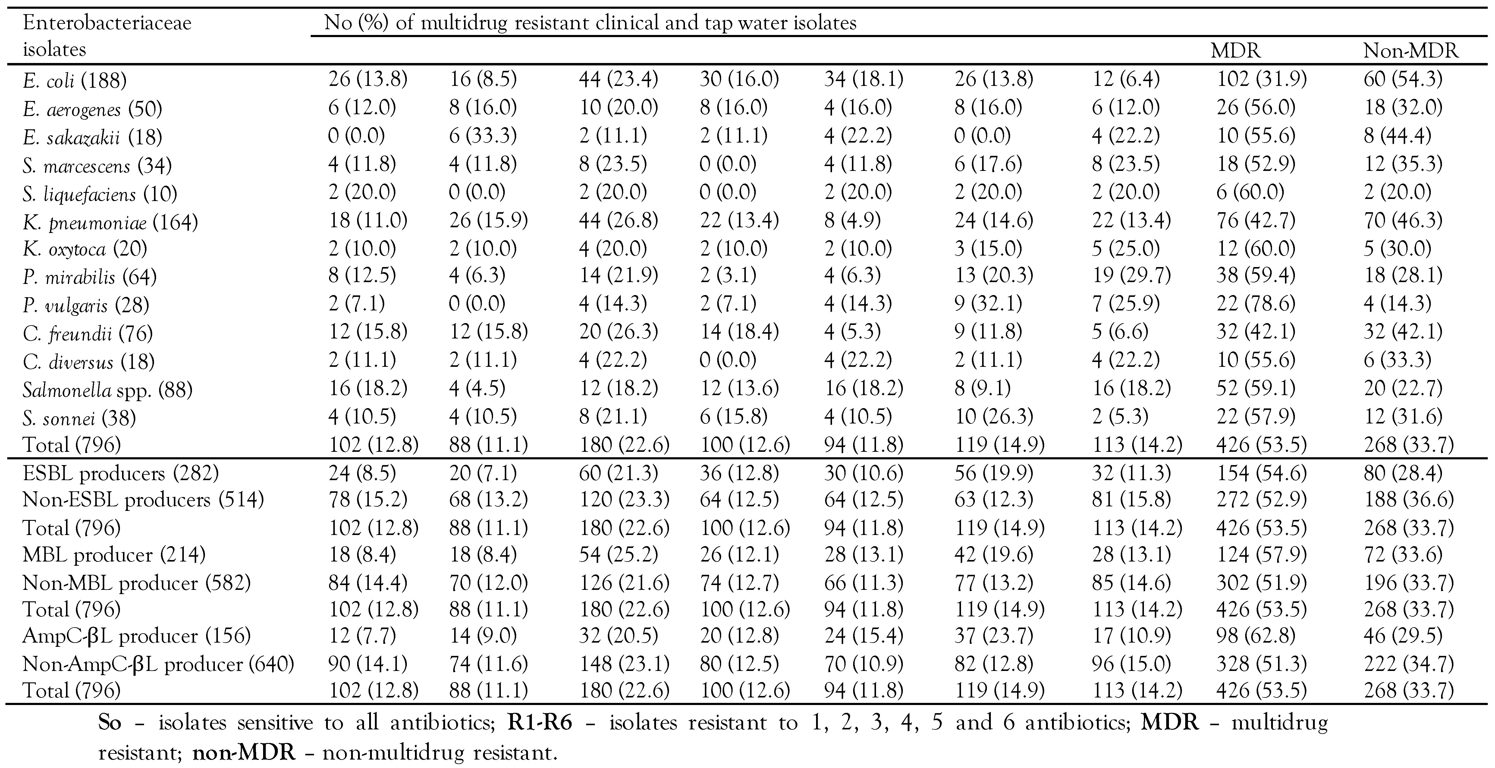
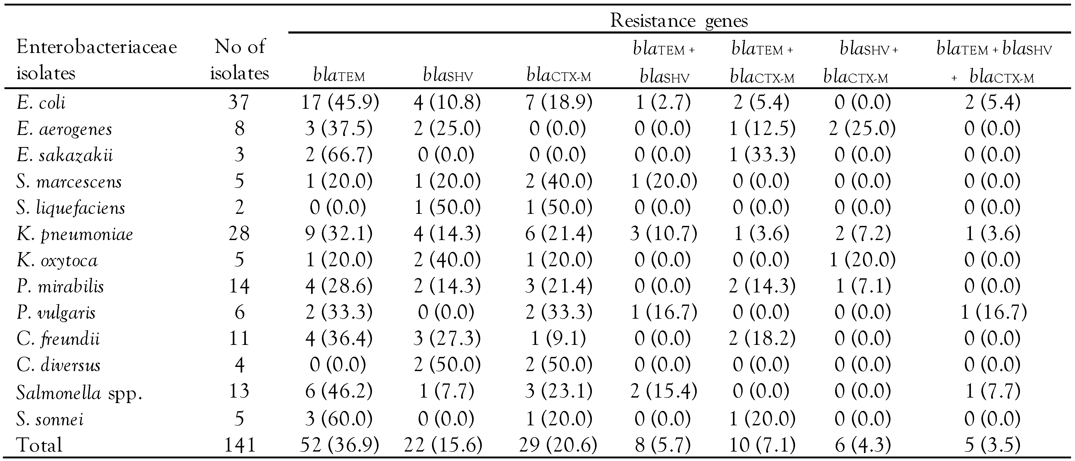
Of the 796 isolates obtained, 53.5% were MDR, 33.7% were non-MDR, and 12.8% were susceptible to all antibiotics tested. Overall, 54.6% (154/282) ESBL producers were MDR, and 28.4% (80/282) were non-MDR. Of the 582 non-MBL producers obtained, 51.9% were MDR whereas 33.7% were non-MDR. The percentage of MDR-AmpC-βL producers (62.8%) was higher than that of non-MDR-AmpC-βL producing strains (51.3%) – Table 4. Of the 141 representative isolates tested, 36.9%, 15.6%, and 20.6% had only blaTEM, blaSHV, and blaCTX-M, respectively. A total of 5.7% (n=8) of the isolates possessed both blaTEM and blaSHV; 7.1% (n=10) of the isolates possessed both blaTEM and blaCTX-M; and 4.3% (n=6) had both blaSHV and blaCTX-M. Five isolates, comprising E. coli (n=2), K. pneumoniae (n=1), P. vulgaris (n=1), and Salmonella spp. (n=1), had all three beta-lactamase genes (blaTEM, blaSHV, and blaCTX-M). Of the 37 E. coli and 28 K. pneumoniae isolates, blaTEM gene targets were detected in 22 (59.5%) and 14 (50.0%), respectively (Table 5).

Discussion

Fourteen bacterial species, in the family Enterobacteriaceae, were recovered from tap water samples, stool, urine, ear discharge, blood, wound swabs in this study. Clinical samples had both E. coli and K. pneumoniae as the most common Enterobacteriaceae isolates and this corroborated the findings of Ibrahim and Hameed [8] who reported E. coli and K. pneumoniae as the most prevalent bacteria from clinical samples in Baghdad, Iraq.
Overall, 36.5% ESBL-PE were obtained from clinical samples in our study and this value was higher than the ESBL-PE prevalence in Austria (0.7%). [9] The predominant ESBL-PE from the clinical samples in this study were C. diversus (57.1%) and K. oxytoca (50.0%) and these results were in dissimilarity with studies carried out in Uganda [10] in which K. pneumoniae and E. coli were the prevalent ESBL-PE. Similarly, this present study detected 29.5% ESBL-PE from household tap water. Of these, S. liquefaciens and K. oxytoca were the most frequent ESBL producers and this finding disagrees with the report in which K. pneumoniae was the most frequent ESBL-PE from drinking water. [11]
The increasing rate of MBL-PE isolates has constituted a global public health problem. In our study, phenotypic detection of MBL production among E coli, K. pneumoniae and Salmonella spp.conformed to the results of Hoang et al. [12] Similarly, 57.1% C. diversus and 20.0% C. freundii were MBL producers in this study and this value was higher than 18.7% for C. diversus and 1.9% for C. freundii obtained by Chaudhary et al. [13] in their study on MBL producing Gram-negative bacteria among patients visiting Shahid Gangalal National Heart Centre in Nepal.
The inhibition-based method employed for screening of AmpC-βL-PE in our study revealed <19.9% prevalence rate of AmpC-βL producing strains in clinical and drinking tap water samples and this result was similar to the findings in which <22% clinical isolates of Gram-negative bacteria were AmpC beta-lactamase producers. [14] Similarly, <30% E. coli from either clinical or tap water samples were AmpC-βL producers and this value was lower than 37.5% obtained in Chennai, South Indian States. [5] In our study, K. oxytoca, C. freundii, and E. aerogenes harbored AmpC-βL and this corroborated the findings of Ratna et al. [15]
In this study, Enterobacteriaceae isolates from clinical and tap water samples showed a high resistance to ceftazidime (47.2%, 40.9%) and cefotaxime (46.6%, 42.6%) and the results of a high prevalence of ceftazidime and cefotaxime resistance in Enterobacteriaceae isolates in our study are in line with the findings of studies conducted in Iran. [16] We observed that E. aerogenes exhibited a high rate of resistance to imipenem and meropenem and this corroborated the report on emergence of E. aerogenes strains with a decreased susceptibility to imipenem. [17] In relation to imipenem, reports have shown that E. aerogenes could more rapidly adapt its regulation of permeability than other enterobacteria. High ceftazidime and cefotaxime resistance rates in S.marcescens were obtained and this is in consonance with the study conducted by Simsek [18] on determination of the antibiotic resistance rates of S. marcescens isolates obtained from various clinical specimens. The K. oxytoca isolates were highly resistant to imipenem, meropenem and ciprofloxacin. These findings corroborated the results of Singh et al. [19] on the antibiotic resistance by K. oxytoca.
Of the 246 ESBL-PE isolates from clinical samples, 34.1% were resistant to ciprofloxacin, 33.3% to gentamicin and 39.8% to amoxicillin-clavulanic acid. This frequency of resistance recorded against ciprofloxacin, gentamicin and amoxicillin-clavulanic acid respectively was lower than results reported in Ethiopia. [20] In our study, MBL-PE isolates from clinical and tap water samples showed a high resistance to ceftazidime, imipenem, cefotaxime and meropenem. This finding is in tandem with Walsh et al. [21] on the resistance of MBL positive isolates to β-lactam antibiotics. The percentages of resistance of AmpC-βL-PE from clinical samples to meropenem (46.3%) and imipenem (46.3%) in this present study were lower than 49.5% for meropenem and 57.2% for imipenem reported in Saudi Arabia. [22] These values are of serious concern as carbapenems are often considered as drugs of choice for treatment of serious AmpC-βL-associated infections.
In our present study 54.6% of the Enterobacteriaceae isolates were MDR and this value is lower than 93.5% reported in Ethiopia. [23] E. coli producing blaCTX-M were detected in the clinical samples in our study, and this corroborated the findings of Pitout et al. [24] that E. coli producing blaCTX-M has become widely distributed. The detection of blaSHV, blaTEM and blaCTX-M among Enterobacteriaceae isolates in our study using a multiplex PCR assay was also in conformity with the reports of Monstein et al. [25] in their study on multiplex PCR amplification assay for the detection of β-lactamase genes in Enterobacteriaceae isolates.

Conclusions

This study demonstrated a high prevalence of ESBL-, MBL-, and AmpC-βL-PE isolates in clinical and tap water samples. Enterobacteriaceae isolates exhibited varied levels of resistance to antibiotics. Similarly, a high rate of MDR isolates was obtained from clinical and tap water samples. We therefore recommend further studies on molecular epidemiology of β-lactamase producing isolates and their impacts in the region.

Author Contributions

OJA, ATO and ESU contributed equally in designing and conducting the experiment. Data analyses and interpretation were made by OOA, CSY and IE. Manuscript was prepared by OJA and IUE. All authors contributed to the editing of the manuscript. All authors read and approved the final version of the manuscript.

Institutional Review Board Statement

Approval (AKSMH/PH/18/2022) from the Ethical Review Committee, Akwa Ibom State Ministry of Health, Uyo, and verbal informed consent of participants were obtained. Permission from the members of the households where tap water samples were collected was likewise sought.

Acknowledgments

The authors greatly acknowledge the support and co-operation of the patients and those whose household tap water samples were obtained. We equally acknowledge the valuable assistance rendered by the technical staff of government and private medical laboratories used.

Conflicts of Interest

All authors – none to declare.

References

  1. Qadi, M.; Alhato, S.; Khayyat, R.; Elmanama, A.A. Colistin resistance among Enterobacteriaceae isolated from clinical samples in Gaza Strip. Can J Infect Dis Med Microbiol 2021, 2021, 6634684. [Google Scholar] [CrossRef]
  2. Kameyama, M.; Yabata, J.; Nomura, Y.; Tominaga, K. Detection of CMY-2 AmpC beta-lactamase- producing enterohemorrhagic Escherichia coli O157:H7 from outbreak strains in a nursery school in Japan. J Infect Chemother 2015, 21, 544–546. [Google Scholar] [CrossRef] [PubMed]
  3. Thenmozhi, S.; Moorthy, K.; Sureshkumar, B.T.; Suresh, M. Antibiotic resistance mechanism of ESBL producing Enterobacteriaceae in clinical field: A review. Int J Pure App Biosci 2014, 2, 207–226. [Google Scholar]
  4. Etukudo, I.U.; Onyeagba, R.A.; Akinjogunla, O.J.; Ibe, C.; Ikpe, M.E. Phenotypic expression of extended spectrum βeta-lactamases and antibiogram of uro-pathogenic bacterial isolates from out- patients attending some private hospitals in Uyo, Nigeria. Sci World Jour 2020, 15, 59–66. [Google Scholar]
  5. Akinjogunla, O.J.; Eghafona, N.O.; Enabulele, I.O. Aetiologic agents of acute otitis media (AOM): Prevalence, antibiotic susceptibility, β-lactamase (βL) and extended spectrum β-lactamase (ESBL) production. J Microbiol Biotechnol Food Sci 2012, 1, 333–353. [Google Scholar]
  6. Biradar, S.; Roopa, C. Prevalence of metallo-β-lactamase producing Pseudomonas aeruginosa and its antibiogram in a tertiary care centre. Int J Curr Microbiol Appl Sci 2015, 4, 952–956. [Google Scholar]
  7. Akinjogunla, O.J.; Divine-Anthony, O. Asymptomatic bacteriuria among apparently healthy undergraduate students in Uyo, South- South, Nigeria. Ann Res Rev Biol 2013, 3, 213–225. [Google Scholar]
  8. Ibrahim, I.A.J.; Hameed, T.A.K. Isolation, characterization and antimicrobial resistance patterns of lactose-fermenter Enterobacteriaceae isolates from clinical and environmental samples. Open J Med Microbiol 2015, 5, 169–176. [Google Scholar] [CrossRef]
  9. Kateregga, J.N.; Kantume, R.; Atuhaire, C.; Lubowa, M.N.; Ndukui, J.G. Phenotypic expression and prevalence of ESBL-producing Enterobacteriaceae in samples collected from patients in various wards of Mulago hospital, Uganda. BMC Pharmacol Toxicol 2015, 16, 14. [Google Scholar] [CrossRef]
  10. Nijssen, S.; Florijn, A.; Bonten, M.J.; Schmitz, F.J.; Verhoef, J.; Fluit, A.C. Beta-lactam susceptibilities and prevalence of ESBL-producing isolates among more than 5000 European isolates. Int J Antimicrob Agents 2004, 24, 585–591. [Google Scholar] [CrossRef]
  11. Abera, B.; Kibret, M.; Mulu, W. Extended-spectrum beta-lactamases and antibiogram in Enterobacteriaceae from clinical and drinking water sources from Bahir Dar city, Ethiopia. PLoS ONE 2016, 11, e0166519. [Google Scholar] [CrossRef]
  12. Hoang, T.H.; Wertheim, H.; Minh, N.B.; et al. Carbapenem-resistant Escherichia coli and Klebsiella pneumoniae strains containing New Delhi metallo-β-lactamase isolated from two patients in Vietnam. J Clin Microbiol 2013, 51, 373–374. [Google Scholar] [CrossRef]
  13. Chaudhary, A.K.; Bhandari, D.; Amatya, J.; Chaudhary, P.; Acharya, B. Metallo-beta-lactamase producing Gram-negative bacteria among patients visiting Shahid Gangalal National Heart Centre. Austin J Microbiol 2016, 2, 1010. [Google Scholar]
  14. Singhal, S.; Mathur, T.; Khan, S.; et al. Evaluation of methods for AmpC beta-lactamase in Gram negative clinical isolates from tertiary care hospitals. Indian J Med Microbiol 2005, 23, 120–124. [Google Scholar] [CrossRef]
  15. Ratna, A.K.; Menon, I.; Kapur, I.; Kulkarni, R. Occurrence and detection of AmpC beta-lactamases at a referral hospital in Karnataka. Indian J Med Res 2003, 118, 29–32. [Google Scholar]
  16. Mansouri, S.; Samaneh, A. Prevalence of multiple drug resistant clinical isolates of extended spectrum beta-lactamase producing Enterobacteriaceae in Southeast Iran. Iran J Med Sci 2010, 35, 101–108. [Google Scholar]
  17. Modakkas, E.M.; Sanyal, S.C. Imipenem resistance in Gram-negative bacteria. J Chemother 1998, 10, 97–101. [Google Scholar] [CrossRef]
  18. Simsek, M. Determination of the antibiotic resistance rates of Serratia marcescens isolates obtained from various clinical specimen. Niger J Clin Pract 2019, 22, 125–130. [Google Scholar] [CrossRef] [PubMed]
  19. Singh, L.; Cariappa, M.P.; Kaur, M. Klebsiella oxytoca: An emerging pathogen? Med J Armed Forces India 2016, 72 (Suppl. 1), S59–S61. [Google Scholar] [CrossRef] [PubMed]
  20. Kibret, M.; Abera, B. Antimicrobial susceptibility patterns of E. coli from clinical sources in Northeast Ethiopia. Afr Health Sci 2011, 11 (Suppl. 1), 40–45. [Google Scholar] [CrossRef] [PubMed]
  21. Walsh, T.R. Emerging carbapenemases: A global perspective. Int J Antimicrob Agents 2010, 36 (Suppl. 3), S8–S14. [Google Scholar] [CrossRef] [PubMed]
  22. Ibrahim, E.M.; Abbas, M.; Al-Shahrai, A.M.; Elamin, B.K. Phenotypic characterization and antibiotic resistance patterns of extended-spectrum β-lactamase- and AmpC β-lactamase producing Gram-negative bacteria in a Referral Hospital, Saudi Arabia. Can J Infect Dis Med Microbiol 2019, 2019, 6054694. [Google Scholar] [CrossRef] [PubMed]
  23. Agersew, A.; Mulat, D.; Meseret, A.; Mucheye, G. Uropathogenic bacterial isolates and their antimicrobial susceptibility patterns among HIV/AIDS patients attending Gondar University specialized hospital Gondar, Northwest Ethiopia. J Microb Res Rev 2013, 1, 42–51. [Google Scholar]
  24. Pitout, J.D.; Church, D.L.; Gregson, D.B.; et al. Molecular epidemiology of CTX producing Escherichia coli in the Calgary Health Region: Emergence of CTX-M-15 producing isolates. Antimicrob Agents Chemother 2007, 51, 1281–1286. [Google Scholar] [CrossRef]
  25. Monstein, H.J.; Ostholm-Balkhed, A.; Nilsson, M.V.; Nilsson, M.; Dornbusch, K.; Nilsson, L.E. Multiplex PCR amplification assay for the detection of blaSHV, blaTEM and blaCTX-M genes in Enterobacteriaceae. APMIS 2007, 115, 1400–1408. [Google Scholar] [CrossRef]
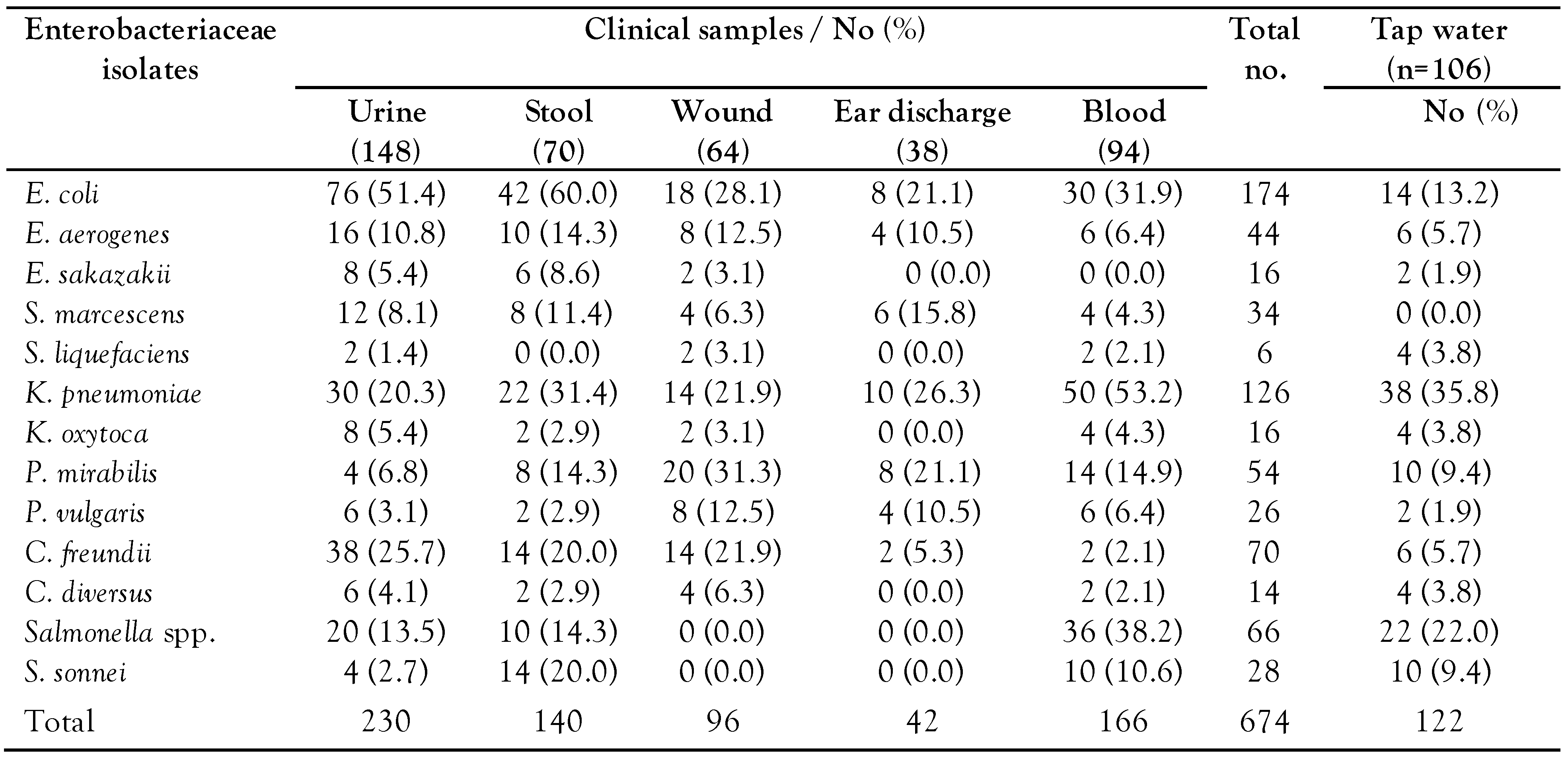
Table 1. Distribution of Enterobacteriaceae isolates in clinical and tap water samples.
Germs 13 00050 i001
Table 2. Prevalence of extended-spectrum, metallo and ampC beta (β)-lactamases producers in clinical and tap water samples.
Germs 13 00050 i002
Table 3a. Antibiotic resistance of clinical isolates, and ESBL, MBL, AmpC beta-lactamases producing Enterobacteriaceae isolates.
Germs 13 00050 i003a
Table 3b. Antibiotic resistance of tap water isolates, and ESBL, MBL, AmpC beta-lactamases producing Enterobacteriaceae isolates.
Germs 13 00050 i003b
Table 4. Multidrug resistant isolates, and ESBL, MBL, AmpC-βL-producing Enterobacteriaceae isolates.
Germs 13 00050 i004
Table 5. Beta-lactamase genes in Enterobacteriaceae isolates from clinical and water samples
Germs 13 00050 i005