Next Article in Journal
Actinotignum schaalii Infection—A Case Series From a Tertiary Hospital in Greece
Previous Article in Journal
Antimicrobial Resistance Pattern in Clinical Escherichia coli and Pseudomonas aeruginosa Isolates Obtained From a Secondary-Care Hospital Prior to and During the COVID-19 Pandemic in Kuwait
 
 
GERMS is published by MDPI from Volume 15 Issue 4 (2025). Previous articles were published by another publisher in Open Access under a CC-BY (or CC-BY-NC-ND) licence, and they are hosted by MDPI on mdpi.com as a courtesy and upon agreement with the former publisher Infection Science Forum.
Article

Association Between Vitamin D Receptor Gene Polymorphisms and Fibrosis Susceptibility in Greek Patients with HCV Infection

by
Angeliki Anna Beka
1,*,
Vasileios Papadopoulos
2,
Theodora Mylopoulou
3,
Maria Panopoulou
4,
Ioannis Karakasiliotis
5,
Penelope Mavromara
1,†,
Konstantinos Mimidis
3,† and
Stavroula Veletza
5,†
1
Laboratory of Biochemistry and Molecular Virology, Department of Molecular Biology and Genetics, Democritus University of Thrace, Dragana, 68100 Alexandroupolis, Greece
2
ENARGEIA Medical Ltd., Michael Karaoli 8 & Elpidos 6, 67131 Xanthi, Greece
3
First Department of Internal Medicine, University General Hospital of Alexandroupolis, Dragana, 68100, Alexandroupolis, Greece
4
Laboratory of Microbiology, Department of Medicine, Democritus University of Thrace, Dragana, 68100 Alexandroupolis, Greece
5
Laboratory of Biology, Department of Medicine, Democritus University of Thrace, Dragana, 68100 Alexandroupolis, Greece
*
Author to whom correspondence should be addressed.
Co-last authors.
GERMS 2022, 12(3), 384-393; https://doi.org/10.18683/germs.2022.1342 (registering DOI)
Submission received: 11 June 2022 / Revised: 18 August 2022 / Accepted: 27 August 2022 / Published: 30 September 2022

Abstract

Introduction Hepatitis C virus (HCV) infection is a prime cause of chronic hepatitis worldwide, that often silently progresses to fibrosis, cirrhosis and hepatocellular carcinoma (HCC). Notably, the majority of individuals infected with HCV develop symptoms at late stages, often associated with liver damage that cannot revert after virus clearance. Thus, current antiviral therapy alone is rather insufficient to eliminate the global burden of HCV in the near future. During the past few years, vitamin D deficiency as well as certain single nucleotide polymorphisms in the vitamin D receptor (VDR) gene have been associated with liver fibrosis. Therefore, the aim of the present study was to investigate the possible correlation between VDR polymorphisms ApaI (rs7975232) and TaqI (rs731236) and the fibrosis stage of patients with HCV infection from Thrace, Greece. Methods Eighty-one patients with HCV infection underwent transient elastography for the assessment of their fibrosis stage, and PCR-restriction fragment length polymorphism (RFLP) genotyping for VDR ApaI and TaqI polymorphisms. VDR genotypes were then statistically associated with the patients’ fibrosis stage using ordinal regression models. Results Non-cirrhotic stages were positively correlated with TaqI TT genotype (p=0.003) and negatively correlated with TaqI TC genotype (p=0.007). In the presence of Hardy-Weinberg equilibrium and linkage disequilibrium between the two VDR polymorphisms, mild fibrosis stages (F0-2) were correlated with ApaI/TaqI GG/TT (p=0.002) and TG/TT (p=0.008) genotypes, while cirrhotic stage F4 was associated with ApaI/TaqI TG/TC genotype (p=0.038). Conclusions TaqI TT and ApaI/TaqI GG/TT, TG/TT and TG/TC genotypes could be explored as prognostic genetic markers for fibrosis susceptibility in HCV patients.

Introduction

Hepatitis C virus (HCV) infection is one of the main causes of chronic hepatitis, which often progresses to liver fibrosis, cirrhosis and hepatocellular carcinoma (HCC) formation. According to the World Health Organization’s (WHO) most recent report [1], 71 million people worldwide suffer from chronic HCV infection. In combination with the nearly 400.000 deaths annually attributed to this disease, HCV infection is undoubtedly a major public health problem [2].
The revolutionary therapy with direct antiviral agents (DAAs) has presented encouraging results, with a cure rate of 95-100% of diagnosed patients. However, the estimated multitude of unaware infected individuals keeps raising a concern on the successful eradication of HCV, as planned by the WHO. Furthermore, it is crucial to point out what multiple studies have shown, i.e., that advanced liver damage caused by HCV could progress to HCC even upon virus clearance achieved by treatment with DAAs [3]. The urgent need of a preventive vaccine has not been met yet [2].
Viral pathogenicity and its effect on patients’ livers are not only influenced by the viral genotype, with HCV genotype 3 being correlated with increased fibrosis progression [4], but also by the host’s genetic variability. Multiple studies have associated the host’s genetic heterogeneity with response to interferon-based therapy, steatosis development and fibrosis progression [5,6].
Over the past years, vitamin D has been assigned many roles, additional to its traditional ones in mineral and skeletal homeostasis, resulting to its pleiotropic theory of action. Vitamin D’s effects are mainly mediated by its receptor (vitamin D receptor, VDR) [7]. Both low levels of the active 1,25(OH)2D3 form of vitamin D and certain VDR polymorphisms have been found to be involved in several diseases, such as cardiovascular, skeletal, autoimmune, neurodegenerative, liver disease and cancer [8]. Notably, VDR polymorphisms affect the receptor’s affinity to the active form of the vitamin [9], whereas there is a debate whether they may also impact vitamin D plasma levels [10].
Two of the most well-studied single nucleotide polymorphisms (SNPs) within VDR are ApaI (rs7975232) and TaqI (rs731236). ApaI (G>T) is situated in the intron area right before exon 9, while TaqI (T>C) is situated in the beginning of exon 9 and is part of VDR’s ligand binding domain [9]. The SNP generates synonymous codons of isoleucine. Due to the close proximity of these polymorphisms, strong linkage disequilibrium (LD) among them has been established early on [11]. Although it is not yet clear how an intron polymorphism and/or a silent polymorphism could be related to a gene’s actions, both ApaI and TaqI VDR polymorphisms have been associated with many diseases, chronic hepatitis C included [5,12,13,14,15].
The direct linking of VDR to liver fibrosis was established with the work of Ding et al. [16], which also shed some light on the underlying mechanism. Upon injury, fibrosis is generally induced by hepatic stellate cells (HSCs) activation, whose fibrotic effect is promoted through the TGFβ1/SMAD pathway, by chromatin remodeling and induction of profibrotic gene expression. That study demonstrated that the TGFβ1/SMAD induced chromatin remodeling increases the accessibility of VDR to adjacent vitamin D response elements. Subsequently, the liganded VDR occupies SMAD Binding Sites and inhibits the acetylation of histone H3, leading to the suppression of SMAD- dependent pro-fibrotic gene expression. Their work also concluded that (i) treatment with VDR ligands inhibits TGFβ induced HSCs activation and abolishes liver fibrosis in vivo, (ii) VDR knockout mice develop liver fibrosis automatically and (iii) both VDR alleles are essential for the preservation of the natural hepatic structure.
However, although both VDR alleles are indispensable in order to avoid irregular fibrogenesis, the genetic composition of these alleles, especially with respect to SNPs, remains unknown. Hence, our aim was to investigate the possible correlation between VDR ApaI and TaqI polymorphisms and fibrosis stage in patients with HCV infection from the northernmost region of Greece, Thrace.

Methods

Patient characteristics

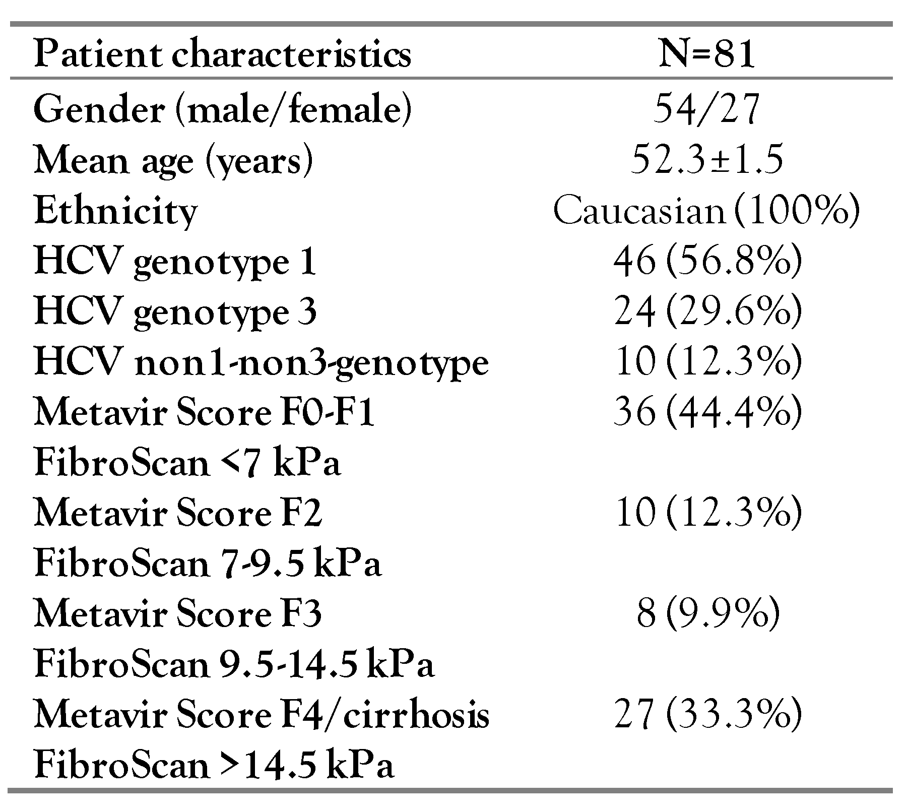
Eighty-one patients with untreated HCV infection were included in this non-invasive retrospective cohort study, all followed at the outpatient liver clinic of the University General Hospital of Alexandroupolis, Thrace (Table 1). All patients, inhabitants of Thrace, were between the ages of 18 and 70 years old and checked for HCV antibody. Only chronically infected patients were included, with the chronicity of the disease and the HCV genotype being checked by detectable HCV RNA by RT-PCR. Fibrosis stage was evaluated by transient elastography (FibroScan, Echosense, France). Mean values (F0-F1 <7 kPa, F2=7-9.5 kPa, F3=9.5-14.5 kPa, F4>14.5kPa) were calculated on the basis of 10 measurements performed on the right liver lobe using an intercostal approach by a trained physician [17]. Patients who had undergone liver transplantation, had exhibited spontaneous clearance, acute HCV infection or co-infection with HBV/HDV and/or HIV were excluded.
The study was approved by the local ethics committee following the guidelines of the Declaration of Helsinki. Written informed consent was obtained from every patient enrolled in this study.

DNA isolation and genotyping of ApaI and TaqI polymorphisms

Patients’ DNA isolation from whole blood samples was performed using the NucleoSpin Blood kit (Macherey-Nagel, Germany). Genotyping was performed by PCR amplification and restriction fragment length polymorphism analysis (PCR-RFLP).
PCR generated multiple copies of a 695 bp sequence, containing both ApaI and TaqI polymorphisms. The primers used were custom designed (forward: 5’-CAGAGCATGGACAGGGAGCAA, reverse: 5’- AAGGGGTTAGGTTGGACAGG) and the annealing temperature was set to 54 °C. Each sample was submitted into two separate restriction reactions, one with ApaI enzyme (New England Biolabs, USA) at 25 °C and another with TaqI enzyme (New England Biolabs) at 65 °C, both for 3 hours. Each patient’s genotype was determined by 2% agarose (agarose for routine use, Sigma-Aldrich, Germany) gel electrophoresis of the digested samples (Supplementary Figure S1). To verify these results, 10% of the samples were re-evaluated by collaborating laboratory personnel and 12% of randomly selected samples were sequenced by an external laboratory. In both cases the results were consistent with those of the original experiment.

Statistical analysis

Chi-square was used for comparison between expected and observed frequencies of ApaI and TaqI genotypes. In case of residuals smaller than 5, the exact Fisher’s test was alternatively used. Student’s t-test was used for comparison of continuous variables between two groups, given that Levene test failed to reject the null hypothesis of equality of variances between groups. ANOVA was used for comparison of continuous variables between three or more groups; in case Levene test was statistically significant, Scheffé test was used to discriminate differences between groups, otherwise the Least Significant Difference (LSD) test was used. Ordinal regression was used to predict an ordinal dependent variable given one or more independent variables. Multinomial regression was preferred to predict the probabilities of the different possible outcomes of a categorical dependent variable with more than two possible discrete outcomes, given a set of independent variables. Mean values are given with at least two significant digits, accompanied by their standard deviations. The level of statistical significance was set to p=0.05. All statistical analysis was performed using the SPSS 26.0 software package (IBM Corp., USA).

Analysis of Hardy-Weinberg Equilibrium

The observed genotype frequencies of ApaI and TaqI polymorphisms were used to compute the allele frequencies and thus the expected genotype frequencies based on Hardy-Weinberg equilibrium (HWE). In all cases the null hypothesis that HWE is met cannot be rejected. ApaI and TaqI polymorphisms are known to be in strong LD [11], which was also evaluated in our cohort.

Results

Patient characteristics

Eighty-one patients with untreated HCV infection, 54 males and 27 females, of mean age 52.3±1.5 years were enrolled in the study. No difference was documented between age, gender and HCV genotype as far as both ApaI (p=0.442, p=0.656 and p=0.392, respectively) and TaqI (p=0.326, p=0.861 and p=0.512, respectively) polymorphisms are concerned (Table 1).
The majority of the patients (86.4%) were infected with genotypes 1 and 3, while the rest of the studied cohort were infected by HCV genotypes 2 and 4. Concerning comorbidities, active alcohol consumption was generally low in the studied cohort (<10 g/d) and only 2 patients consumed >20 gr/d, while only 3 patients were diagnosed with type II diabetes with good glycemic control.

Hardy-Weinberg Equilibrium and Linkage Disequilibrium

Genotypic and allelic frequencies can be found in Supplementary Table S1. ApaI/TaqI haplotype frequencies in heterozygotes and double heterozygotes were estimated using maximum probability derived from observed frequencies of GG/TT (14/81 or 0.1728 instead of expected: 0.0619, p=0.0276), TT/CC (21/81 or 0.2593 instead of expected: 0.1088, p=0.0122), and TT/TT (2/81 or 0.0247 instead of expected: 0.1503, p<0.001). Thus, the observed haplotype frequencies were counted to be 0.0751 for TT (expected 0.3425, p<0.001), 0.5089 for TC (expected 0.2809, p=0.001), 0.4160 for GT (expected 0.2069, p<0.001) and 0 for GC (expected 0.1697, p<0.001). Based on the above-mentioned data, LD parameter D approaches its maximum absolute value of 0.25 (D= −0.2117, p<0.001). Therefore, as HWE is met, ApaI and TaqI are closely linked and intense LD exists in our group of subjects as well.

VDR genotype association with fibrosis stage

Using an ordinal regression model incorporating fibrosis level (F0-F4, F0-F1<7 kPa, F2=7-9.5 kPa, F3=9.5-14.5 kPa, F4>14.5 kPa) as ordinal dependent variable along with ApaI or TaqI genotype, age, gender and HCV genotypes as independent ones, F0-F1 (p=0.019), F2 (p=0.007) and F3 (p=0.003) were positively associated with TT TaqI genotype (p=0.003) and the male gender (p=0.019) and negatively associated with TC TaqI genotype (p=0.007) and increased age (p<0.001). No statistically significant association was observed with the other genotypes although a trend was detected associating ApaI GG genotype with low fibrosis stages (Figure 1).
Therefore, an ordinal regression model incorporating mild (F0-F2) and intense (F3-F4) fibrosis as ordinal dependent variable along with the absence of T allele in ApaI genotype or with the absence of C allele in TaqI genotype as independent ones, mild fibrosis was positively associated with absence of T allele in ApaI (p=0.028) and the absence of C in TaqI (p=0.012) genotype (Figure 2).
As strong LD is met between ApaI and TaqI, joint data regarding fibrosis can be used for further evaluation. Using an ordinal regression model incorporating fibrosis level (F0-F4) as ordinal dependent variable along with ApaI/TaqI paired genotypes as independent ones, both F0-F1 (p=0.002) and F2 (p=0.043) were found to be positively associated with presence of GG/TT (p=0.002) and TG/TT p=0.008) ApaI/TaqI genotypes. Chi-square testing further revealed an association of ApaI/TaqI double heterozygotes (TG/TC) with the cirrhotic stage, F4 (p=0.038)–Figure 3.
Furthermore, ApaI/TaqI polymorphisms are differently distributed (p=0.010) between patients with mild and those with extended fibrosis (F0-F2 versus F3-F4). Applying an ordinal regression model incorporating mild and intense fibrosis as ordinal dependent variable and ApaI/TaqI genotypes, age, gender and HCV genotypes as independent ones, mild fibrosis was negatively correlated with increased age (p<0.001) and positively correlated with presence of GG/TT (p=0.007) and TG/TT (p=0.022) ApaI/TaqI genotypes.
Notably, applying multinomial logistic regression, neither ApaI nor TaqI polymorphisms were demonstrated to be independently associated with excess alcohol intake (p=0.474 for ApaI; p=0.140 for TaqI), BMI≥30 (p=0.062 for ApaI; p=0.159 for TaqI), and diabetes (p=0.513 for ApaI; p=0.973 for TaqI). Similarly, as far as the combined ApaI/TaqI genotypes are concerned, no independent association was revealed with either alcohol intake (p=0.559), or BMI≥30 (p=0.209), or diabetes (p=0.588). Moreover, multinomial logistic regression was applied to test the null hypothesis that HCV genotypes are comparable between ApaI/TaqI polymorphisms and their haplotypes; indeed, HCV genotypes were not associated with ApaI polymorphism (p=0.309), TaqI polymorphism (p=0.380), or ApaI/TaqI haplotypes (p=0.383).

Discussion

During the past few years, VDR gene polymorphisms have been associated both with fibrosis and HCC development in chronic HCV patients. VDR GG ApaI genotype and GGT bAt (BsmI, ApaI, TaqI) haplotype have been found to be predisposing factors of HCC development in Egyptian, Taiwanese and Italian populations [14,15,18,19]. Baur et al. have associated the same genotype and haplotype with rapid fibrosis progression in Swiss patients with HCV infection [5,13], while Scalioni et al. found that BsmI and TaqI polymorphisms induce fibrosis in a cohort of Rio De Janeiro citizens [6]. On the contrary, Thanapirom et al. examined the possible association of ApaI and TaqI genotypes [20], as well as the bAt haplotype and found no correlation with advanced fibrosis.
In the present study, we found a strong association between (i) TaqI TT genotype and non-cirrhotic fibrosis stages, (ii) the absence of ApaI or TaqI alternative alleles and low fibrosis stages F0 and F2 independently, (iii) ApaI/TaqI GG/TT & TG/TT genotypes and mild fibrosis stages F0-F2 and (iv) both TaqI TC genotype alone and together with ApaI TG genotype (ApaI/TaqI double heterozygote, TG/TC) and cirrhotic stages of liver damage. In the studied cohort, advanced age and male gender were found to be predisposing factors of increased fibrosis. Importantly, the observed correlations are independent of confounding factors, such as alcohol intake, BMI and diabetes, further supporting our results.
The reference allele genotypes, which we found to be associated with low fibrosis stages, were associated with rapid fibrosis progression in the studies of Baur et al. and Scalioni et al [5,6]. To support our results, the alternative allele genotypes (ApaI TT, TaqI CC) were associated with severe cirrhosis within a Greek population presenting cirrhosis of various etiology, studied by Triantos et al [12]. Furthermore, the distinct ancestry of the studied population within each study should be taken into account, especially since the Thracian population is composed of people originated from the Balkan Peninsula and the Black Sea region. Moreover, an interesting aspect of El-Edel’s study is the distribution on ApaI alleles and genotypes between cirrhotic and HCC patients [14]. ApaI reference allele and genotype outbalance T allele and TT genotype in the HCC group, while the exact opposite trend is observed in the group of cirrhotic patients. Similar results were obtained by Rowida et al. in a study performed in Egypt [21]. The low representation of TaqI CC genotype in cirrhotic patients, which is consistent with our results, and its strong association with HCC development, could suggest a role of the mutated VDR in triggering HCC formation, while on the other hand being protective of advanced fibrosis and cirrhosis.
The molecular mechanisms of vitamin D mediated liver protection against fibrosis are very elegantly explained in a review by W. Udomsinprasert and J. Jittikoon [22]. Although the role of VDR in the liver is also thoroughly described, no mechanism explaining the association of VDR polymorphisms with fibrosis is presented. How could a silent polymorphism, such as VDR TaqI, affect fibrogenesis? Agliardi et al. discovered that peripheral blood mononuclear cells (PBMCs) treated with myelin basic protein (MBP) carrying TaqI TT or TC genotype express VDR mRNA significantly more than CC MBP treated PBMCs [23]. Similar results were also presented by Gisbert-Ferrándiz et al [24], who studied the VDR protein and mRNA levels in patients with Crohn’s disease and found them decreased in patients with VDR TaqI CC genotype.
Based on the above-mentioned data and the findings of our study, we suggest the following theory to biologically explain our results (Figure 4). In patients with HCV infection with VDR TaqI TT genotype, VDR expression is enhanced, as suggested by Agliardi et al [23]. The elevated VDR levels could more sufficiently antagonize the TGFβ1/SMAD pathway, as suggested by Ding et al. [16], inhibiting fibrosis. In case this hypothesis is correct, the VDR agonist compound Ding has claimed as potential antifibrotic treatment would probably be less efficient in patients with VDR TaqI CC genotype [25].
Our study presents two main limitations: the small, although statistically sufficient, number of patients enrolled and its retrospective nature, with the latter only allowing us an estimation of duration of the chronic HCV infection.

Conclusions

In conclusion, we have identified TaqI TT genotype alone and ApaI/TaqI GG/TT & TG/TT double genotypes as protective of advanced fibrosis and the TaqI or double ApaI/TaqI heterogeneity as a predisposing factor of advanced fibrosis/cirrhosis in a cohort of patients with HCV infection from Thrace, Greece. Nonetheless, the biological pathways disturbed by VDR, expressed from a gene barring either of these SNPs, and their repercussions call for further investigation, to become understood to a greater degree and to possibly allow the utilization of these SNPs as genetic markers for fibrosis susceptibility in patients with HCV infection in the future.

Supplementary Materials

The following supporting information can be downloaded at: https://www.mdpi.com/article/doi/s1.

Author Contributions

Conception and design: AAB, MP, PM, KM, SV. Informed consent collection/interaction with patients: TM, KM. Data collection: AAB, TM, KM. Analysis and interpretation of the data: AAB, VP, TM, MP, IK, PM, KM, SV. Drafting of the article: AAB, VP. Critical revision of the article for important intellectual content: IK, PM, KM, SV. Final approval of the article: PM, KM, SV. All authors read and approved the final version of the manuscript.

Funding

This research received no external funding.

Acknowledgments

The authors would like to thank Dr. Elisavet Gatzidou for her contribution in the organization of the biobank of patients’ blood samples. We devote a great thank you to Eftychia Filippopoulou and Emmanouil. Cheilas for their assistance in part of the experimental procedures, as well as to all the members of the Laboratory of Biochemistry and Molecular Virology and the Laboratory of Biology for their help.

Conflicts of Interest

The authors declare no conflict of interest.

References

  1. World Health Organization. Global Hepatitis Programme. 2017. Global hepatitis report, 2017. Available online: http://apps.who.int/iris/bitstream/10665/255016/1/9 789241565455-eng.pdf?ua=1 (accessed on 15 May 2019).
  2. Bartenschlager, R.; Baumert, T.F.; Bukh, J.; et al. Critical challenges and emerging opportunities in hepatitis C virus research in an era of potent antiviral therapy: Considerations for scientists and funding agencies. Virus Res. 2018, 248, 53–62. [Google Scholar] [CrossRef]
  3. Aleman, S. The hurdle with remaining risk for hepatocellular carcinoma in cirrhotic patients after a hepatitis C cure. Hepatol Med Policy 2016, 1, 11. [Google Scholar] [CrossRef]
  4. Rüeger, S.; Bochud, P.Y.; Dufour, J.F.; et al. Impact of common risk factors of fibrosis progression in chronic hepatitis C. Gut. 2015, 64, 1605–1615. [Google Scholar] [CrossRef]
  5. Baur, K.; Mertens, J.C.; Schmitt, J.; et al. Combined effect of 25-OH vitamin D plasma levels and genetic vitamin D receptor (NR 1I1) variants on fibrosis progression rate in HCV patients. Liver Int. 2012, 32, 635–643. [Google Scholar] [CrossRef] [PubMed]
  6. Scalioni, L.P.; Santos, B.R.D.; Spritzer, P.M.; et al. Impact of vitamin D receptor and binding protein gene polymorphisms in clinical and laboratory data of HCV patients: Cross sectional study. Medicine 2018, 97, e9881. [Google Scholar] [CrossRef] [PubMed]
  7. Haussler, M.R.; Whitfield, G.K.; Haussler, C.A.; et al. The nuclear vitamin D receptor: Biological and molecular regulatory properties revealed. J Bone Miner Res. 1998, 13, 325–349. [Google Scholar] [CrossRef] [PubMed]
  8. Pludowski, P.; Holick, M.F.; Pilz, S.; et al. Vitamin D effects on musculoskeletal health, immunity, autoimmunity, cardiovascular disease, cancer, fertility, pregnancy, dementia and mortality-A review of recent evidence. Autoimmun Rev. 2013, 12, 976–989. [Google Scholar] [CrossRef]
  9. Gezen-Ak, D.; Dursun, E.; Ertan, T.; et al. Association between vitamin D receptor gene polymorphism and Alzheimer’s disease. Tohoku J Exp Med. 2007, 212, 275–282. [Google Scholar] [CrossRef]
  10. Wang, T.J.; Zhang, F.; Richards, J.B.; et al. Common genetic determinants of vitamin D insufficiency: A genome-wide association study. Lancet. 2010, 376, 180–188. [Google Scholar] [CrossRef]
  11. Morrison, N.A.; Qi, J.C.; Tokita, A.; et al. Prediction of bone density from vitamin D receptor alleles. Nature 1994, 367, 284–287. [Google Scholar] [CrossRef]
  12. Triantos, C.; Aggeletopoulou, I.; Kalafateli, M.; et al. Prognostic significance of vitamin D receptor (VDR) gene polymorphisms in liver cirrhosis. Sci Rep. 2018, 8, 14065. [Google Scholar] [CrossRef]
  13. Rau, M.; Baur, K.; Geier, A. Host genetic variants in the pathogenesis of hepatitis C. Viruses 2012, 4, 3281–3302. [Google Scholar] [CrossRef]
  14. El-Edel, R.; Mostafa, M.; Montaser, B.; El-Hag Ali, Y. Study of Apa-I vitamin D receptor gene polymorphism in patients with hepatocellular carcinoma. Menoufia Med J. 2017, 30, 619. [Google Scholar] [CrossRef]
  15. Hung, C.H.; Chiu, Y.C.; Hu, T.H.; et al. Significance of vitamin D receptor gene polymorphisms for risk of hepatocellular carcinoma in chronic hepatitis C. Transl Oncol. 2014, 7, 503–507. [Google Scholar] [CrossRef] [PubMed]
  16. Ding, N.; Yu, R.T.; Subramaniam, N.; et al. A vitamin D receptor/SMAD genomic circuit gates hepatic fibrotic response. Cell 2013, 153, 601–613. [Google Scholar] [CrossRef] [PubMed]
  17. de Lédinghen, V.; Vergniol, J. Transient elastography (FibroScan). Gastroenterol Clin Biol. 2008, 32, 58–67. [Google Scholar] [CrossRef] [PubMed]
  18. Mohammed, M.A.; Omar, N.M.; Mohammed, S.A.; Deiab, A.G. The significance of vitamin D receptor gene polymorphisms for susceptibility to hepatocellular carcinoma in subjects infected with hepatitis C virus. Gastroenterol Hepatol. 2017, 7, 00246. [Google Scholar] [CrossRef]
  19. Cusato, J.; Boglione, L.; De Nicolò, A.; et al. Vitamin D pathway gene polymorphisms and hepatocellular carcinoma in chronic hepatitis C-affected patients treated with new drugs. Cancer Chemother Pharmacol. 2018, 81, 615–620. [Google Scholar] [CrossRef]
  20. Thanapirom, K.; Suksawatamnuay, S.; Sukeepaisarnjaroen, W.; et al. Genetic associations of vitamin D receptor polymorphisms with advanced liver fibrosis and response to pegylated interferon-based therapy in chronic hepatitis C. PeerJ 2019, 7, e7666. [Google Scholar] [CrossRef]
  21. Raafat Rowida, I.; Eshra, K.; El-Sharaby, R.; et al. Apa1 (rs7975232) SNP in the vitamin D receptor is linked to hepatocellular carcinoma in hepatitis C virus cirrhosis. Br J Biomed Sci. 2020, 77, 53–57. [Google Scholar] [CrossRef]
  22. Udomsinprasert, W.; Jittikoon, J. Vitamin D and liver fibrosis: Molecular mechanisms and clinical studies. Biomed Pharmacother. 2019, 109, 1351–1360. [Google Scholar] [CrossRef]
  23. Agliardi, C.; Guerini, F.R.; Saresella, M.; et al. Vitamin D receptor (VDR) gene SNPs influence VDR expression and modulate protection from multiple sclerosis in HLA-DRB1*15-positive individuals. Brain Behav Immun. 2011, 25, 1460–1467. [Google Scholar] [CrossRef]
  24. Gisbert-Ferrándiz, L.; Salvador, P.; Ortiz-Masiá, D.; et al. A single nucleotide polymorphism in the vitamin D receptor gene is associated with decreased levels of the protein and a penetrating pattern in Crohn’s disease. Inflamm Bowel Dis. 2018, 24, 1462–1470. [Google Scholar] [CrossRef]
  25. Ding, N.; Downes, M.; Liddle, C.; Evans, R.M.; Ding, N.; Downes, M.; et al. Vitamin D receptor/SMAD genomic circuit gates fibrotic response. US20160106762A1, 2016. Available online: https://patents.google.com/patent/US20160106762/en?oq=VITAMINDRECEPTOR%2fSMADGENOMIC+C IRCUIT+GATES+FIBROTC+RESPONSE (accessed on 16 May 2019).
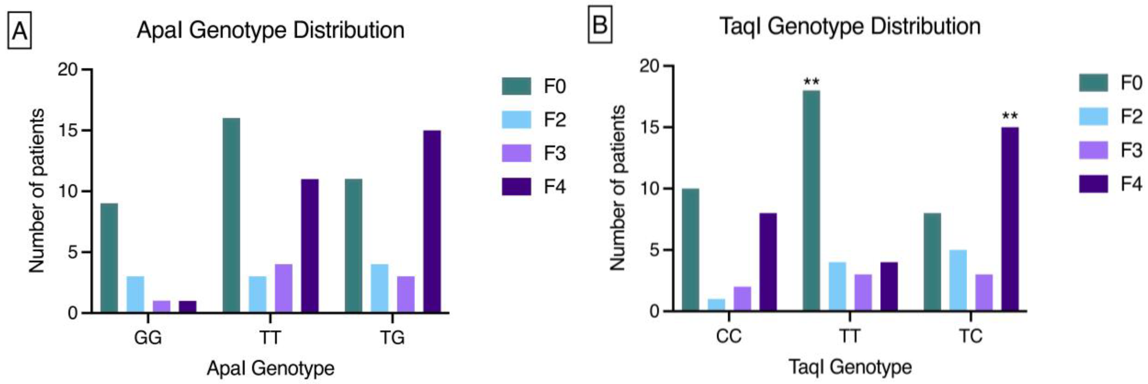
Figure 1. Distribution of ApaI genotype (Panel A) and TaqI genotype (Panel B) among patients with different levels of liver fibrosis (F0-F4). TaqI TT genotype is correlated with low fibrosis stage F0, while TaqI TC genotype is correlated with high fibrosis stage F4 (p=0.003 and p=0.007, respectively). F0<7 kPa, F2=7-9.5 kPa, F3=9.5-14.5 kPa, F4>14.5 kPa. **p<0.01.
Germs 12 00384 g001
Figure 2. Distribution of ApaI T allele (Panel A) and TaqI C allele (Panel B) among patients with low (F0-2) and high (F3-F4) liver fibrosis. ApaI GG and TaqI TT genotypes independently are correlated with low fibrosis stages F0-2 (p=0.016 and p=0.010, respectively). F0<7 kPa, F2=7-9.5 kPa, F3=9.5-14.5 kPa, F4>14.5 kPa. *p<0.05.
Germs 12 00384 g002
Figure 3. Distribution of ApaI/TaqI genotypes among patients with different stages of liver fibrosis (F0-4). Only pairs of genotypes with significant association with fibrosis stage are illustrated. ApaI/TaqI GG/TT and TG/TT genotypes are correlated with the lowest fibrosis stage F0 (p=0.002 and p=0.008, respectively), while double heterozygotes are correlated with cirrhosis (F4, p=0.038). F0<7 kPa, F2=7-9.5 kPa, F3=9.5-14.5 kPa, F4>14.5 kPa. **p<0.01.
Germs 12 00384 g003
Figure 4. Schematic representation of proposed mechanism of action of VDR gene and protein in fibrosis inhibition. As found by Ding et al. [16], upon liver injury, TGF-β binding to its receptor promotes phosphorylation of SMAD2 and SMAD3, further promoting hetero-trimerization with SMAD4, translocation to the nucleus and chromatin remodeling, increasing the accessibility of liganded VDR/Retinoid X Receptor (RXR) heterodimer to adjacent vitamin D response elements (VDREs). Subsequently, the liganded VDR also occupies SMAD Binding Sites (SBS) and inhibits the acetylation of histone H3, leading to the suppression of SMAD-dependent pro-fibrotic gene expression. The top left corner within the nucleus summarizes the findings of Agliardi et al. [23] and Gisbert-Ferrándiz et al. [24] concerning the increased VDR mRNA levels in the presence of TT or TC TaqI polymorphisms and the decreased VDR mRNA levels in the presence of TT or TC TaqI polymorphism, respectively. The combination of these findings is the mechanism we propose to explain the association we found between these VDR TaqI polymorphisms and lower fibrosis levels. Some illustrations used were derived from Servier Medical Art by Servier (smart.servier.com), licensed under a Creative Commons Attribution 3.0 Unported License.
Germs 12 00384 g004
Table 1. Cohort characteristics.
Germs 12 00384 i001