Introduction
Multisystem inflammatory syndrome in children (MIS-C) is a severe complication following the new coronavirus disease-2019 (COVID-19) occurring in childhood. The pathophysiology is not well understood, and it is suggested that it associates an abnormal immune
1response with certain clinical similarities to Kawasaki disease, macrophage activation syndrome, or cytokine release syndrome [
1]. The exact mechanisms whereby SARS-CoV-2 triggers this type of abnormal immune response are not clear. It has been hypothesized that patients with severe MIS-C present persistent immunoglobulin G (IgG) antibodies and a propensity for activation of monocytes and CD8+ T cells, coupled with persistent cytopenia, that differ from the findings regularly seen in acute COVID-19 [
2].
Although all perceptions and perspectives have shifted to COVID-19 inevitably during the pandemic, it is imperative to keep in mind other infectious diseases in the differential diagnosis. Careful examination of the patients for the signs and symptoms of other infectious diseases is crucial to excluding other plausible diagnoses. One of these signs is costovertebral angle (CVA) tenderness. Here, two cases admitted with MIS-C-like clinical features and one of whom showed COVID-19 enzyme-linked immunosorbent assay (ELISA) test positivity for IgG are presented. They were misdiagnosed as MIS-C in the first place. However, both cases had flank pain with CVA tenderness, and on further investigation, they were both diagnosed with acute focal bacterial nephritis (AFBN), an upper urinary tract infection (UTI).
Case reports
Case-1
An 8-year-old girl presented with fever for three days, headache, flank pain, vomiting, lethargy. There was no history of cough, difficulty in breathing, diarrhea, dysuria. Before presenting, she had experienced alteration in consciousness, not recognizing her parents for an hour. She had had contact with a COVID-19 patient about three weeks previously. She was a previously healthy child without medication, except for the history of enuresis and frequent lower UTI. Her family history was unremarkable. The physical examination revealed fever (39.2°C, axillary) and tachycardia (135 beats/minute). Her blood pressure was 105/64 mmHg (below the 90th percentile according to the age, gender and height of the patient). Respiratory sounds and rate were normal (20 breaths/minute). She had normal skin turgor, moist oral mucosa. No mucocutaneous skin lesion or edema was noted. There was no rigidity, rebound tenderness, or organomegaly, but bilateral CVA tenderness was remarkable in the abdominal examination. She had no growth retardation (height of 1.40 m with standard deviation score (SDS) 1.45 and weight of 30 kg with SDS 0.26).
On laboratory analyses, there was leukocytosis, thrombocytopenia, neutrophilia with high inflammatory markers (elevated C-reactive protein, fibrinogen, procalcitonin, and D-dimer levels) –
Table 1. The estimated glomerular filtration rate was calculated as 70 mL/min/1.73m [
2] by the Schwartz formula [
3]. Also, she had a high brain-natriuretic peptide (BNP) with normal troponin I levels. Urinalysis showed microscopic hematuria and (2+) proteinuria without nitrite or leukocyte esterase positivity. After obtaining urine and blood cultures, vancomycin (60 mg/kg/day in four divided doses), cefotaxime (150 mg/kg/day in three divided doses), and acyclovir (30 mg/kg/day in three divided doses) were started with intravenous route. Abdominal ultrasonography and echocardiography were found to be normal.
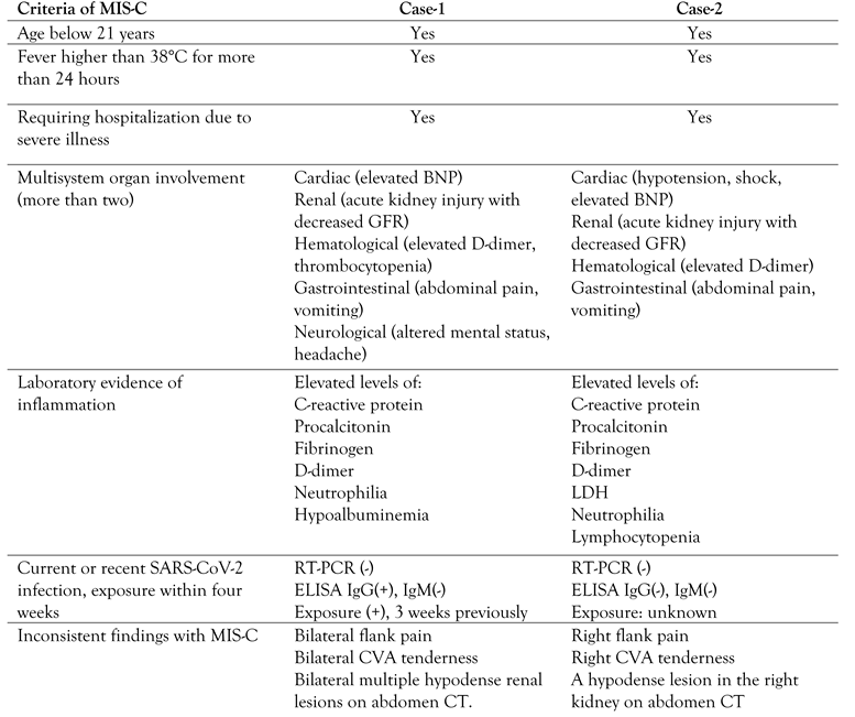
Meanwhile, the result of the SARS-CoV-2 RT-PCR test came back negative. However, the COVID-19 ELISA was positive for IgG and negative for IgM. Due to fever duration, clinically severe illness requiring hospitalization, multisystem organ involvement (cardiac, hematological, renal, gastrointestinal, neurological), she was diagnosed with MIS-C (
Table 2). Intravenous immunoglobulin (IVIG) (2 g/kg/day), enoxaparin sodium (1 mg/kg/day) and prednisolone (2 mg/kg/day) were started.
Case-2
A 16-year-old female patient presented to the hospital with fever for four days, right flank pain, headache, and vomiting. There was no cough, difficulty in breathing, diarrhea, dysuria. She had no known chronic disease, history of urinary tract infection. She had no known COVID-19 contact. Her family history was irrelevant. On physical examination, she had a fever (38.8°C, axillary), tachycardia (124 beats/minute), and hypotension (73/37 mmHg). Auscultation of the lungs was normal. Remarkable rebound tenderness and guarding in the right lower quadrant indicated acute abdomen. In addition, right CVA tenderness was found.
On laboratory analysis, she had leukocytosis, neutrophilia, lymphocytopenia, and high inflammatory markers (elevated C-reactive protein, procalcitonin, fibrinogen, lactate dehydrogenase, and D-dimer levels) –
Table 1. The estimated glomerular filtration rate was calculated as 82.5 mL/min/1.73m
2 by the Schwartz formula [
3]. Slightly elevated BNP with normal troponin I levels were also found. In urinalysis, there was microscopic hematuria, 2+ proteinuria, leukocyte esterase reaction. After obtaining urine and blood cultures, vancomycin (60 mg/kg/day in four divided doses), meropenem (60 mg/kg/day in three divided doses), and amikacin (15 mg/kg/day) were started. Due to hypotension, intravenous sodium chloride 0.9% bolus (20 cc/kg) was given twice. However, hypotension persisted, and noradrenalin infusion was started. Systolic functions were found as normal on echocardiography.
Since the patient had acute abdomen, oral intake was stopped entirely after pediatric surgery consultation. Abdominal ultrasonography did not reveal any pathology.
Meanwhile, SARS-CoV-2 RT-PCR and ELISA test results came back negative. Due to fever duration, clinically severe illness requiring hospitalization, multisystem organ involvement (cardiac, hematological, renal, gastrointestinal), she was diagnosed with MIS-C (
Table 2).
Clinical course of both cases
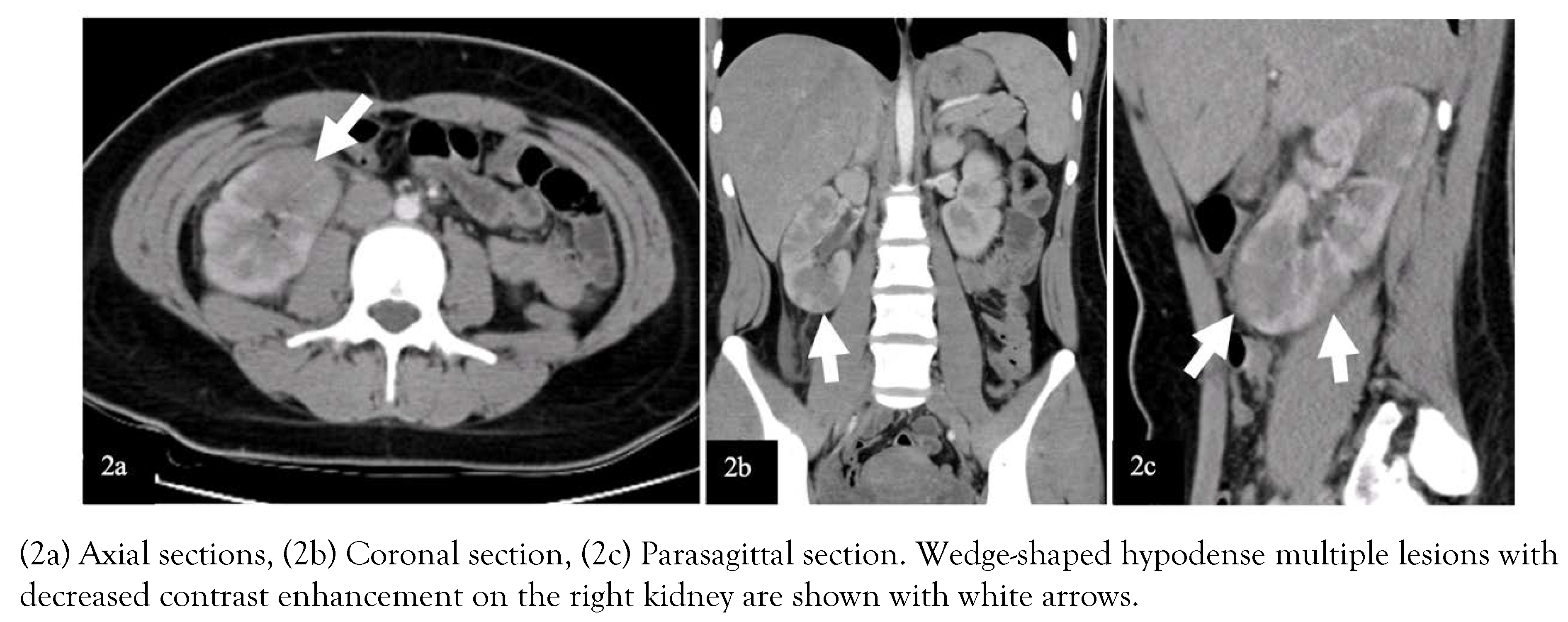
Due to the acute abdomen clinical finding in Case-2 and the significant CVA tenderness in both cases (bilateral in Case-1 and unilateral in Case-2), contrast-enhanced abdominal computed tomography (CT) was performed for differential diagnosis. In Case-1, nonhomogeneous hypodense mass-like lesions with decreased contrast enhancement in both kidneys were detected (
Figure 1). In Case-2, a similar lesion was seen in the lower pole of the right kidney (
Figure 2). Both cases were diagnosed with AFBN since these lesions were specific for it.
Case-1 was admitted to the pediatric nephrology inpatient clinics. The treatment was continued with intravenous cefotaxime and acyclovir at the doses mentioned above. Her general condition improved, and her fever regressed on follow-up. The midstream urine culture on admission resulted in no bacterial growth. Neurological impairment did not repeat during follow-up. Electroencephalogram (EEG) performed on the 7th day of hospitalization was found normal. After 14 days of hospitalization, she was discharged to continue oral cefixime (8 mg/kg/day) treatment for seven more days. The urodynamic study resulted in overactive bladder with decreased functional bladder capacity. The treatment was continued with antibiotic prophylaxis and oxybutynin. Voiding cystourethrography (VCUG) revealed grade 3 vesicoureteral reflux in the right kidney. Four months after the infection, a radionuclide scan with dimercaptosuccinic acid (DMSA) was performed showing no hypoactive scar lesions on both kidneys.
Case-2 was treated in the pediatric intensive care unit for two days. After resolution of hypotension and cessation of inotrope infusion, she was transported to the pediatric nephrology inpatient clinics. A DMSA scan was performed to support the diagnosis of AFBN, and it showed hypoactive lesions on the lower pole of the left kidney and the upper and lower poles of the right kidney. The midstream urine culture on admission showed Escherichia coli growth. The treatment was continued with only intravenous meropenem at the dose mentioned above during hospitalization for 14 days and peroral cefixime (8 mg/kg/day) treatment for 7 days. Then, antibiotic prophylaxis was started. The control DMSA and VCUG are planned, and waiting for the results.
Discussion
Some of the first reports of MIS-C emerged from the UK, of children with an unexplained multisystemic inflammation which has similar characteristics with Kawasaki disease and toxic shock syndrome [
4]. These cases are reported across the world with the spread of the COVID-19 pandemic. Most children affected had negative RT-PCR tests with high IgG antibody levels, indicating past infection [
5].
The diagnostic criteria of MIS-C have been described by the Centers for Disease Control and Prevention (CDC) as age below 21 years old, fever lasting more than 24 hours, high inflammatory markers, more than two system involvement, no plausible alternate diagnosis, COVID-19 exposure in the last four weeks, or evidence of past infection [
6]. Gastrointestinal and cardiac involvements are reported as common presentations [
7,
8]. Kidney involvement is reported between 10% to 46% in MIS-C patients [
9,
10]. Both presented cases had gastrointestinal and cardiac involvement in addition to renal and hematological involvement. Case-1 also had neurological system involvement (
Table 2). Therefore, Case-1 fulfilled the diagnostic criteria of MIS-C with clinical and laboratory aspects and with positive serology of SARS-CoV-2. However, Case-2 had no known COVID-19 contact, although clinical and laboratory characteristics suited MIS-C (
Table 2). In countries like Turkey, where COVID-19 incidence rates are very high, anyone can be considered in a suspected position about COVID-19 contact. Therefore, Case-2 had also been misdiagnosed with MIS-C.
Since the pandemic has spread all around the world with high contagion rates, uncertain prognosis, and newly isolated virus variants, the healthcare providers may become hyperalert on the diagnosis of MIS-C. However, MIS-C is a diagnosis of exclusion, and other severe infections may present similar clinical features. Therefore, clinicians should focus on unexpected findings and the findings incompatible with MIS-C to avoid overdiagnosis and delay in correct diagnosis [
11]. Careful evaluation of each case is mandatory. In the presented cases, flank pain with CVA tenderness was the unexpected finding, suggesting the possibility of a plausible alternative diagnosis (
Table 2).
Costovertebral angle tenderness is one of the specific indicators for kidney pathology and is often seen in acute pyelonephritis [
12]. However, it may present in other diseases such as nephrolithiasis, kidney abscess, vesicoureteral reflux, obstructive pathologies of the urinary tract, retrocecal appendicitis, retroperitoneal abscess [
13]. In a patient presenting with fever and CVA tenderness, the clinician should consider first upper UTI, among other diagnoses. In selected cases, imaging techniques can be used for differential diagnosis. Herein, due to the presence of CVA tenderness, we performed contrast-enhanced CT imaging, which showed hypodense wedge-shaped kidney lesions indicating AFBN (
Figure 1 and 2) [
14].
Acute focal bacterial nephritis (AFBN) or acute lobar nephronia is a rare bacterial UTI that causes non-liquefactive inflammatory lesions localized to the cortex of the kidneys and is considered to be located in a spectrum between acute pyelonephritis and renal abscess. It mainly occurs as ascending infection in children with urinary tract anomalies, although reported also in healthy children. The incidence is 8.6% in children with febrile UTI and 19.2% in healthy children with first febrile UTI [
14,
15].
Persistent fever, severe flank pain/ abdominal pain, and rapid deterioration of the general condition are remarkable symptoms of AFBN. Laboratory analysis shows intense inflammation with leukocytosis, neutrophilia, and high acute phase reactant levels [
16]. These clinical and laboratory findings are all compatible with the criteria of MIS-C and may cause the misdiagnosis with MIS-C. However, a careful anamnesis indicating predisposing features to UTI (like enuresis and frequent UTI in Case-1) and physical examination revealing specific symptoms of upper UTI (like CVA tenderness) help the differential diagnosis.
Pyuria, bacteriuria, or bacterial isolation in urine culture may not be detected in 5-10% of AFBN patients since the infection does not involve the calyceal system [
17]. Therefore, these laboratory findings may not help in differential diagnosis with MIS-C, as Case-1 presented in this report.
Early diagnosis and effective treatment are essential in terms of preventing renal scars and morbidity such as hypertension, proteinuria, and renal failure [
18]. The diagnosis of AFBN is based on radiologic examinations. In many cases, there is no finding on kidney ultrasonography (USG), although it may show nephromegaly, focal lesions with poorly defined irregular margins. Contrast-enhanced abdomen CT is the gold standard imaging technique for the diagnosis of AFBN. Herein, both cases had no finding on kidney USG. The lesions were observed on CT as the typical wedge-shaped, poorly defined hypodense lesions after contrast-medium administration [
14]. The DMSA scan of the kidneys may help in the diagnosis of AFBN and the detection of renal scarring during follow-up [
19]. In Case-1, the diagnosis of AFBN was supported with the DMSA scan. In Case-2, the DMSA scan in follow-up shows no scars indicating the effective treatment.
In the treatment of AFBN it is recommended to continue with intravenous antibiotics until the fever response is seen. The duration of antibiotic therapy can be planned as two or three weeks, depending on the presence of complicated lesions (microabscess formation, liquefaction areas) on CT imaging [
14,
19]. Renal abscess formation develops in approximately 25% of patients who are not given appropriate and adequate antibiotic therapy [
20]. In the presented cases, we preferred to continue the antibiotic treatment for three weeks (intravenously for two weeks and peroral for one week). We didn’t see progression to renal abscess.


