Next Article in Journal
Treatment Initiation or Switch to Bic/FTC/Taf – Real-World Safety and Efficacy Data from Two HIV Centers in Romania
Previous Article in Journal
COVID-19 Health Literacy, Coping Strategies and Perception of COVID-19 Containment Measures Among Community Members in a Southwestern State in Nigeria
 
 
GERMS is published by MDPI from Volume 15 Issue 4 (2025). Previous articles were published by another publisher in Open Access under a CC-BY (or CC-BY-NC-ND) licence, and they are hosted by MDPI on mdpi.com as a courtesy and upon agreement with the former publisher Infection Science Forum.
Article

Prevalence of Antimicrobial Resistance in Gram-Negative Clinical Isolates from a Major Secondary Hospital in Kuwait: A Retrospective Descriptive Study

by
Walid Q. Alali
1,*,
Wadha AlFouzan
2,3 and
Rita Dhar
2
1
Department of Epidemiology & Biostatistics, Faculty of Public Health, Kuwait University, Hawalli 13060, Kuwait
2
Microbiology Unit, Department of Laboratories, Farwaniya Hospital, Farwaniya 85000, Kuwait
3
Department of Microbiology, Faculty of Medicine, Kuwait University, Jabriya 85000, Kuwait
*
Author to whom correspondence should be addressed.
GERMS 2021, 11(4), 498-511; https://doi.org/10.18683/germs.2021.1285
Submission received: 8 August 2021 / Revised: 23 October 2021 / Accepted: 23 October 2021 / Published: 29 December 2021

Abstract

Introduction: Building an antimicrobial resistance (AMR) surveillance system in a country requires analysis of available data on AMR in clinical isolates. This study’s objective was to determine the AMR prevalence of Gram-negative bacterial (GNB) isolates cultured from clinical specimens at a major general hospital in Kuwait. Methods: A retrospective descriptive study was conducted on AMR profiles of GNB clinical isolates (n=5290) between January and December 2018. Data were extracted from the laboratory information system in the hospital. The GNB organisms (i.e., Escherichia coli, Klebsiella pneumoniae, Pseudomonas aeruginosa, and Acinetobacter baumannii) were isolated from five main locations at the hospital (i.e., intensive care units [ICUs], surgical wards, medical wards, pediatric wards, and outpatient polyclinics). Results: Overall, high AMR prevalence (>50%) against ampicillin, cefuroxime, cefotaxime, ceftazidime, ciprofloxacin, and trimethoprim/sulfamethoxazole, were observed across the GNB organisms. However, low resistance prevalence (<10%) were observed against amikacin, ertapenem, meropenem, and piperacillin/tazobactam. In general, AMR prevalence in E. coli isolates from ICU, medical and surgical wards was significantly (p<0.05) higher compared to other locations, whereas, AMR prevalence in P. aeruginosa isolates from pediatric ward was higher (p<0.05) compared to other locations. The overall multidrug resistance (MDR) prevalence was 38.7% (95% CI: 37.4-40.0). The highest MDR prevalence was among E. coli isolates from respiratory specimens (48%); wounds, bones, or other tissues (47.7%); and body fluids (47.1%). Similarly, MDR prevalence in K. pneumoniae, P. aeruginosa, and A. baumannii isolated from respiratory specimens was significantly (p<0.05) higher compared to other specimen types. The most frequent MDR phenotypes in the four GNB organisms and across the different specimen types included three antimicrobial drug classes: penicillins, cephalosporins, and fluroquinolones. Conclusions: Our findings demonstrate high AMR prevalence among common Gram-negative bacteria at this major hospital. Monitoring data on antimicrobial susceptibility of common bacterial organisms is critical for assessing trends in AMR at hospitals and for informing policy decisions.

Introduction

Antimicrobial resistance (AMR) is a major public health challenge in both developed and developing countries. Infections caused by AMR bacteria can lead to health complications such as extended hospitalization, treatment failure, and deaths. The misuse of antibiotics in healthcare facilities the inadequate compliance with infection control programs; the lack of surveillance systems to track both unique and common resistant phenotypes have contributed to development and emergence of AMR bacteria. The World Health Organization (WHO) has initiated the Global Action Plan on Antimicrobial Resistance which included, among other pillars, the increased knowledge about AMR through surveillance and data collection [1]. Therefore, one of the first steps in building an AMR surveillance system in a country is to explore and analyze available data resistance in clinical isolates from hospital settings as is the case in Kuwait.
Infections with AMR bacteria in the Gulf Cooperation Council (GCC) countries are prevalent [2]. For instance, infections caused by extended spectrum beta-lactamase (ESBL) producing bacteria, carbapenemase-producing bacteria, and multidrug resistant (MDR) Gram- negative organisms (GNB) have been reported [3]. Moreover, Yezli et al [4]. found that 60% to 90% of Klebsiella pneumoniae isolates were ESBL producers. Additionally, other studies reported that the majority of Acinetobacter spp. isolates associated with pneumonia were MDR and caused high morbidity and mortality [5].
In Kuwait, there are few studies that reported data on the prevalence, incidence, and/or trends in GNB clinical isolates from hospital settings [6,7,8,9]. These studies were limited by the number and diversity of the isolates, information on specimen type, location of the patient, and the number of antibiotics to which the organisms were tested for. The identification of unique and common resistance phenotypes in bacterial isolates obtained from patients is a step forward toward better understanding of epidemiology and distribution of AMR bacteria in hospital settings in Kuwait. Moreover, monitoring antimicrobial susceptibility data of common GNB organisms is needed to: 1) assess trends and changes in AMR bacteria, 2) determine risk factors related to AMR distribution, 3) evaluate existing and new intervention strategies, and 4) select effective antibiotic treatment. The objective of this retrospective study was to determine the prevalence and distribution of AMR patterns among clinical GNB isolates in relation to specimen type and patient location based on available data from Farwania hospital microbiological Laboratory Information System (LIS).

Methods

A retrospective descriptive study was conducted between January and December 2018. The data on AMR profiles of GNB clinical isolates were extracted from the microbiology LIS at Farwania hospital (a secondary care facility) in Kuwait. Antimicrobial susceptibility profiles of GNB isolated from patients across five main locations at the hospital (i.e., intensive care units [ICUs], surgical wards, medical wards, pediatric wards, and outpatient polyclinics) were analyzed. This general hospital is one of the major secondary care hospitals in Kuwait that offers healthcare services to nearly one-fourth of the country’s population. The hospital has 900 beds with multiple medical and surgical specialties including outpatient polyclinics. During the study period, all consecutive non-duplicated GNB isolates tested for antimicrobial susceptibility were used in this study. In order to differentiate hospital-acquired infection (HAI) from contamination, CDC protocols (for bloodstream, respiratory, and urinary tract infection) were adopted for different types of HAIs by the infection control team [10,11,12].
Isolates of presumed clinical significance (i.e., any patient case presented to the treating physician whether accompanied by signs/symptoms or at an early stage of a suspected infection) and as determined by the hospital infection control program were included. One isolate per bacterial species per patient was used for the analysis. The clinical information extracted from the LIS included patient identification number, patient’s location at the time of isolation, specimen type a) body fluids, b) blood, c) respiratory samples including sputum, tracheal aspirate, or bronchoalveolar lavage, d) wound, bone, or other tissues (such as external tissue infections around the eye, ear, and mouth), and e) urine; name of GNB isolates (Escherichia coli, Klebsiella pneumoniae, Pseudomonas aeruginosa, and Acinetobacter baumannii), and the antimicrobial resistance profile. The identity of the patients was removed prior to sharing the data with the authors. The demographic information (e.g., age, sex, nationality) and clinical diagnosis were not available. All the clinical information was confirmed by the laboratory technician and reconfirmed by the laboratory manager. The four common GNB bacteria were identified using the standard operational procedures used by the laboratory. Generally, the clinical specimens such as blood, respiratory samples, urine, and other tissues were cultured on 5% sheep blood agar and MacConkey agar. The isolates were identified by Vitek 2 (bioMérieux, France) or Phoenix (Becton and Dickinson, USA). Antimicrobial susceptibility testing was performed using Vitek 2 or Phoenix automated systems per manufacturer’s instructions. Briefly, for Vitek 2 testing, Vitek cards were used for identification of the GNB isolates. Antimicrobial susceptibility testing was then conducted using appropriate cards (bioMérieux, France). For each isolate, the card was inoculated according to the manufacturer’s instruction. The results of the testing were interpreted according breakpoints provided by the Clinical and Laboratory Standards Institute (CLSI) [13] using the Vitek software version VTK-R01.02. For the Phoenix instrument, the identification (ID) broth was inoculated with the GNB isolate colony from a pure culture that was adjusted to McFarland 0.5-0.6 standard using a Phoenix spectrophotometric instrument. Then, 25 µL of the standardized ID broth suspension was transferred to the Phoenix broth to first detect the organism growth before being added to the antimicrobial susceptibility panel. The panel was then sealed and loaded into the Phoenix device. The results of antimicrobial testing were interpreted according to the breakpoints of CLSI [13] using the device software (BD Diagnostic). For both instruments, the outcomes of the antimicrobial susceptibility testing were reported as susceptible, intermediate, or resistant. The GNB isolates were tested against 13 antibiotics (except for those intrinsically resistant) that are grouped by the antimicrobial classes as follows: penicillins (ampicillin, amoxicillin/clavulanic acid, and piperacillin/tazobactam), cephalosporins (ceftazidime, cefotaxime, cefuroxime, and cefepime), fluroquinolones (ciprofloxacin), carbapenems (ertapenem, meropenem), aminoglycosides (amikacin and gentamicin), and sulfonamides (trimethoprim/sulfamethoxazole). Quality control testing was performed at the laboratory using ATCC quality control strains (i.e., P. aeruginosa 27853, E. coli 25922, and K. pneumoniae 12883) to check the consistency of the antimicrobial susceptibility testing. For the purpose of this study’s analysis, intermediate antimicrobial susceptibility test results were reclassified as susceptible. Pseudomonas aeruginosa and A. baumannii isolates were not tested for antimicrobial susceptibility against ertapenem.

Ethical considerations

This study was approved by the Standing Committee for the Coordination of Health and Medical Research (i.e., the Research Ethics Committee) of Ministry of Health, Kuwait City, Kuwait (approval number: 1236/2019).

Statistical analysis

The 13 individual antimicrobial resistance outcomes (binary) as well as the multidrug resistance totals (resistance to three or more antibiotics belonging to different drug classes) for each organism were cross tabulated by location and type of sample. The prevalence of individual antimicrobial resistance as well as the multidrug resistance were compared by location and type of sample using the Chi-square statistic in STATA statistical software version 16.1 (STATA corp., USA).

Results

A total of 5290 GNB isolates were analyzed for antimicrobial susceptibility. Overall, E. coli isolates were most frequently isolated (51.9%), followed by K. pneumoniae (21.3%), P. aeruginosa (15.7%), and A. baumannii (11.2%). Most of the isolates were cultured from urine specimens (66.0%), followed by respiratory specimens (13.5%), wound, bone, or other tissue (10.7%), blood (8.6%), and body fluids (1.2%).

Individual antibiotic resistance

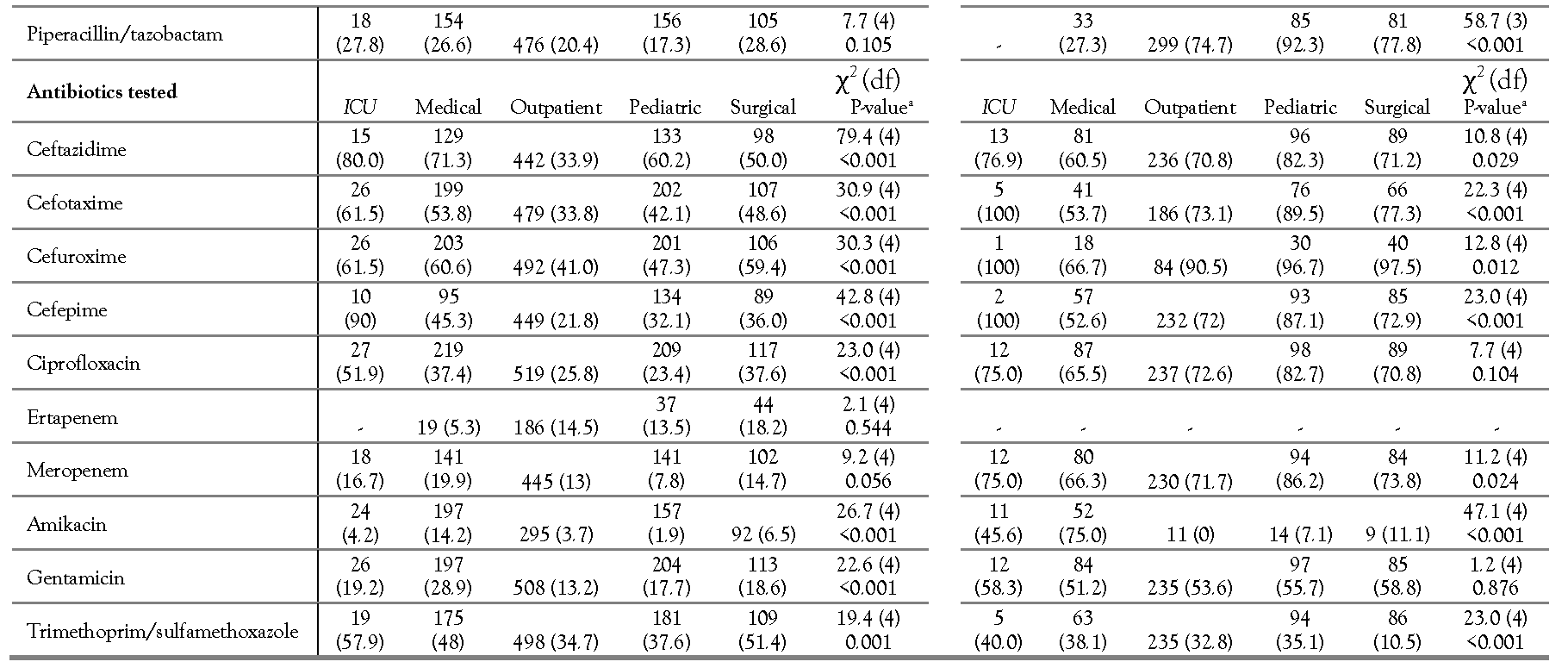
The individual AMR among the four GNB phenotypes cross-tabulated by isolate location is shown in Table 1. Overall, the prevalence of individual AMR among E. coli and K. pneumoniae isolates to cephalosporins, ciprofloxacin and trimethoprim/sulfamethoxazole was high, whereas the lowest AMR prevalence was to ertapenem, meropenem, and amikacin. When comparing across different hospital locations, there were significant differences (p<0.05) in AMR prevalence for most of the antibiotics across the locations (Table 1). Moreover, when comparing prevalence of AMR P. aeruginosa across different hospital locations (Table 1), there were significant differences (p<0.05) for all antibiotics (except for ceftazidime and ciprofloxacin) by location (Table 1). As for A. baumannii, the AMR prevalence among the isolates was high (>50%) to all tested antibiotics except for amikacin and trimethoprim/sulfamethoxazole. When comparing across different hospital locations, there were significant differences (p<0.05) in A. baumannii AMR prevalence for all antibiotics (except for ciprofloxacin and gentamicin) (Table 1).

Multidrug resistance

The distribution of AMR phenotypes, including total MDR isolates, cross-tabulated by location, are shown in Table 2. Furthermore, the distribution of resistant phenotypes including MDR totals and the frequency of MDR phenotypes, cross-tabulated by specimen type, are shown in Table 3. Moreover, the frequency distribution of MDR phenotypes, not collapsed into the 3+ categories, to better show the maximum multidrug resistance phenotype for each isolate, is shown in Figure 1.
Overall, the MDR prevalence was 38.7% (95% CI: 37.4-40.0) across all the isolates. In Table 2, we revealed that the prevalence of MDR E. coli and K. pneumoniae isolates from inpatients (ICU, medical and surgical wards) was significantly (p<0.05) higher than that from pediatric wards and outpatient clinics. On the contrary, MDR prevalence for P. aeruginosa and A. baumannii isolates from pediatric wards was significantly (p<0.05) higher than that from ICU, medical, and surgical wards, and outpatient clinics.
In Table 3, we revealed that MDR prevalence among E. coli isolates from three specimen types (i.e., respiratory specimens (48%), wounds, bones, or other tissues (47.7%) and body fluids (47.1%)) was significantly (p<0.05) higher than that from other specimen types. Furthermore, the most frequent MDR phenotype among E. coli isolates included three antimicrobial drug classes: penicillins, cephalosporins, and fluroquinolones. However, the MDR prevalence among K. pneumoniae isolates from respiratory specimens was not significantly (p>0.05) different between specimen types. The most frequent MDR phenotypes among K. pneumoniae isolates included the following three antimicrobial drug classes: cephalosporins, fluroquinolones, and sulfonamides. Interestingly, 10.3% of K. pneumoniae isolates from respiratory samples were resistant to five different antibiotic classes (Table 3). The MDR prevalence among P. aeruginosa isolates from respiratory specimens (25.8%) was significantly (p<0.05) higher than that from other specimen types. Furthermore, the most frequent P. aeruginosa MDR across the different specimen types (except from body fluids) included a distinct phenotype: penicillins, cephalosporins, fluroquinolones, carbapenems, and aminoglycosides. The MDR prevalence among A. baumannii isolates from respiratory specimens (82.7%) was significantly (p<0.05) higher compared to that from other specimen types. Moreover, the most frequent MDR phenotype among A. baumannii isolates included five antimicrobial drug classes: penicillins, cephalosporins, fluroquinolones, carbapenems, and aminoglycosides.

Discussion

We reported the prevalence and distribution of AMR in clinical GNB isolates obtained from one of the largest secondary governmental/public hospitals in Kuwait. Overall, high resistance prevalence (>50%) across the four GNBs to ampicillin, cefuroxime, cefotaxime, ceftazidime, ciprofloxacin, and trimethoprim/sulfamethoxazole was detected. In addition, MDR prevalence was high (i.e., 38.7%). A lower resistance prevalence (<10%) was observed to amikacin, ertapenem, meropenem, and piperacillin/tazobactam. The AMR prevalence by location and specimen type varied by the GNB organisms. Interestingly, MDR prevalence was significantly higher in isolates from respiratory specimens than other types. Furthermore, the most frequent MDR phenotype among the four GNB organisms and across the different specimen types included penicillins, cephalosporins, and fluroquinolones antimicrobial classes. These findings demonstrate the common presence of GNB AMR (non-MDR and MDR) from this hospital and their variable distribution by location and specimen type. Therefore, monitoring data on antimicrobial susceptibility of common bacterial organisms is critical for assessing AMR trends in hospitals and informing policy decisions.
For E. coli and K. pneumoniae, there were moderate to high AMR prevalence to penicillins (ampicillin [except for K. pneumoniae due to intrinsic resistance]) and amoxicillin/clavulanic acid), cephalosporins (ceftazidime, cefotaxime, cefuroxime, and cefepime), fluoroquinolones (ciprofloxacin), and trimethoprim/sulfamethoxazole and a relatively lower resistance to penicillins (piperacillin/tazobactam), carbapenems (ertapenem and meropenem), and aminoglycosides (amikacin, and gentamicin). In 2005, a study was conducted to assess AMR in uropathogens isolated from inpatient and outpatient settings in Kuwait [14]. The authors found that E. coli (the most frequent uropathogen) AMR prevalence to ampicillin, amoxicillin-clavulanic acid, trimethoprim- sulfamethoxazole, piperacillin was very high (40- 70%) with a greater AMR proportion in isolates from inpatient than that from outpatient settings. Furthermore, they reported lower resistance to amikacin, cefotaxime, ciprofloxacin. Their findings are in partial in agreement with AMR levels in E. coli isolates from inpatient and outpatients in our study. Furthermore, the findings from the 2005 study and ours indicates poor activities of these antibiotics against clinical E. coli isolates. In this study, we found lower AMR prevalence to piperacillin/tazobactam, but higher resistance to cefotaxime and ciprofloxacin compared to Al-Sweih et al [14]. Furthermore, while most of our isolates originated from urine (n=2326 isolates), we reported findings from other specimens. The overall AMR in E. coli isolates from urine specimens to at least one antibiotic class in our study was 17.2% and the MDR prevalence was 37.7%. Moreover, the most frequent phenotype (at 9%) was penicillins, cephalosporins, and fluroquinolones, and sulfonamides. Interestingly, we found higher MDR prevalence in the three GNB organisms isolated from urine samples other than E. coli. This may indicate that while AMR E. coli is a concern in the treatment of urinary tract infections (UTIs), other organisms are also a major health concern and should be addressed by health authorities in Kuwait. Al Sweih et al [14]. reported that E. coli isolates were more resistant to some of the beta-lactam antibiotics such as ampicillin and amoxicillin-clavulanic acid compared to other beta-lactam antibiotics (the 2nd and 3rd generation cephalosporins (e.g., cefotaxime, cefuroxime and cephalothin). However, our E. coli isolates were highly resistant to most beta-lactam antibiotics (including 2nd, 3rd, and 4th generation cephalosporins) with exception of carbapenems. Therefore, both antibiotic stewardship action plan and a surveillance program to monitor AMR trends in hospitals are necessary to reduce antibiotic use and AMR in different bacterial spp.
K. pneumoniae isolates were both prevalent across different specimens and locations (inpatient and outpatient) with the highest percentage of isolates from urine samples (most likely associated with UTIs. Other studies from the region and worldwide have reported high MDR prevalence among clinical K. pneumoniae isolates collected from different specimen types [3,15] For instance, in a study from Kuwait between 2005 and 2007, authors reported that 12% (n=7129 E. coli isolates) and 17% (n=1709 K. pneumoniae) from UTI specimens were resistant to ≥4 antibiotics with lower prevalence in those from the community than those from the hospital [15]. Moreover, in a study conducted at an Iraq’s teaching hospital in 2018, authors reported high AMR prevalence to penicillins, cephalosporins, and ciprofloxacin in E. coli and K. pneumoniae isolates and lower prevalence to piperacillin/tazobactam which agrees with our findings. In addition, they reported MDR in 67.3% and 65.7% of the E. coli and K. pneumoniae isolates, respectively, which is higher than our findings. Nonetheless, the authors did not state/clarify the MDR definition in their article which may explain the very high prevalence they reported. In another study from the region, Khanfar et al [16]. in Saudi Arabia revealed that ESBL E. coli and K. pneumoniae isolates were more frequently detected in hospital settings compared to outpatient settings. Moreover, they found that these isolates were most frequently detected from urine specimens than from other types such as skin, blood, sputum, and wounds [16]. The resistance prevalence was higher in inpatients compared to outpatients; this agrees with our findings, however, we reported higher resistance from respiratory specimens compared to other specimen types. A lower AMR percentage was observed for some of the antibiotics in some of the hospital’s locations. This might be explained by the higher antibiotic use in inpatient compared to outpatient settings. For instance, ceftazidime use at the hospital is through parenteral route (intravenous only); hence, it is mostly used for inpatient wards. Similarly, piperacillin/tazobactam is used intravenously only for inpatient wards. Previous studies have also shown higher AMR prevalence in isolates from inpatient location compared to outpatients. Furthermore, the difference was partially linked to variability in antibiotic use [17]. Klebsiella pneumoniae is a major Gram-negative opportunistic pathogen that is associated with a range of infections such as UTIs, cystitis, pneumonia, surgical wound infections, and septicemia causing high morbidity and mortality. Therefore, it is essential for departments of public health to monitor and report changes in AMR isolates.
Patients in ICU receive empirical treatment with antibiotics due to the condition that necessitates faster intervention. We have generally observed high AMR prevalence in the four GNB isolated from the ICU patients. Nonetheless, lower AMR prevalence was only present to carbapenem drugs and amikacin in E. coli and K. pneumoniae isolates. Interestingly, AMR prevalence was higher for these antibiotics in P. aeruginosa and A. baumannii from the ICU than the other two organisms. Similar findings were observed in Jamal et al [18]. from Kuwait. The authors mentioned that the trend in AMR prevalence has been increasing in ICUs particularly for cephalosporin drugs, which was due to higher antibiotic usage in these units.
P. aeruginosa showed AMR to multiple antibiotics including cephalosporins and carbapenem (meropenem in this study). This pathogen exhibits both intrinsic and acquired resistance to various antimicrobials including β- lactam antibiotics. Therefore, we excluded those antibiotics to which this organism is intrinsically resistant to as well as those not used for clinical treatment. It has been found that MDR P. aeruginosa is frequent in different countries including Kuwait [19]. In the Middle East, there has been a significant increase in the prevalence of MDR carbapenem-resistant P. aeruginosa in the past decade [20]. In our study, MDR P. aeruginosa was more frequently isolated from outpatient clinics and pediatric wards compared to medical and surgical wards, which may indicate that this organism is widespread in both hospital and the community. Moreover, this pathogen with high AMR prevalence was present in all specimen types. Moreover, AMR P. aeruginosa was found in urine, wounds, respiratory samples, and blood with variable AMR percentages [21,22] In a study from Saudi Arabia carried out at a teaching hospital in Riyadh, authors reported that P. aeruginosa isolates from both ICU and non-ICU locations was highly resistant (>50%) [23]. Additionally, the MDR P. aeruginosa isolates were most frequently identified from skin, wound, and urine specimens (except from body fluids). This organism is ubiquitous in nature and found in normal intestinal flora. P. aeruginosa uses both intrinsic and acquired resistance mechanisms causing low susceptibility to many antibiotics. Since this organism is responsible for a wide range of nosocomial infections including UTI, blood stream infection, and gastrointestinal infection, AMR P. aeruginosa continues to be a major public health threat. Strong AMR surveillance systems are needed to curb the spread of this pathogen in healthcare facilities.
In our study, A. baumannii isolates were highly resistant to almost all tested antibiotics (except amikacin) with high AMR prevalence in different specimen types and locations. A. baumannii is known to be a major pathogen associated with hospital-acquired infections (HAIs). We isolated this organism from both outpatient and in-patient samples with various AMR percentages. Both carbapenem-resistant and MDR A. baumannii are major public health issues in hospital settings and within the community in Kuwait, other GCC countries [24] and around the globe. Furthermore, moderate to high resistant A. baumannii in different specimen types (blood, sputum, swabs, urine, respiratory specimens and body fluids) have been reported [22,24] which is partially similar to our findings of high AMR prevalence across specimens. In a study from a hospital in Italy, the authors reported MDR prevalence of 54% among 57 A. baumannii isolates indicating high resistance levels [22]. In our study, the MDR A. baumannii was found more often in outpatient clinics as well as pediatric and surgical wards compared to that from ICU and medical wards. This may indicate circulation of this pathogen in both the hospital and the community. Acinetobacter baumannii has a large capacity to acquire and transfer AMR makers. This enables the organism to resist the action of most current available antibiotics, hence, lowering treatment effectiveness. The presence of MDR organisms in the hospital poses a substantial risk for treatment failure as well as for transmission risk to susceptible patients. In this study, we revealed that MDR prevalence was relatively high (>50%) in isolates from the surgical ward (except for P. aeruginosa [34.1%]). This might be explained due to the post-surgical antibiotic usage and patients’ readmission. The MDR phenotypes were frequently identified in respiratory specimens. Hence, efforts are needed to address this issue to control possible treatment failure associated with respiratory infections [25]. Implementation of infection control and prevention programs are necessary to curb the development and maintenance of MDR organisms in general and AMR A baumannii.
The study has several limitations. First, the study was based on retrospective AMR data from one hospital in Kuwait which might not represent other public hospitals in the country. However, this participating hospital is a major health institution that serves about a quarter of Kuwait’s population. Second, data on sociodemographics and historical antibiotic use were not available. Nonetheless, we believe the study findings reveal the magnitude of the AMR problem in this hospital. Furthermore, the findings are important for decision makers to improve AMR control and prevention as well as to establish a surveillance program to monitor trends and changes. Third, the available data represent one year only. Hence, we could not establish yearly or monthly AMR trends.

Conclusions

We reported a high prevalence of MDR organisms isolated from several locations and specimen types at a major hospital in Kuwait. These findings provide important information to hospital management and healthcare professionals to enhance the infection control programs and improve antibiotic stewardship. Importantly, the establishment of an integrated AMR surveillance program at the hospital to monitor AMR prevalence and changes in trends is needed to reduce the burden of AMR bacterial infection and to improve public health.

Author Contributions

All authors (WQA, WA, RD) were involved in the scholarly creativity and design of the study. WA and RD were involved in data extraction and data quality check. WQA conducted the statistical analysis and write up. All authors were involved in reviewing and approving the manuscript.

Funding

None to declare.

Conflicts of Interest

All authors—none to declare.

References

  1. World Health, Organization. Global action plan on antimicrobial resistance. Geneva, Switzerland. 2015. Available online: http://apps.who.int/iris/bitstream/10665/193736/1/9 789241509763eng.pdf?ua=1 (accessed on 14 July 2019).
  2. Al Johani, S.M.; Akhter, J.; Balkhy, H.; El-Saed, A.; Younan, M.; Memish, Z. Prevalence of antimicrobial resistance among gram-negative isolates in an adult intensive care unit at a tertiary care center in Saudi Arabia. Ann Saudi Med. 2010, 30, 364–369. [Google Scholar] [CrossRef] [PubMed]
  3. Zowawi, H.M.; Forde, B.M.; Alfaresi, M.; et al. Stepwise evolution of pandrug-resistance in Klebsiella pneumoniae. Sci Rep. 2015, 5, 15082. [Google Scholar] [CrossRef] [PubMed]
  4. Yezli, S.; Shibl, A.M.; Livermore, D.M.; Memish, Z.A. Prevalence and antimicrobial resistance among Gram- negative pathogens in Saudi Arabia. J Chemother. 2014, 26, 257–272. [Google Scholar] [CrossRef] [PubMed]
  5. El-Saed, A.; Balkhy, H.H.; Al-Dorzi, H.M.; Khan, R.; Rishu, A.H.; Arabi, Y.M. Acinetobacter is the most common pathogen associated with late-onset and recurrent ventilator-associated pneumonia in an adult intensive care unit in Saudi Arabia. Int J Infect Dis. 2013, 17, e696–701. [Google Scholar] [CrossRef] [PubMed]
  6. Al-Sweih, N.A.; Al-Hubail, M.A.; Rotimi, V.O. Emergence of tigecycline and colistin resistance in Acinetobacter species isolated from patients in Kuwait hospitals. J Chemother. 2011, 23, 13–16. [Google Scholar] [CrossRef] [PubMed]
  7. Jamal, W.; Shahin, M.; Rotimi, V.O. Surveillance and trends of antimicrobial resistance among clinical isolates of anaerobes in Kuwait hospitals from 2002 to 2007. Anaerobe. 2010, 16, 1–5. [Google Scholar] [CrossRef] [PubMed]
  8. Vali, L.; Dashti, K.; Opazo-Capurro, A.F.; Dashti, A.A.; Al Obaid, K.; Evans, B.A. Diversity of multi-drug resistant Acinetobacter baumannii population in a major hospital in Kuwait. Front Microbiol. 2015, 6, 743. [Google Scholar] [CrossRef] [PubMed]
  9. Bonnin, R.A.; Rotimi, V.O.; Al Hubail, M.; et al. Wide dissemination of GES-type carbapenemases in Acinetobacter baumannii isolates in Kuwait. Antimicrob Agents Chemother. 2013, 57, 183–188. [Google Scholar] [CrossRef] [PubMed]
  10. Centers for Disease Control and Prevention. Bloodstream infection event (central line-associated bloodstream infection and non-central line associated bloodstream infection). 2020. Available online: https://www.cdc.gov/nhsn/pdfs/pscmanual/4psc_clabs current.pdf (accessed on 28 November 2020).
  11. Centers for Disease Control and Prevention. Pneumonia (ventilator-associated [VAP] and nonventilator-associated pneumonia [PNEU]) event. 2020. Available online: https://www.cdc.gov/nhsn/pdfs/pscmanual/6pscvapcur rent.pdf (accessed on 28 November 2020).
  12. Centers for Disease Control and Prevention. Urinary tract infection (catheter-associated urinary tract infection [CAUTI] and non-catheter-associated urinary tract infection [UTI]) events. 2020. Available online: https://www.cdc.gov/nhsn/pdfs/pscmanual/7psccautic urrent.pdf (accessed on 28 November 2020).
  13. Clinical and Laboratory Standards Institute. Performance Standards for Antimicrobial Susceptibility Testing, CLSI supplement M100, 27th Ed. ed; CLSI: Wayne, PA, 2017. [Google Scholar]
  14. Al Sweih, N.; Jamal, W.; Rotimi, V.O. Spectrum and antibiotic resistance of uropathogens isolated from hospital and community patients with urinary tract infections in two large hospitals in Kuwait. Med Princ Pract. 2005, 14, 401–407. [Google Scholar] [CrossRef] [PubMed]
  15. Al Benwan, K.; Al Sweih, N.; Rotimi, V.O. Etiology and antibiotic susceptibility patterns of community- and hospital-acquired urinary tract infections in a general hospital in Kuwait. Med Princ Pract. 2010, 19, 440–446. [Google Scholar] [CrossRef] [PubMed]
  16. Husam, S.K.; Khalid, M.B.; Abiola, C.S.; Giuseppe, A.B. Extended spectrum beta-lactamases (ESBL) in Escherichia coli and Klebsiella pneumoniae: Trends in the hospital and community settings. J Infect Dev Ctries. 2009, 3, 295–299. [Google Scholar] [CrossRef]
  17. Saperston, K.N.; Shapiro, D.J.; Hersh, A.L.; Copp, H.L. A comparison of inpatient versus outpatient resistance patterns of pediatric urinary tract infection. J Urol. 2014, 191 Suppl. 5, 1608–1613. [Google Scholar] [CrossRef] [PubMed]
  18. Jamal, W.Y.; El-Din, K.; Rotimi, V.O.; Chugh, T.D. An analysis of hospital-acquired bacteraemia in intensive care unit patients in a university hospital in Kuwait. J Hosp Infect. 1999, 43, 49–56. [Google Scholar] [CrossRef]
  19. Sievert, D.M.; Ricks, P.; Edwards, J.R.; et al. Antimicrobial- resistant pathogens associated with healthcare-associated infections: Summary of data reported to the National Healthcare Safety Network at the Centers for Disease Control and Prevention, 2009-2010. Infect Control Hosp Epidemiol. 2013, 34, 1–14. [Google Scholar] [CrossRef] [PubMed]
  20. Zowawi, H.M.; Balkhy, H.H.; Walsh, T.R.; Paterson, D.L. β- lactamase production in key gram-negative pathogen isolates from the Arabian Peninsula. Clin Microbiol Rev. 2013, 26, 361–380. [Google Scholar] [CrossRef] [PubMed]
  21. Mittal, R.; Aggarwal, S.; Sharma, S.; Chhibber, S.; Harjai, K. Urinary tract infections caused by Pseudomonas aeruginosa: A minireview. J Infect Public Health. 2009, 2, 101–111. [Google Scholar] [CrossRef] [PubMed]
  22. De Francesco, M.A.; Ravizzola, G.; Peroni, L.; Bonfanti, C.; Manca, N. Prevalence of multidrug-resistant Acinetobacter baumannii and Pseudomonas aeruginosa in an Italian hospital. J Infect Public Health. 2013, 6, 179–185. [Google Scholar] [CrossRef] [PubMed]
  23. Babay, H.A. Antimicrobial resistance among clinical isolates of Pseudomonas aeruginosa from patients in a teaching hospital, Riyadh, Saudi Arabia, 2001-2005. Jpn J Infect Dis. 2007, 60, 123–125. [Google Scholar] [CrossRef] [PubMed]
  24. Zowawi, H.M.; Sartor, A.L.; Sidjabat, H.E.; et al. Molecular epidemiology of carbapenem-resistant Acinetobacter baumannii isolates in the Gulf Cooperation Council States: Dominance of OXA-23-type producers. J Clin Microbiol. 2015, 53, 896–903. [Google Scholar] [CrossRef] [PubMed]
  25. Rodrigo-Troyano, A.; Sibila, O. The respiratory threat posed by multidrug resistant Gram-negative bacteria. Respirology. 2017, 22, 1288–1299. [Google Scholar] [CrossRef] [PubMed]
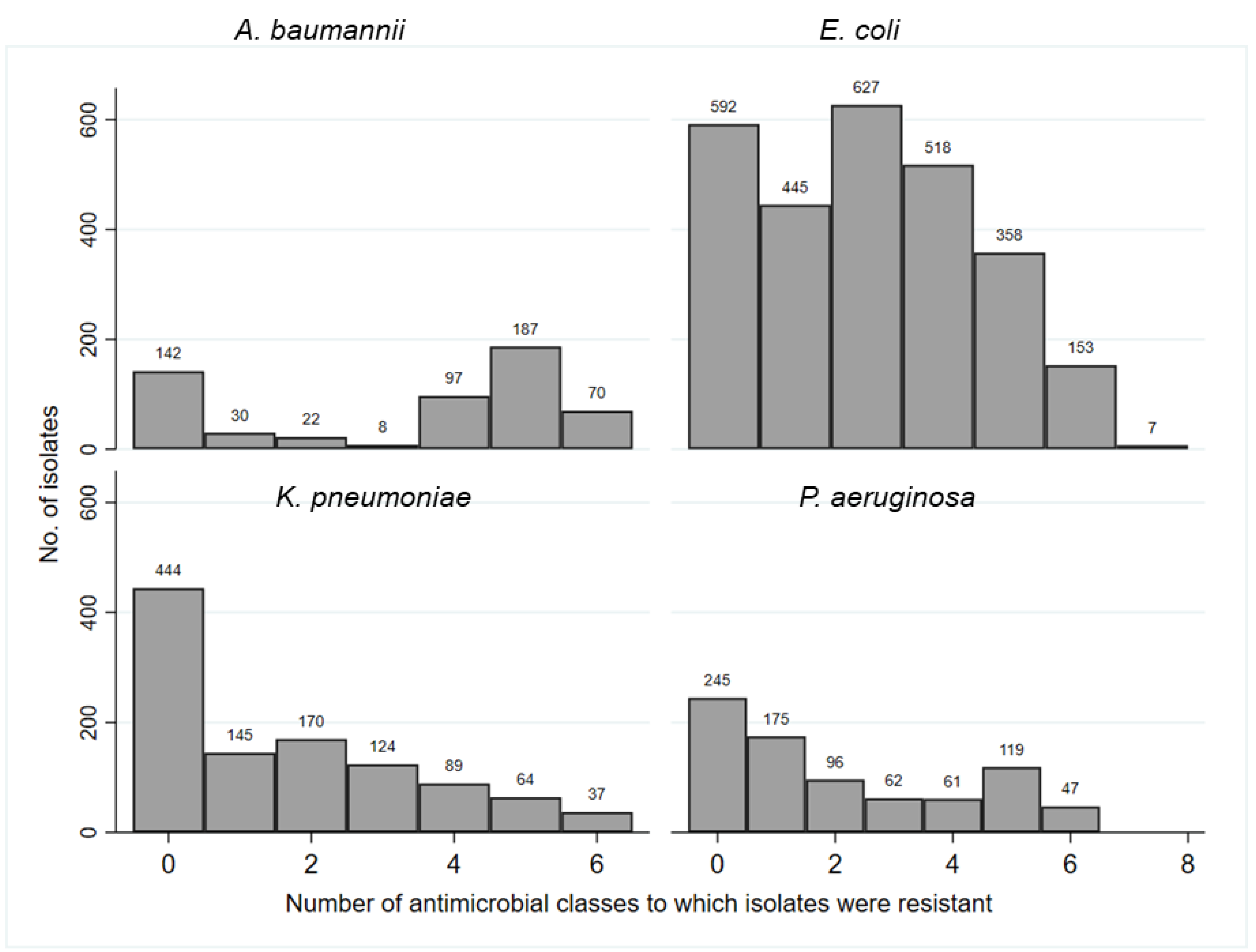
Figure 1. Frequency bar chart illustrating the distribution of phenotypic resistance to up to 7 antimicrobial classes among four Gram-negative bacteria isolates.
Germs 11 00498 g001
Table 1. Comparison of phenotypic resistance of four Gram-negative bacteria (GNB) sampled across different hospital locations.
Germs 11 00498 i001Germs 11 00498 i002
aP-value is based on comparison of percentage of antimicrobial resistant per antibiotic across the location using likelihood ratio Chi-square statistic (χ2) in STATA software version 15.1 (STATA corp., USA). P-value <0.05 indicates dependency between the location and resistant isolates per antibiotic tested. “df” indicates degrees of freedom. b”-“ indicates no isolates were tested.
Table 2. Distribution of resistant phenotypes of four Gram-negative bacteria (GNB) isolates by number of resistant antimicrobial classes and different hospital locations.
Percentage of resistance per GNB and location
E. coli2 = 110.2 (12), p<0.001)a P. aeruginosa χ2 = 38.2 (12), p=0.001)a

Location/MDR
Total no. of isolates
0

1

2

3+b
Total no. of isolates
0

1

2

3+b
ICU5213.715.417.353.9 1947.421.15.326.3
Medical44816.38.719.955.1 16943.217,212.427.2
Outpatient109623.816.224.635.3 39425.024.411.938.6
Pediatric100723.120.625.031.3 16225.316.711.047.0
Surgical12419.412.113.754.8 8536.527.111.824.7
K. pneumoniae χ2 = 22.5 (12), p=0.033)aA. baumannii2 = 68.6 (12), p<0.001)a

Location/MDR
Total no. of isolates
0

1

2

3+b
Total no. of isolates
0

1

2

3+b
ICU2938.56.917.241.4 3669.42.82.825.0
Medical23036.110.919.633.5 12138.87.48.345.5
Outpatient52545.314.515.624.7 24420.93.74.171.3
Pediatric21543.714.016.326.1 10014.02.03.081.0
Surgical12033.313.313.340.0 9020.05.64.470.0
aDifferences in resistant prevalence by each GNB and location were significant (p<0.05) using likelihood ratio Chi-square statistic (χ2 [degrees of freedom—df)]) in STATA. A significant p-value indicates dependency between the location and resistance phenotypes. b0: pan-susceptible; 1: resistant to one antimicrobial class; 2: resistant to two antimicrobial classes; 3+: multidrug resistance to greater than or equal to three antimicrobial classes was collapsed into a single upper category.
Table 3. Distribution of resistant phenotypes of four Gram-negative bacteria (GNB) isolates by number of resistant antimicrobial classes and specimen type.
Percentage of resistance per microorganism and specimen typea
Specimen type/MDRTotal no. of isolates0123+dMost frequent MDR phenotype (%)e
E. coliBody fluids3429.48.814.747.1PEN-CEP-FLO-SUL (16.7)
Blood16019.416.927.536.3PEN-CEP-FLO-SUL (10.2)
χ2=23.7 (12),
Respiratoryb5211.513.526.948.0PEN-CEP-SUL (17.4)
p=0.022
Wound, bone, or other tissuec12821.17.024.247.7PEN-CEP-FLO-AMN-SUL (14.9)
Urine232622.217.222.937.7PEN-CEP-FLO-SUL (9.5)
Specimen type/MDRTotal no. of isolates0123+Most frequent MDR phenotype (%)
K. pneumoniaeBody fluids1369.215.4.7.77.7CEP-FLO-SUL (25.0)
Blood12642.911.99.535.7CEP-FLO-SUL (16.7)
χ2=20.4 (12),
Respiratory14037.712.112.937.1PEN-CEP-FLO-SUL-CAR (10.3)
p=0.060
Wound, bone, or other tissue10142.611.913.931.7PEN-CEP-SUL-FLO-CAR (6.9)
Urine69341.114.318.026.6PEN-CEP-SUL-FLO-CAR-AMN (5.9)
Specimen type/MDRTotal no. of isolates0123+Most frequent MDR phenotype (%)
P. aeruginosaBody fluids966.722.211.10None
Blood8426.231.010.732.1PEN-CEP-FLO-CAR-AMN (11.5)
χ2=32.1 (12),
Respiratory27530.216.710.242.9PEN-CEP-FLO-CAR-AMN (25.8)
p=0.001
Wound, bone, or other tissue18738.019.311.231.6PEN-CEP-FLO-CAR-AMN (14.2)
Urine25025.226.014.834.0PEN-CEP-FLO-CAR-AMN (11.8)
Specimen type/MDRTotal no. of isolates0123+Most frequent MDR phenotype (%)
A. baumanniiBody fluids6500050.0PEN-CEP-FLO-CAR-AMN (100)
Blood7324.76.96.961.6PEN-CEP-FLO-CAR-AMN (35.2)
χ2=73.2 (12),
Respiratory22611.53.12.782.7PEN-CEP-FLO-CAR-AMN (39.6)
p<0.001
Wound, bone, or other tissue13345.12.33.049.6PEN-CEP-FLO-CAR-AMN (38.4)
Urine11827.17.610.255.1CEP-FLO-CAR-AMN (31.4)
aDifferences in resistant prevalence by each GNB and specimen type were significant (p<0.005) using likelihood ratio Chi-square test in STATA. A significant p- value indicates dependency between the specimen type and multidrug resistance (MDR) phenotypes per organism. bRespiratory specimens included sputum, tracheal aspirate, or bronchial. cWound, bone, or other tissue: The ‘other tissue’ included other external tissue infections around the eye, ear, and mouth. d0: pan-susceptible; 1: resistant to one antimicrobial class; 2: resistant to two antimicrobial classes; 3+: multidrug resistance to greater than or equal to three antimicrobial classes was collapsed into a single upper category. ePEN—penicillins; CEP—cephalosporins; FLO—fluroquinolones; SUL—sulfonamides; CAR—carbapenems; AMN—aminoglycosides.