Next Article in Journal
Across Regions: Are Most COVID-19 Deaths Above or Below Life Expectancy?
Previous Article in Journal
Microbial Profile, Antimicrobial Resistance, and Molecular Characterization of Diabetic Foot Infections in a University Hospital
 
 
GERMS is published by MDPI from Volume 15 Issue 4 (2025). Previous articles were published by another publisher in Open Access under a CC-BY (or CC-BY-NC-ND) licence, and they are hosted by MDPI on mdpi.com as a courtesy and upon agreement with the former publisher Infection Science Forum.
Article

The Prevalence of HCV RNA Positivity in Anti-HCV Antibodies-Negative Hemodialysis Patients in Thrace Region. Multicentral Study

by
Eleni I. Konstantinidou
1,†,
Eftychia G. Kontekaki
1,2,†,
Aristidis Kefas
1,†,
Theocharis Konstantinidis
2,3,*,
Gioulia Romanidou
4,
Eleni Fotiadou
3,
Viki Rekari
5,6,
Eleni Triantafyllidou
6,
Stavroula Zisaki
2,
Evi Kasmeridou
4,
Mariana Andreadou
4,
Konstantina Kantartzi
7,
Konstantinos Mavromatidis
4,
George Martinis
2,
Dimitrios Cassimos
8,
Elias Thodis
7,
Maria Panopoulou
3 and
Konstantinos Mimidis
9
1
“Infectious Diseases—International Medicine: From Bench to Bedside”, Democritus University of Thrace, Dragana Campus, 68100 Alexandroupolis, Greece
2
Blood Transfusion Center, University General Hospital of Alexandroupolis Dragana Campus, 68100 Alexandroupolis, Greece
3
Laboratory of Microbiology, Democritus University of Thrace, University General Hospital of Alexandroupolis Dragana Campus, 68100 Alexandroupolis, Greece
4
General Hospital “Sismanoglio”, Sismanoglou 45, 69133 Komotini, Greece
5
General Hospital of Xanthi, Neapoli, 67100 Xanthi, Greece
6
General Hospital of Didimoticho, 25thMay, 141, 683 00 Didimoticho, Greece
7
Department of Nephrology, Democritus University of Thrace, University General Hospital of Alexandroupolis Dragana Campus, 68100 Alexandroupolis, Greece
8
Democritus University of Thrace, Pediatric Department, Alexandroupolis, Greece
9
First Department of Internal Medicine, Democritus University of Thrace, Alexandroupolis, Greece
*
Author to whom correspondence should be addressed.
These authors contributed equally to this work.
GERMS 2021, 11(1), 52-58; https://doi.org/10.18683/germs.2021.1240
Submission received: 6 August 2020 / Revised: 7 January 2021 / Accepted: 12 January 2021 / Published: 15 March 2021

Abstract

Introduction HCV infection in patients under hemodialysis for end stage chronic kidney disease (ESCKD) may exist despite the absence of anti-HCV antibodies. Molecular methods are widely accepted as “gold standard” techniques for the detection of viral RNA. However, the molecular methods are more expensive in comparison to conventional methods and their replacement is not cost-effective. The aim of this study was to estimate the prevalence of HCV RNA positivity in anti-HCV negative hemodialysis patients and evaluate new diagnostic methods for the detection and the monitoring of hepatitis C in ESCKD patients. Methods The study was performed in four hospitals of Thrace region of Greece and 233 patients with no history of hepatitis C were enrolled. Measurement of anti-HCV antibodies and HCV core antigen was performed by microparticle chemiluminescence immunoassay. Molecular detection of viral RNA was performed by the real-time RT PCR. Results The mean age of the patients was 64.9 ± 23.3 years. HCV-Ag was positive in 2/233 patients (0.86%). Nevertheless, viral RNA was negative in those patients. Conclusions The results of the present study showed that the incidence of HCV-RNA in patients with negative anti-HCV Abs, in hemodialysis patients in Thrace region of Greece was negligible (0/233).

Introduction

The prevalence of hepatitis C virus (HCV) according to the World Health Organization is about 3% worldwide [1]. However, the estimates published in medical literature widely vary. The prevalence of hepatitis C in Greece ranges from 0.5% to 2% [2]. In developed countries, most HCV infections occur among drug abusers. However, in lower to middle-income settings HCV infection is mostly associated with inadequate infection control and viral transmission from one person to other [3].
Patients on hemodialysis (HD) belong to the high-risk group for HCV infection, with recorded transmission of HCV during dialysis. Akhtar S. et al., in a metanalysis study reported that the HCV prevalence in hemodialysis patients ranged from 1.4-2.3% in developed countries to 4.7-41.9% in developing countries [4,5]. The dialysis units must implement effective infection prevention and control programs to prevent HCV transmission. However, small-scale HCV outbreaks were reported and HCV transmission in the dialysis unit (DU) represents a public health problem, especially in developing countries [6]. HCV infection has been proven as an independent risk factor for fatal liver complications. The diagnosis of HCV infection is currently based on the detection of anti-HCV antibodies by EIA and is confirmed by the presence of HCV ribonucleic acid (HCV RNA). According to Kidney Disease Improving Global Outcomes (KDIGO) guidelines of 2018, the diagnosis of HCV infection in end stage chronic kidney disease (ESCKD) is based on the detection of anti-HCV antibodies [7]. Measurement of anti-HCV antibodies is recommended every 6 to 12 months for all HCV negative patients. In contrast, previous studies concluded that anti-HCV antibody testing has less reliability for detecting HCV infection in hemodialysis patients [8,9]. The new clinical guidelines have suggested to search for HCV-RNA after anti-HCV antibodies have been detected, but molecular methods carry several limitations such as cost and high technical skill requirement. The availability of new automated assays such as hepatitis C core antigen (HCVAg) enabled us to propose this kit for the diagnosis of HCV [10]. Furthermore, Moini M. et. al., reported their experiences for prognostic value of HCV core antigen assay for detection and its special application for hemodialysis patients [11].
The aim of the present study was to evaluate the prevalence of HCV infection using HCV core antigen and HCV RNA tests among anti-HCV antibodies-negative HD patients from four DU in Thrace, Greece, and asses the efficacy of these two methods.

Methods

The study was approved by the Bioethics Committee of the University General Hospital of Alexandroupolis, Greece (Ethics Committee 13619).

Study population

All dialysis centers of National Health System (NHS) in the Thrace region of Greece were invited and accepted to engage in this study. The study population consisted of >18 years old patients on dialysis program (three times a week), without history of HCV infection. The total number of patients that agreed to participate in the study and signed an informed consent form was 233. More specifically, 41 from General Hospital of Xanthi (Hospital 1), 75 from General Hospital of Komotini (Hospital 2), 74 from General University Hospital of Alexandroupolis (Hospital 3) and finally, 43 patients from General Hospital of Didymoteicho (Hospital 4). The exclusion criteria for this study were as following:
  • Patients who did not give a written consent for participation in this study or were unable to do so.
  • Age under 18 years.
  • Patients with a history of chronic hepatitis C.
  • Patients treated for chronic hepatitis C.
  • Patients on peritoneal dialysis.
A questionnaire with clinical and epidemiological data was completed from an interview with the patients, considering the following information: age, gender, duration of hemodialysis (in months), etiology of chronic renal failure, frequency of blood transfusion and jaundice in the past six months, previous renal transplant. All clinical and epidemiological data were registered. The causes of ESCKD were recorded according to the personal folder of each patient. Blood samples were obtained from the patients before the initiation of the hemodialysis session. The amount of blood that was drawn was 10 mL. Consequently, it was collected in two tubes, and finally centrifuged. Thereafter, one tube was used for routine laboratory investigations, completed by automated methods. The serum from the second tube was collected and stored at -80 °C for HCV core antigen testing and RNA extraction.

HCV core antigen test

The HCV core Ag assay was performed using the Microparticle Immunochemical Luminescence (CMIA) method (Architect, Abbott Diagnostics Division, USA). The cut-off point for positive result was ≥3.00 fmol/L. The method has a specificity of ≥99.5% and a sensitivity of 97.8% [12].

Molecular detection of HCV RNA

Viral RNA from the serum samples was extracted by using the Viral RNA/DNA Nucleospin Virus kit, (Macherey-Nagel, Germany), 200 μL of serum sample were used for RNA extraction. One step real time PCR kit for the qualitative detection of HCV in human plasma (Sacace Biotechnologies, Como, Italy) was used. This qualitative assay was run by SaCycler-96 Real Time PCR System (Sacace Biotechnologies, Italy) with detection limit of 50 IU/mL. Moreover, positive samples were examined by quantitative RT real-time PCR assays with detection limit: 6.00±0.9 IU/mL with Cobas AmpliPrep/Cobas TaqMan HCV v2.0 (Roche Molecular Systems, USA). For each run, positive and negative controls as well as internal control for extraction were included.

Dialysis process

Patients were dialyzed for 4 hours thrice weekly. All patients, except one, were dialyzed in one center only. The anti-HCV positive patients were given dialysis in isolated rooms. A monitoring of the hepatic enzymes such as aminotransferase and anti-HCV testing, on a regular basis, were performed for early detection of HCV acquisition in HD patients. All participants gave their written informed consent to be included into the study.

Statistical analysis

For quantitative variables like age, duration of hemodialysis and number of blood transfusions we calculated mean ± standard deviation (SD). The data for qualitative variables like gender, cause of kidney failure, prior renal transplant is presented as percentages. Nonparametric tests (Mann-Whitney and Kruskal–Wallis) and the Chi-square (χ2) test were performed. A value of p <0.05 was considered as statistically significant.

Results

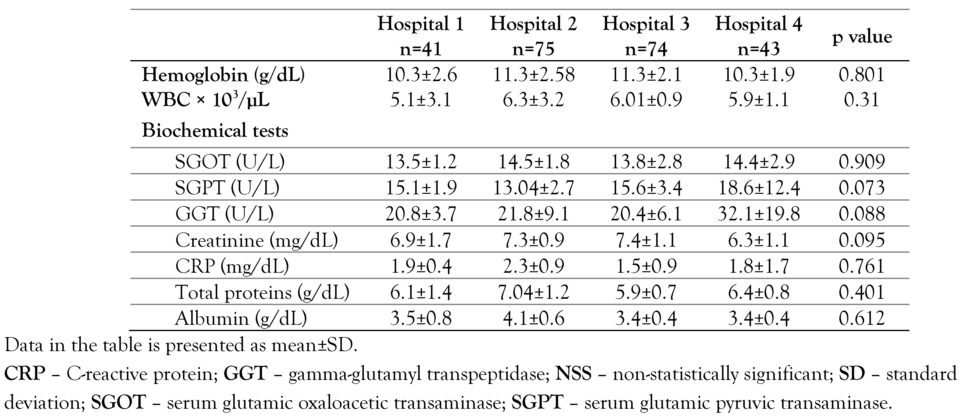
In the present study 233 patients who met the inclusion criteria were enrolled. The mean age of the patients was 64.9±23.3 years (range 18 to 89); men were 59.7% (n= 139). The most common causes of ESCKD were hypertension and diabetes mellitus. The main demographic and epidemiological characteristics of the study groups are presented in Table 1 and laboratory findings of the cohort in Table 2.

HCV core antigen

The measurement of HCV core antigen was positive in two patients. The first patient was from Hospital 1 (2.4%) and the second one from Hospital 3. There were no positive patients from Hospitals 2 and 4.
The first case was a 49-year-old male with ESCKD, secondary to diabetic renal disease and polycystic nephropathy. From his past medical history, he suffered from multiple sclerosis and coronary heart disease treated with coronary artery bypass grafting and angioplasty. In addition, he also suffered from myelodysplastic syndrome, for which he had required blood transfusions the last year. Τhe HCV-Ag value was 14.49 fmol/L the first time and 15.11 fmol/L the second time. Unexpectedly, the RT PCR was twice negative.
The second case was a 62-year-old female, with ESCKD secondary to diabetic renal disease. The HCV-Ag value was 4.49 fmol/L, and 3.69 fmol/L, in two measurements. In this patient as well, the RT PCR was twice negative.

HCV RNA test

All patients were negative for HCV RNA with qualitative real-time RT PCR. Even for the two patients who were found positive for HCV core antigen the test could not be confirmed either with highly sensitive quantitative real-time RT PCR, or qualitative RT PCR.

Discussion

Previous studies have demonstrated that patients on HD had an over 10-fold higher risk of HCV infection compared to other chronic patients [13−15]. Several factors such as longer duration on HD and transfusion of blood components are incriminated as predisposing factors for HCV transmission [15]. In addition, older age, dialysis treatment in more than one different center, history of transplantation, co-infections with hepatitis B and/or HIV, and diabetes mellitus as comorbidity are all an independent risk factors [13,16]. Furthermore, HCV infection is associated with the progression of chronic kidney disease and higher mortality in HD patients. Nosocomial transmission of HCV infection can occur through hemodialysis procedures, surgical manipulations, dental treatment, and even by environmental contamination [17]. Previous studies have reported that viremia in anti-HCV negative patients is 0-16% [9,18]. Dalekos GN et al. recently concluded that routine HCV RNA testing is not necessary, because all patients were negative.18 However, in a subsequent study the same authors reported that HCV RNA assay increased their diagnostic accuracy by more than 33% [19]. Sakamoto N et al. postulated that HD patients may have prolonged period of HCV seronegativity, secondary to immunosuppression [20]. Furthermore, anti-HCV antibody negative patients usually have normal liver function tests and they are not routinely further investigated for the possibility of HCV infection [21]. It was reported in previous studies that HD patients have lower viremia levels, through membrane-dependent absorption of HCV RNA. According to Etik OD et al., blood specimens for HCV RNA testing should be drawn prior to a dialysis procedure for two reasons: (1) the presence of heparin in the sample can induce a false-positive PCR result; (2) a patient’s HCV viral load can decrease during the HD session and return to baseline within 48 h. A patient’s HCV RNA could be falsely low due to several reasons. Firstly, the restraint of HCV particles to the dialysis membrane, since HCV virions are larger (30-40 nm) than the membrane’s pores (10-20 nm). Finally, with the destruction of HCV particles during the HD sessions these particles rarely escape into the dialysate [22]. Studies failed to detect HCV-RNA in the dialysis ultrafiltrate. The previously reported results by R Hubmann et al. indicate that no HCV RNA passed through the dialysis membranes [23]. Nevertheless, the impact of the HD procedure, during hemofiltration and hemodiafiltration, on HCV viral kinetics requires further investigation. In any case, to avoid false negative results, all samples were obtained prior to the HD sessions.
Previous studies suggest that HCV Ag testing is a sensitive and specific method that can be used for HCV diagnosis in immunodeficient patients [10]. HCV core Ag is a trustworthy marker of viral replication which correlates with HCV RNA. Moreover, Mederacke I et al. reported that HCV core Ag correlates with HCV RNA in HCV-infected patients undergoing hemodialysis [24]. Our results are in accordance with previously published studies. A multicentric HD study in France found that only 2 patients out of 4357 anti-HCV negative (0.05%) were positive for HCV RNA [25]. Papadopoulos N et al. reported that none out of 41 anti-HCV-negative hemodialysis patients were proven to be viremic [9]. The presence of two cases with positive HCV antigen and negative HCV RNA tests could be due to the low detection limit of qualitative PCR analysis. We hypothesize that these two patients had low viremic load that could not be detected by our used test. Another explanation is that these are false positive results.
The above mentioned is the main limitation of the present study, and more specifically the detection limit of 50 IU/mL in qualitative RT real-time PCR for detection of HCV RNA.

Conclusions

In conclusion, in this study, in HD patients in Thrace region, we found an exceptionally low level of occult HCV infection. In patients with negative anti-HCV Ab, HCV core Ag as well as HCV RNA was not detected. Therefore, HCV core Ag testing of dialysis patients with negative anti-HCV antibodies is adequate for early detection of HCV infection and subsequently elimination of HCV transmission in DU.
Molecular detection of HCV RNA could be limited only to HCV core Ag positive patients with quantitative RT PCR.

Author Contributions

EIK, EGK and AK contributed to conceptualization, methodology and validation. EF, VR, EK, MK and SZ contributed to laboratory investigation. GR, ET, KK and ET contributed to clinical data curation. TK contributed to writing – original draft preparation. GM, DC and KM contributed to writing – review and editing. MP and KM contributed to supervision. All authors read and approved the final version of the manuscript.

Funding

None to declare.

Conflicts of interest

All authors – none to declare.

References

  1. World Health Organization. Hepatitis C. 2020. Available online: https://www.who.int/news-room/fact-sheets/detail/hepatitis-c (accessed on 25 September 2020).
  2. Triantos, C.; Konstantakis, C.; Tselekouni, P.; Kalafateli, M.; Aggeletopoulou, I.; Manolakopoulos, S. Epidemiology of hepatitis C in Greece. World J Gastroenterol. 2016, 22, 8094–8102. [Google Scholar] [CrossRef]
  3. Leblebicioglu, H.; Arends, J.E.; Ozaras, R.; et al. Availability of hepatitis C diagnostics and therapeutics in European and Eurasia countries. Antiviral Res. 2018, 150, 9–14. [Google Scholar] [CrossRef]
  4. Akhtar, S.; Nasir, J.A.; Usman, M.; Sarwar, A.; Majeed, R.; Billah, B. The prevalence of hepatitis C virus in hemodialysis patients in Pakistan: A systematic review and meta-analysis. PLoS One. 2020, 15, e0232931. [Google Scholar] [CrossRef]
  5. Akabayashi, A.; Nakazawa, E.; Ino, H.; Ozeki-Hayashi, R.; Jecker, N.S. Sacrificing the Fukushima 50 again? J Public Health (Oxf). 2020, 42, 194–197. [Google Scholar] [CrossRef]
  6. Jakupi, X.; Mlakar, J.; Lunar, M.M.; et al. A very high prevalence of hepatitis C virus infection among patients undergoing hemodialysis in Kosovo: a nationwide study. BMC Nephrol. 2018, 19, 304. [Google Scholar] [CrossRef]
  7. Kidney Disease: Improving Global Outcomes (KDIGO) Hepatitis C Work Group. KDIGO 2018 clinical practice guideline for the prevention, diagnosis, evaluation, and treatment of hepatitis C in chronic kidney disease. Kidney Int Suppl (2011) 2018, 8, 91–165. [CrossRef]
  8. Kalantar-Zadeh, K.; Miller, L.G.; Daar, E.S. Diagnostic discordance for hepatitis C virus infection in hemodialysis patients. Am J Kidney Dis. 2005, 46, 290–300. [Google Scholar] [CrossRef]
  9. Papadopoulos, N.; Griveas, I.; Sveroni, E.; et al. HCV viraemia in anti-HCV-negative haemodialysis patients: do we need HCV RNA detection test? Int J Artif Organs. 2018, 41, 168–170. [Google Scholar] [CrossRef]
  10. Galli, C.; Julicher, P.; Plebani, M. HCV core antigen comes of age: a new opportunity for the diagnosis of hepatitis C virus infection. Clin Chem Lab Med. 2018, 56, 880–888. [Google Scholar] [CrossRef]
  11. Moini, M.; Ziyaeyan, M.; Aghaei, S.; et al. Hepatitis C virus (HCV) infection rate among seronegative hemodialysis patients screened by two methods; HCV core antigen polymerase chain reaction. Hepat Mon. 2013, 13, e9147. [Google Scholar] [CrossRef]
  12. Chevaliez, S.; Soulier, A.; Poiteau, L.; Bouvier-Alias, M.; Pawlotsky, J.M. Clinical utility of hepatitis C virus core antigen quantification in patients with chronic hepatitis. J Clin Virol. 2014, 61, 145–148. [Google Scholar] [CrossRef]
  13. Ali, N.; Hussain, W.; Hayat, A.; et al. Prevalence and risk factors of hepatitis B and C viruses among haemodialysis patients: a multicentric study. Eur J Gastroenterol Hepatol. 2019, 31, 29–33. [Google Scholar] [CrossRef]
  14. Kataruka, M.; Gupta, S.; Ramchandran, R.; Singh, M.; Dhiman, R.K.; Lal Gupta, K. Incidence and risk factors for hepatitis C virus and hepatitis B virus seroconversion in end-stage renal failure patients on maintenance hemodialysis. J Clin Exp Hepatol. 2020, 10, 316–321. [Google Scholar] [CrossRef]
  15. Carneiro, M.A.; Martins, R.M.; Teles, S.A.; et al. Hepatitis C prevalence and risk factors in hemodialysis patients in Central Brazil: a survey by polymerase chain reaction and serological methods. Mem Inst Oswaldo Cruz. 2001, 96, 765–769. [Google Scholar] [CrossRef]
  16. Hwang, J.C.; Jiang, M.Y.; Lu, Y.H.; Weng, S.F. Impact of HCV infection on diabetes patients for the risk of end-stage renal failure. Medicine (Baltimore). 2016, 95, e2431. [Google Scholar] [CrossRef]
  17. Johannessen, I.; Danial, J.; Smith, D.B.; et al. Molecular and epidemiological evidence of patient-to-patient hepatitis C virus transmission in a Scottish emergency department. J Hosp Infect. 2018, 98, 412–418. [Google Scholar] [CrossRef]
  18. Dalekos, G.N.; Boumba, D.S.; Katopodis, K.; et al. Absence of HCV viraemia in anti-HCV-negative haemodialysis patients. Nephrol Dial Transplant. 1998, 13, 1804–1806. [Google Scholar] [CrossRef]
  19. Rigopoulou, E.I.; Stefanidis, I.; Liaskos, C.; et al. HCV-RNA qualitative assay based on transcription mediated amplification improves the detection of hepatitis C virus infection in patients on hemodialysis: results from five hemodialysis units in central Greece. J Clin Virol. 2005, 34, 81–85. [Google Scholar] [CrossRef]
  20. Sakamoto, N.; Enomoto, N.; Marumo, F.; Sato, C. Prevalence of hepatitis C virus infection among long- term hemodialysis patients: detection of hepatitis C virus RNA in plasma. J Med Virol. 1993, 39, 11–15. [Google Scholar] [CrossRef]
  21. Kiani, I.G.; Khan, A.N.; Butt, B.; Sabir, S.; Ejaz, S.; Perveen, A.; Ghani, E. HCV-RNA PCR positivity in HCV antibody negative patients undergoing haemodialysis. J Ayub Med Coll Abbottabad. 2018, 30, 397–400. [Google Scholar]
  22. Ozer Etik, D.; Ocal, S.; Boyacioglu, A.S. Hepatitis C infection in hemodialysis patients: A review. World J Hepatol. 2015, 7, 885–895. [Google Scholar] [CrossRef] [PubMed]
  23. Hubmann, R.; Zazgornik, J.; Gabriel, C.; Garbeis, B.; Blauhut, B. Hepatitis C virus – does it penetrate the haemodialysis membrane? PCR analysis of haemodialysis ultrafiltrate and whole blood. Nephrol Dial Transplant. 1995, 10, 541–542. [Google Scholar] [CrossRef] [PubMed]
  24. Mederacke, I.; Potthoff, A.; Meyer-Olson, D.; et al. HCV core antigen testing in HIV- and HBV-coinfected patients, and in HCV-infected patients on hemodialysis. J Clin Virol. 2012, 53, 110–115. [Google Scholar] [CrossRef] [PubMed]
  25. Sauné, K.; Kamar, N.; Miédougé, M.; et al. Decreased prevalence and incidence of HCV markers in haemodialysis units: a multicentric French survey. Nephrol Dial Transplant. 2011, 26, 2309–2316. [Google Scholar] [CrossRef]
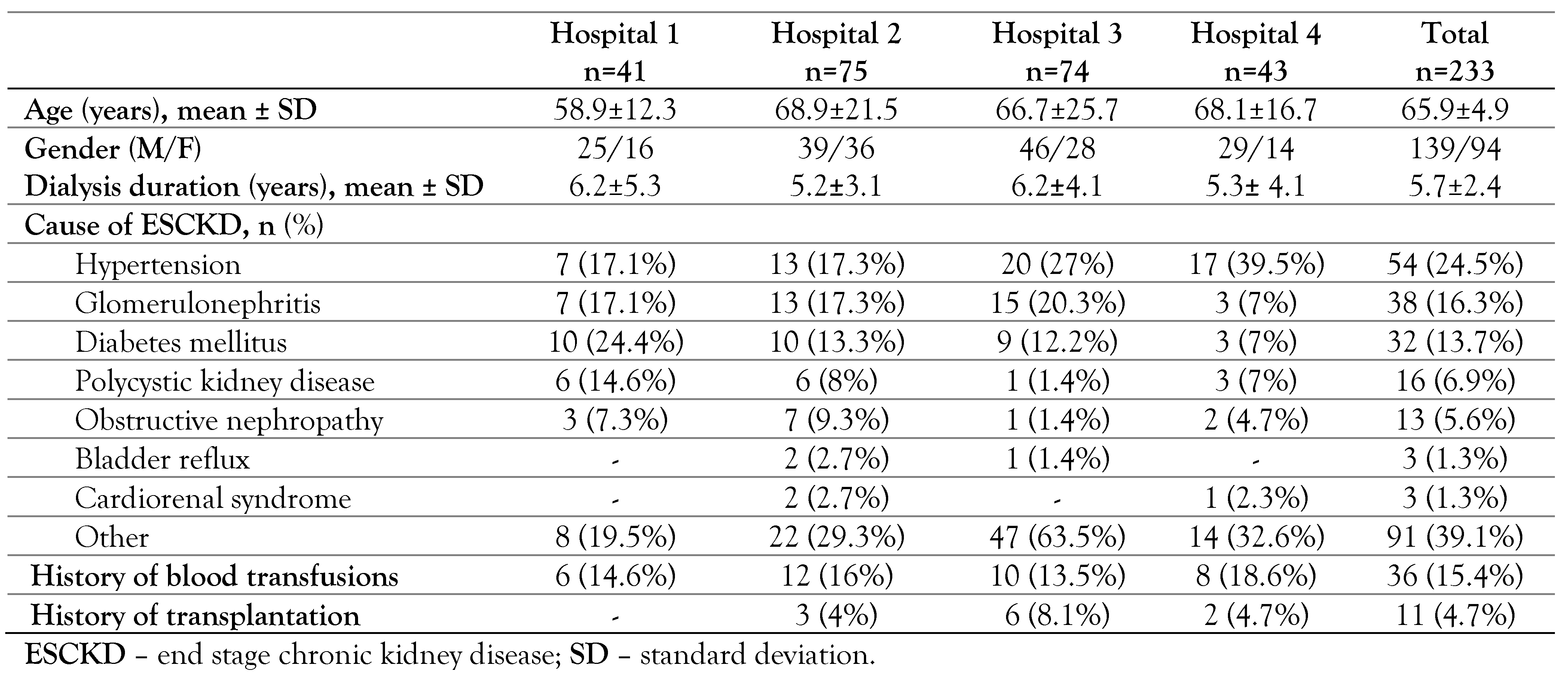
Table 1. Demographic characteristics of patients.
Germs 11 00052 i001
Table 2. Laboratory characteristics of the cohort.
Germs 11 00052 i002