Abstract
Introduction: The etiopathogenesis of purulent pericarditis has changed significantly in modern antibiotic era with the emergence of community-acquired methicillin-resistant Staphylococcus aureus (CA-MRSA) in the last few decades. Pericarditis due to MRSA is rarely reported in the literature without risk factors like immunosuppression, thoracic surgery, chest trauma or pre-existing pericardial diseases. Case report: We describe an 18-year-old male who presented with 5 days history of fever, chest pain and shortness of breath. Echocardiogram and thorax CT showed significant pericardial effusion. The patient underwent pericardiocentesis, MRSA was isolated from blood and pericardial fluid. The patient improved with intravenous antibiotics (linezolid). Follow-up echocardiography at 3 months was unremarkable, without any residual fluid or features of constrictive pericarditis. Discussion: In the absence of known risk factors, MRSA is an extremely rare cause of pericarditis in modern antibiotics era. The possibility of MRSA pericarditis should be sought in every case of pericarditis to achieve prompt diagnosis and treatment. Conclusions: Our case highlights the role of aggressive pericardiocentesis and appropriate antibiotic therapy in purulent pericarditis.
Introduction
Methicillin-resistant Staphylococcus aureus (MRSA) infection is a leading cause of hospital-acquired infections and causes significant mortality, morbidity and cost burden. Most of the MRSA infections are healthcare-associated, commonly associated with skin and soft tissue infections, osteomyelitis, pneumonia, endocarditis and life-threatening bacteremia [1]. Purulent pericarditis due to community-acquired MRSA is rarely reported in the literature. In developing countries, tuberculosis remains the most common cause of purulent pericarditis [2]. The other common bacteria associated with pericarditis are Staphylococcus aureus, Streptococcus pneumoniae, and Gram-negative bacilli in patients exposed to healthcare facilities [2]. Herein we describe a case of community-acquired MRSA infection presenting as purulent pericarditis, in an 18-year-old male with a recent history of dengue infection. To the best of our knowledge, this is the first report of a case of purulent pericarditis caused by community-acquired MRSA from India. The advent of the modern antibiotic era has led to a significant reduction in MRSA in the setting of pericarditis. However, the possibility of MRSA should be kept as an important differential in patients presenting with pericarditis, even without known risk factors.
Case report
An 18-year-old male presented to our hospital with complaints of chest pain, rapidly progressive shortness of breath and fever for the last 5 days. There was no history of cough, rash, joint pain, jaundice or decreased urine output. He had recovered from dengue illness 10 days before hospitalisation. He initially had fever 15 days previously for 2-3 days, he was diagnosed with dengue based on serology and became asymptomatic in 4-5 days. His past history was unremarkable, with no history of recent antibiotics use or illicit drug intake. On examination, he was alert, febrile (temperature 101.4 °F/38.6 °C), with a pulse rate of 108/min, blood pressure of 102/74 mmHg and respiratory rate of 22/min. Systemic examination revealed raised jugular venous pulse and muffled heart sounds. Complete blood count showed raised total leukocyte count (31 × 103/µL, 88% neutrophils), his platelet count was 354 × 103/µL and hemoglobin was 13.4 g/dL. Further biochemical analysis revealed elevated alanine aminotransferase and aspartate aminotransferase (ALT—112 IU/L, AST—88 IU/L). His C-reactive protein and procalcitonin levels were also elevated (CRP 98 mg/L, normal < 1 mg/L, procalcitonin 23 ng/mL, normal < 0.1 ng/mL). The rest of the biochemical investigations (urea—24 mg/dL, creatinine 0.83 mg/dL, electrolytes, thyroid function tests, D-dimer, and viral markers such as HBsAg, anti-HCV, HIV) were normal and negative, respectively. Blood cultures were sent and patient was treated with intravenous piperacillin/tazobactam. His electrocardiogram showed diffuse ST-segment elevation and sinus tachycardia. Chest X-ray was suggestive of cardiomegaly and bilateral minimal pleural effusions. On further evaluation, computed-tomography imaging of thorax revealed moderate to large pericardial effusion with bilateral pleural effusions (Figure 1a,b). In addition, echocardiogram also revealed moderate pericardial effusion. There was preserved left ventricular ejection fraction with no evidence of vegetation in the echocardiogram. On 7th day of illness (day 3 of hospitalisation) his condition worsened with the development of severe respiratory distress and a fall in blood pressure and oxygen saturation. Due to worsening symptoms (shortness of breath), muffled heart sounds and presence of pulsus paradoxus, the possibility of cardiac tamponade was considered. We decided for emergency pericardiocentesis after cardiologist consultation. A total 375 mL of purulent fluid was drained from the pericardial space and sent for cytological evaluation and culture. As tuberculosis is the most common cause of pericardial effusion in India, the pericardial fluid was also sent for GeneXpert MTB/RIF assay [2]. After pericardiocentesis, the patient improved symptomatically. Cytological analysis was consistent with purulent pericardial effusion (11,623 nucleated cells/µL with 88% neutrophils, proteins 6 g/dL, glucose 38 mg/dL and lactate dehydrogenase 205 mg/dL). A concomitant blood sample showed glucose of 99 mg/dL, serum protein of 7.6 g/dL, lactate dehydrogenase of 264 mg/dL. Blood and pericardial fluid cultures were positive for MRSA, which was susceptible to linezolid, co-trimoxazole, clindamycin, and resistant to levofloxacin, cefoxitin. He was treated with intravenous linezolid (600 mg every 12 h for 2 weeks). Pericardial fluid analysis for Ziehl-Neelsen stain and Cartridge based nucleic acid amplification testing were negative. In addition, pericardial mycobacterial culture was negative for Mycobacterium tuberculosis. We also performed RT-PCR for dengue virus (from the pericardial fluid) due to the patient’s recent history of dengue illness, and the results came back negative. Our patient showed significant improvement after the initiation of antibiotic therapy with resolution of fever by the end of the week. Repeat blood cultures were also negative. After receiving i.v. antibiotics for 2 weeks, we decided to continue oral linezolid for 1 more week. Follow-up echocardiogram at the end of 3 weeks and at 2 months did not show any sign of reappearance of fluid or constrictive pericarditis.
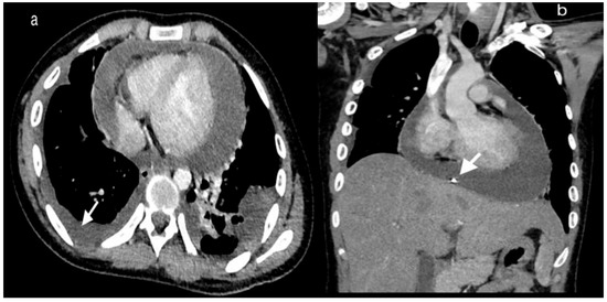
Figure 1.
1(a) Axial post contrast CT thorax shows moderate pericardial effusion without significant enhancement of the pericardium. Note the associated moderate right sided pleural effusion (white arrow) and minimal left sided pleural effusion, 1(b) Coronal post-contrast images show the pericardial effusion with pericardial drain in situ (white arrow).
Discussion
The evaluation of Staphylococcus aureus has identified a new lineage of MRSA defined as community-acquired (CA)-MRSA, which has the propensity to infect young healthy individuals without any risk factors. According to a meta-analysis report, the worldwide pooled prevalence of CA-MRSA is 39% (range 30.8-47.8%) [3]. Purulent pericarditis is a fatal disease with a mortality rate of around 40% in treated patients [4]. Prolonged or recent hospitalization, HIV infection, recent antibiotic use, invasive surgical procedures, stay in long-term care facility or presence of indwelling hemodialysis catheter are the common risk factors associated with MRSA infection. Our patient had none of these risk factors.
Pericarditis can present as an initial presentation of systemic disease or an isolated condition. More than 90% of cases of acute pericarditis are related to viral or idiopathic causes. Bacterial pericarditis is a rare disease, which comprises about less than 1% of all pericardial pathology [2]. In the modern antibiotic era, most cases of purulent pericarditis are associated with nosocomial infections, thoracic surgery or immunosuppression [5]. Our patient did not show any sign of overt immunodeficiency. Features related to pre-existing pericardial injury (malignancy, uremia or connective tissue disorders) were also absent. Pericarditis due to MRSA is a rare event, only a handful of cases (12 patients) have been reported in the literature (Table 1) [6,7,8,9,10,11,12,13,14,15].

Table 1.
Clinical, demographic characteristics and outcome of reported cases of CA-MRSA pericarditis.
Our review of literature revealed that patients across all age groups developed MRSA pericarditis (ranging from 7 months to 75 years). Direct spread from intrathoracic focus of infection (myocardial foci, thoracic surgery or contamination from penetrating trauma), hematogenous spread and extension from subdiaphragmatic suppurative focus are the various mechanisms described for the development of purulent pericarditis. Previously reported cases had some predisposing factors for MRSA, like an axillary abscess, lower limb skin infection, diabetes, underlying malignancy (lung cancer, breast cancer) and steroid therapy (Table 1). Out of 13 patients, 3 patients had no predisposing factors (including our case). Among complications, 1 patient developed a pseudoaneurysm [6], which was successfully repaired, another patient underwent pericardiectomy for fibrinous loculated effusion [11]. Cardiac tamponade was documented in a total of 7 patients [6,7,8,9,10,12,15]. Our search results revealed that vancomycin was the most common antibiotic used for the treatment of MRSA pericarditis. Duration for antibiotics therapy was variable (ranging from 2 to 6 weeks, Table 1). Early therapeutic pericardiocentesis was key to recovery of all patients with MRSA pericarditis. Daily intrapericardial instillation of physiologic saline and fibrinolytic agents (streptokinase, urokinase) has been successfully used in some patients in the setting of loculated fibrinous collection [11]. Our review highlights the pivotal role of aggressive antibiotic therapy and pericardiocentesis, which were crucial for patient survival.
Conclusions
Our case illustrates community-acquired MRSA as an important cause of purulent pericarditis. Duration of antibiotic therapy should be individualised depending upon antimicrobial sensitivity, adequate pericardial drainage and improvement in clinical signs and symptoms related to bacteremia. A high index of clinical suspicion, prompt initiation of antibiotics and aggressive pericardial drainage are vital to reduce the significant morbidity and mortality associated with MRSA pericarditis.
Author Contributions
DSM, DK, MG and GKB contributed to concept, design and drafting of the manuscript. PV, ST and NM contributed to analysis, image collection and critical revision. All authors read and approved the final version of manuscript.
Funding
None to declare.
Informed Consent Statement
Written informed consent was obtained from the patient for publication of this case report and images.
Conflicts of Interest
All authors—none to declare.
References
- Klevens, R.M.; Morrison, M.A.; Nadle, J.; et al. Invasive methicillin-resistant Staphylococcus aureus infections in the United States. JAMA 2007, 298, 1763–1771. [Google Scholar] [CrossRef] [PubMed]
- Imazio, M.; Gaita, F.; LeWinter, M. Evaluation and treatment of pericarditis a systematic review. JAMA 2015, 314, 1498–1506. [Google Scholar] [CrossRef] [PubMed]
- Li, S.; Li, J.; Qiao, Y.; Ning, X.; Zeng, T.; Shen, X. Prevalence and invasiveness of community-acquired methicillin-resistant Staphylococcus aureus: A meta-analysis. Indian J. Microbiol. 2014, 57, 418–422. [Google Scholar] [CrossRef]
- Pankuweit, S.; Ristić, A.D.; Seferović, P.M.; Maisch, B. Bacterial pericarditis: Diagnosis and management. Am. J. Cardiovasc. Drugs. 2005, 5, 103–112. [Google Scholar] [CrossRef] [PubMed]
- Adler, Y.; Charron, P.; Imazio, M.; et al. 2015 ESC Guidelines for the diagnosis and management of pericardial diseases: The Task Force for the Diagnosis and Management of Pericardial Diseases of the European Society of Cardiology (ESC) Endorsed by: The European Association for Cardio-Thoracic Surgery (EACTS). Eur. Heart J. 2015, 36, 2921–2964. [Google Scholar] [CrossRef] [PubMed]
- Patel, S.; Maves, R.; Barrozo, C.P.; et al. Mycotic pseudoaneurysm and purulent pericarditis attributable to methicillin-resistant Staphylococcus aureus. Mil. Med. 2006, 171, 784–787. [Google Scholar] [CrossRef] [PubMed]
- Hussam, M.A.; Ragai, M.F.; Iman, M.F.; Zakaria, A. Community-acquired methicillin-resistant Staphylococcus aureus pericarditis presenting as cardiac tamponade. South. Med. J. 2010, 103, 834–836. [Google Scholar] [CrossRef] [PubMed]
- Arora, N.P.; Kottam, A.; Mahajan, N.; et al. Purulent pericardial effusion from community-acquired methicillin-resistant Staphylococcus aureus. Am. J. Med. Sci. 2012, 344, 160–162. [Google Scholar] [CrossRef] [PubMed]
- Kurahara, Y.; Kawaguchi, T. Cardiac tamponade with community-acquired methicillin-resistant Staphylococcus aureus pericarditis. Intern. Med. 2013, 52, 1753. [Google Scholar] [CrossRef] [PubMed]
- Lutmer, J.E.; Yates, A.R.; Bannerman, T.L.; Marcon, M.J.; Karsies, T.J. Purulent pericarditis secondary to community-acquired, methicillin-resistant Staphylococcus aureus in previously healthy children. A sign of the times? Ann. Am. Thorac. Soc. 2013, 10, 235–238. [Google Scholar] [CrossRef] [PubMed]
- Terada, M.; Watanabe, H.; Kobukai, Y.; et al. Successful treatment of a patient with purulent pericarditis by daily intrapericardial washouts. Ann. Thorac. Surg. 2014, 98, 1451–1454. [Google Scholar] [CrossRef] [PubMed]
- Mada, P.K.; Cady, B.; De Silva, A.; Alam, M. Disseminated MRSA infection with purulent pericarditis. BMJ Case Rep. 2017, 2017, bcr2016218463. [Google Scholar] [CrossRef] [PubMed]
- Sanchez, J.; Schneider, A.; Tretter, J.T.; et al. Community-acquired MRSA pericarditis and mediastinitis in a previously healthy infant. J. Pediatr. Intensive Care 2018, 7, 97–101. [Google Scholar] [CrossRef] [PubMed]
- Abdel-Haq, N.; Moussa, Z.; Farhat, M.H.; Chandrasekar, L.; Asmar, B.I. Infectious and noninfectious acute pericarditis in children: An 11-year experience. Int. J. Pediatr. 2018, 2018, 5450697. [Google Scholar] [CrossRef] [PubMed]
- Ganji, M.; Ruiz, J.; Kogler, W.; Lung, J.; Hernandez, J.; Isache, C. Methicillin-resistant Staphylococcus aureus pericarditis causing cardiac tamponade. IDCases 2019, 18, e00613. [Google Scholar] [CrossRef] [PubMed]


© GERMS 2020.