Article

A Molecularly Imprinted Fluorescence Sensor Based on Upconversion-Nanoparticle-Grafted Covalent Organic Frameworks for Specific Detection of Methimazole

State Key Laboratory of Food Nutrition and Safety, Tianjin University of Science and Technology, Tianjin 300457, China
*
Author to whom correspondence should be addressed.
Processes 2024, 12(3), 626; https://doi.org/10.3390/pr12030626
Submission received: 2 March 2024 / Revised: 19 March 2024 / Accepted: 20 March 2024 / Published: 21 March 2024
(This article belongs to the Section Materials Processes)

Abstract

:
Rapid detection and sensitive analysis of MMZ is of great importance for food safety. Herein, a fluorescent molecularly imprinted sensor based on upconversion nanoparticles (UCNPs) grafted onto covalent organic frameworks (COFs) was designed for the detection of MMZ. COFs with a high specific surface area and excellent affinity serve as substrates for grafting of UCNPs, which can inhibit the aggregation burst of UCNPs and improve the mass transfer rate of the sensor. Through a series of characterizations, it was found that the proposed UCNP-grafted COFs@MIP-based sensor had good optical stability, high adsorption efficiency, strong anti-interference ability, and high sensitivity owing to the integration of the advantages of UCNPs, COFs and MIPs. Under the optimal conditions, a good linear relationship was presented between the fluorescence intensity of UCNP-grafted COFs@MIPs and the methimazole concentration in the range of 0.05–3 mg L−1, and the detection limit was 3 μg L−1. The as-prepared UCNP-grafted COFs@MIPs were successfully applied for the detection of MMZ in actual samples, and the results were relevant with those determined by high-performance liquid chromatography. The sensor has good sensitivity, reusability, and high selectivity, which are highly valuable in the rapid analysis and detection of food safety.

Graphical Abstract

1. Introduction

As a kind of imidazole clinical drug, methimazole (MMZ) has been applied in the remedy of hyperthyroidism and other diseases by regulating animal endocrine [1]. Currently, MMZ is illegally used as a feed additive that can improve protein metabolism and increase lean meat rate [2]. The residue of MMZ in food accumulates in the human body and can produce toxic or other side effects including nephritis, liver cirrhosis, or leukopenia in the blood. Hence, it is exceptionally vital to establish a reliable, fast, and accurate method for the detection of MMZ.
To date, some analytical methods [3] have been utilized for the quantificational determination of MMZ, such as chromatography [4,5], electrochemical analysis [2,6], surface-enhanced Raman scattering (SERS) [7,8], and colorimetric detection [9]. Although these methods can achieve accurate detection of MMZ, most of them usually require high instrumental costs, time-consuming operation procedures, and complicated sample pretreatment procedures. In this respect, fluorescence analytical methods, with the obvious advantages of fast response, easy operation, low analytical cost, high selectivity, and sensitivity, have been widely regarded as relatively economical and rapid analytical methods [10,11].
Lanthanide-doped upconversion nanoparticles (UCNPs) have emerged as promising candidates in the field of fluorescent detection owing to their intriguing characteristics [12]. Compared to carbon dots and semiconductor quantum dots, UCNPs feature unique photophysical properties that can launch high energy, short wavelength visible light under low-energy near-infrared (NIR) light excitation [13]. UCNPs exhibit unique merits such as weak auto-fluorescence background, narrow emission spectrum, high photochemical stability, high signal-to-noise ratio, high fluorescence quantum yield, no photobleaching phenomenon, and high detection sensitivity, which make them more suitable for the detection of analytes in complex matrices [14,15]. Moreover, UCNPs and other emerging advanced materials are combined to construct the multifunctional composite fluorescent sensors, which are of great utilitarian value for the development of exceedingly sensitive sensors.
Covalent organic frameworks (COFs)are an emerging highly crystalline porous materials [16] with uniformly ordered pore channels and exact topological structure, which are basically consist of C, H, N, O, S, B, and other light-weight elements through reversible covalent bonds. COFs can be covalently synthesized by various organic building units to form two-dimensional (2D) or three-dimensional (3D) spatial structure [17]. In addition, COFs possess the excellent features of low density, high specific surface area, ordered pore channel, high crystallinity, and easy surface modification, which enables it to demonstrate great prospects in the application fields of gas storage and separation, catalysis, drug delivery, photoelectric materials, and sensors [18,19]. Moreover, compared with the previously reported COFs that were synthesized via the solvothermal method, COFs synthesized based on the room temperature method are relatively straightforward, easy to operate, and do not require harsh experimental conditions [20,21]. To date, COFs have been grafted onto diverse fluorescent materials and have been generally utilized [22]. Owing to the large surface area of COFs, the obtained luminescent COFs (LCOFs) have the advantages of strong adsorption capacity, great sensitivity, and excellent sensing performance. However, LCOFs still have inadequacies such as poor selectivity.
Molecularly imprinted polymers (MIPs) are a variety of polymers with tailor-made spatial structure that is completely matched with the template in shape, size, and functional groups [23]. As the identification elements of sensors, MIPs have the characteristics of structure predictability, recognition specificity, application versatility, easy preparation, and low cost [24,25]. Considering the excellent selective adsorption capacity, MIPs have been widely used in various analytical fields of purification and enrichment of target molecules, and some remarkable progress has been made [26,27]. At present, molecular imprinting recognition technology has been combined with LCOF sensors to realize the recognition and detection of target molecules, obtaining highly selective and sensitive sensors. In this case, a novel quantum dots (QDs) fluorescent sensor, QDs-doped COFs@MIPs [28], was constructed and employed for the determination of nereistoxin-related insecticide.
Based on the abovementioned research, a novel fluorescence sensor for high selectivity and sensitivity detection of MMZ was constructed via an efficient one-pot polymerization, which combined the advantages of UCNPs, COFs and MIPs. Herein, UCNPs with strong green luminescence were introduced and acted as a signal converter. Then, TFPB-PA COFs with a high specific surface area were synthesized at room temperature and used to construct COFs-based fluorescence sensors for the first time, which can improve the adsorption capacity and binding affinity of the sensing system. Finally, MIPs were imprinted on the surface of UCNP-grafted COFs through the Schiff base reaction between COFs and functional monomers, which endowed the sensor with the specific adsorption recognition sites of the target. The double recognition function based on imprinting layer and UCNPs enabled the sensor to display satisfactory selective binding ability for target analytes. To verify the feasibility of the sensor, the constructed UCNP-grafted COFs@MIPs were applied to the detection of MMZ in milk and pork. Meanwhile, the properties of UCNP-grafted COFs@MIPs and their application in MMZ-selective sensing were described in detail.

2. Materials and Methods

2.1. Materials and Characterization

Y(CH3COO)3·4H2O, Yb(CH3COO)3·4H2O, Er(CH3COO)3·4H2O were obtained from Sigma-Aldrich (St.Louis, MO, USA). 1-octadecene (ODE), oleic acid (OA), tetraethyl orthosilicate (TEOS), 3-aminopropyl-triethoxysilane (APTES) were applied by J&K Scientific Ltd. (Bejing, China). Ammonium fluoride (NH4F), methacrylic acid (MAA), acrylamide (AM), ethylene glycol dimethacrylate (EGDMA), azodiisobutyronitrile (AIBN), sodium hydroxide (NaOH), dichloromethane (DCM) and acetic acid (Hac) were purchased from TCI chemicals (Shanghai, China). Methimazole (MMZ), 2-mercaptoimidazole (MZY), N-methylthiourea (Metu) and 2-Thiouracil (2-TU) were bought from Aladdin Industrial Co., Ltd. (Shanghai, China). 1,3,5-Tri-(4-formylphenyl) benzene (TFPB), 1,4-P-phenylenediamine (PA) were supplied by Shanghai Macklin Biochemical Co., Ltd. (Shanghai, China).
The fluorescence Intensity of UCNP-grafted COFs@MIPs to MMZ was recorded on an F7100 fluorescence spectrophotometer (Hitachi Co., Ltd., Tokyo, Japan) coupled to an external 980 nm diode laser. Ultraviolet–visible (UV-Vis) absorption spectra of MMZ were measured using a UV-Cary100 spectrophotometer (Agilent, Santa Clara, CA, USA) over the scan range of 200–800 nm. SEM (Hitachi, Ltd., Tokyo, Japan) and transmission electron microscope (JEOL, Tokyo, Japan) were used to observe the surface morphologies of UCNP-grafted COFs@MIPs, UCNP-grafted COFs@NIPs, TFPB-PA COFs and UCNPs. Fourier transform infrared spectra (FT-IR) over the range 4000–500 cm−1 was acquired using KBr pellets in a FT-IR spectrophotometer (Thermo Scientific, Waltham, MA, USA). Thermo gravimetric analysis (TGA) between room temperature and 800 °C was performed (TA, New castle, DE, USA) at a heating rate of 10 °C min−1 in N2. The crystal structure of the synthesized materials was characterized using an X-ray power diffractometer (XRD; Bruker, Karlsruhe, Germany).

2.2. Preparation of UCNPs

UCNP nanoparticles (NaYF4: Yb3+, Er3+) were prepared via the thermal decomposition method [29]. Yttrium hydrate (0.78 mmol), ytterbium acetate (0.20 mmol), and erbium acetate (0.02 mmol) were first mixed with octadecene (17 mL) and oleic acid (6 mL) and vigorously stirred. Additionally, the temperature was adjusted to 160 °C in an argon atmosphere. Within 30 min, a yellowish solution was obtained. After cooling, 10 mL of methanol consisting of NaOH (100 mg) and NH4F (148 mg) was dropped to the reaction system and shaken for half an hour. Subsequently, methanol was removed by raising the temperature. Then, the mixture was adjusted to 300 °C, and maintained for an hour. UCNPs were collected by washing three times with ethanol, then dried at 60 °C for 12 h.

2.3. Preparation of UCNPs@NH2

Overall, 20 mg of UCNPs was completely scattered in 60 mL isopropanol, and 2.5 mL ammonia water and 20 mL ultra-pure water were dripped into the system under stirring conditions. Then, 20 mL isopropyl alcohol with 25 μL TEOS was injected into the reaction. After mixing vigorously for 4 h in the dark, 30 mL isopropanol with 200 μL APTES was dropped and stirred for 1 h. The precipitates were cleaned with absolute ethanol, and dried at 50 °C for 12 h.

2.4. Preparation of TFPB-PA COFs

The TFPB-PA-COFs were prepared by a reported Schiff-base reactions [20,30]. In a 20 mL borosilicate glass, TFPB (10.5 mg) and PA (6.5 mg) were dissolved in 4 mL of a mixed solution containing mesitylene/dioxane (v:v, 1:1). After that, 0.45 mL of 12M aqueous acetic acid was added into the abovementioned solution. The system was allowed to react at room temperature for 72 h. The precipitates were purified several times with tetrahydrofuran (THF) and acetone, and dried at 60 °C overnight under vacuum.

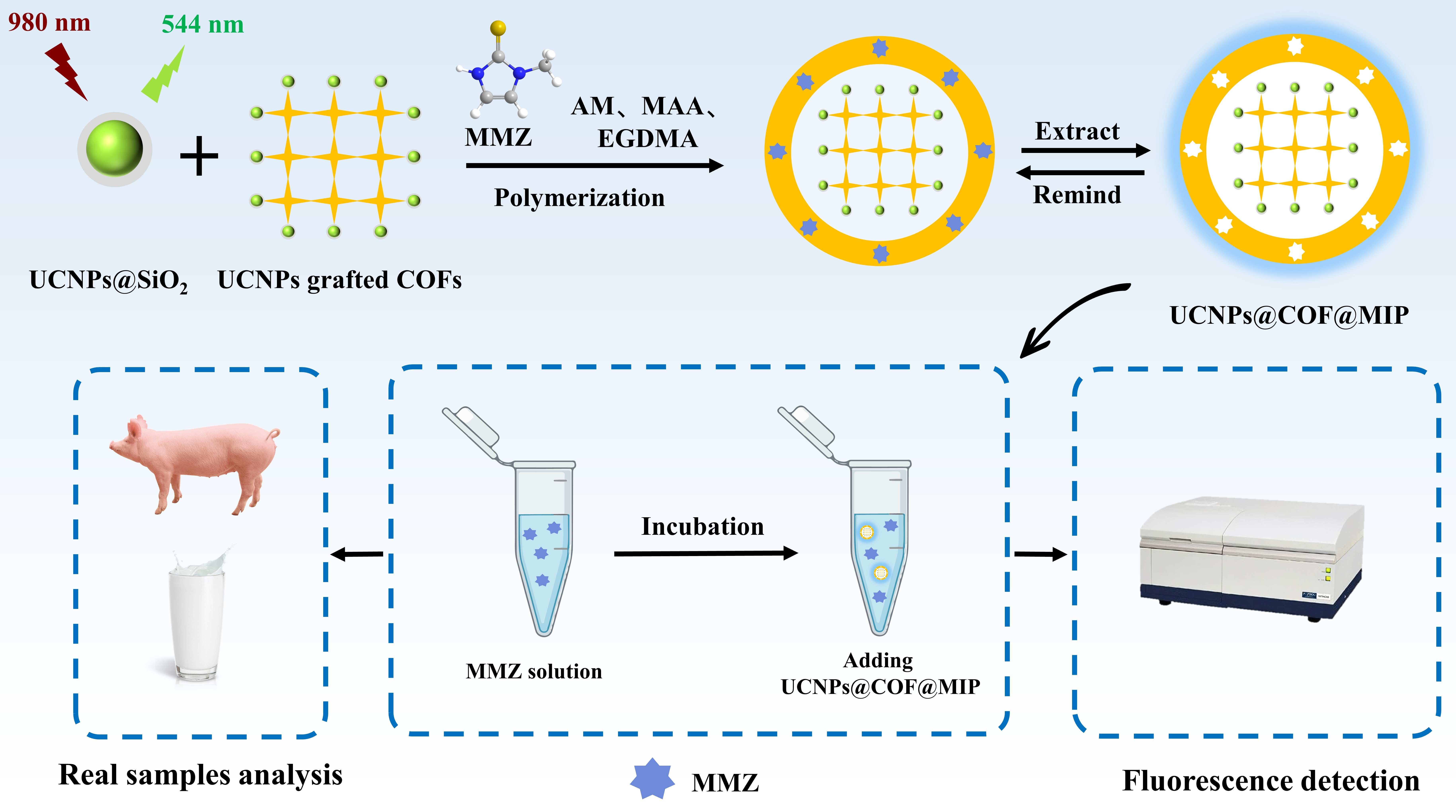
2.5. The Preparation of MIPs Based on UCNP-Grafted COFs

The MIPs based on UCNP-grafted COFs were fabricated by one-pot surface-imprinting synthesis strategy [31,32]. UCNPs (20 mg) were dissolved in 20 mL ethanol by ultrasound, blended with COFs (1 mg), and continuously stirred for 3 h at room temperature within the dim. At that point, MMZ (0.1 mmol) blended with MAA (0.4 mmol) and AM (0.4 mmol) were immersed in the reaction. After stirring for 30 min, AIBN (20 mg) and EGDMA (0.8 mmol) were moreover introduced into the reaction. Subsequently, the reaction system was submerged in a water bath at 60 °C for 18 h. The imprinted MMZ were extricated with methanol-acetic acid (v:v, 9:1). MIPs with cavities were obtained by drying in a vacuum stove at 50 °C for 4 h.
For comparison, NIPs were prepared utilizing the same strategy without the template molecules.

2.6. Fluorescent Determination

The fluorescence response of MMZ was carried out employing a Hitachi FL-7100 fluorescence spectrophotometer by mixing UCNP-grafted COFs@MIPs (1 mg) and different concentrations of MMZ (2 mL). After mixing at room temperature for 60 min, the fluorescence intensity was recorded at the emission wavelength of 544 nm and excitation wavelength of 980 nm.

2.7. The Preparation of Samples

Taking fresh pork and milk purchased from supermarket as samples, these samples were used to investigate the practical application of MIPs based on UCNP-grafted COFs by recovery experiment. Pork should be homogenized before the spiking experiment. The different concentrations of MMZ solution were added into 5 g samples to make the spiked concentration to be 0.1, 0.2 and 0.5 mg kg−1, respectively. After standing for 10 h in the dark, 20 mL ethylacetate was added for extraction and shaken vigorously for 30 min. The supernatant was obtained by centrifugation (4000 rpm, 5 min) at 4 °C. The residues followed the abovementioned procedure again. Combining the two supernatants, 30 mL acetonitrile-saturated hexane was added to remove fat, and the acetonitrile phase was separated by oscillation for 3 min. The acetonitrile phase was consistently dissipated to dry under the condition of 40 °C. The residues were dissolved with 10 mL 0.0025 mol L−1 phosphoric acid solution.

3. Results

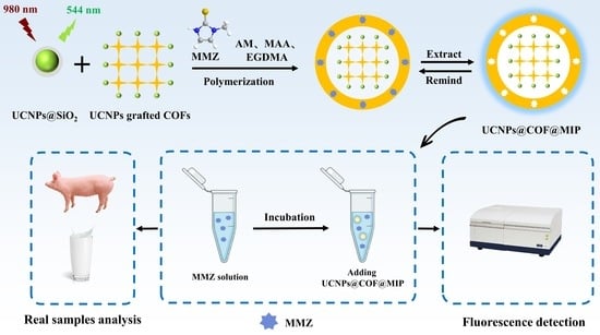
3.1. Synthesis of MIPs Based on UCNP-Grafted COFs

In this study, a fluorescent molecularly imprinted sensor of MMZ was prepared based on UCNPs and COFs. The preparation process was schematically shown in Scheme 1. First, NaYF4:Yb3+, Er3+ with good dispersibility and luminescence properties was prepared via the thermal decomposition method. The amino-functionalized UCNPs were successfully prepared using TEOS and APTES, which made UCNPs surface-rich in amino groups. Meanwhile, TFPB-PA COFs was prepared by Schiff base reactions between the PA and TFPB. Then, the amino-functionalized UCNPs and TFPB-PA COFs were added to the one-pot synthesis process of MIPs. During the imprinting process, UCNPs were successfully grafted onto COFs due to the Schiff base reaction between the amino groups of UCNPs and the aldehyde groups of COFs. The synthesized UCNP-grafted COFs were used as the supporting material and the fluorescent signal source, and the MAA and AM were used as functional monomers with EGDMA as a cross-linking agent. Among them, MAA and MMZ were connected through a hydrogen bond. AM was combined with COFs by Schiff base reaction. Under the action of the initiator AIBN, the polymerization occurred through carbon–carbon double bonds, and the specific recognition sites for MMZ were formed on the surface of UCNP-grafted COFs@MIPs. Finally, methanol/acetic acid (v:v, 9:1) was used to remove MMZ by disrupting the hydrogen bond, and MIPs with specific recognition sites were obtained.
The imprinting process dominated the sensing performance of the constructed nanosensors. To obtain UCNP-grafted COFs@MIPs with specific binding ability and sensitive fluorescence response to MMZ, the corresponding synthesis conditions were optimized in Tables S1 and S2.

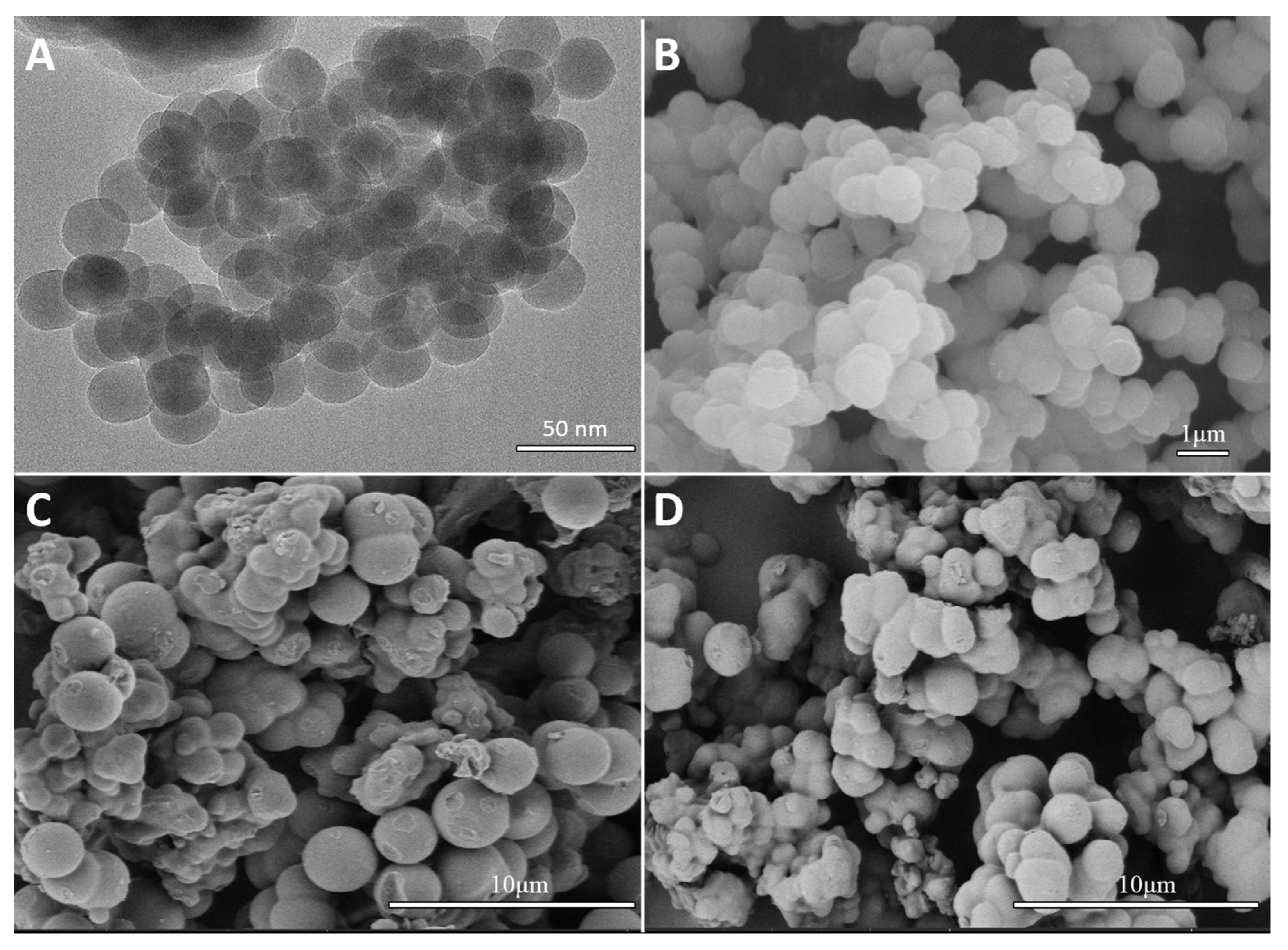
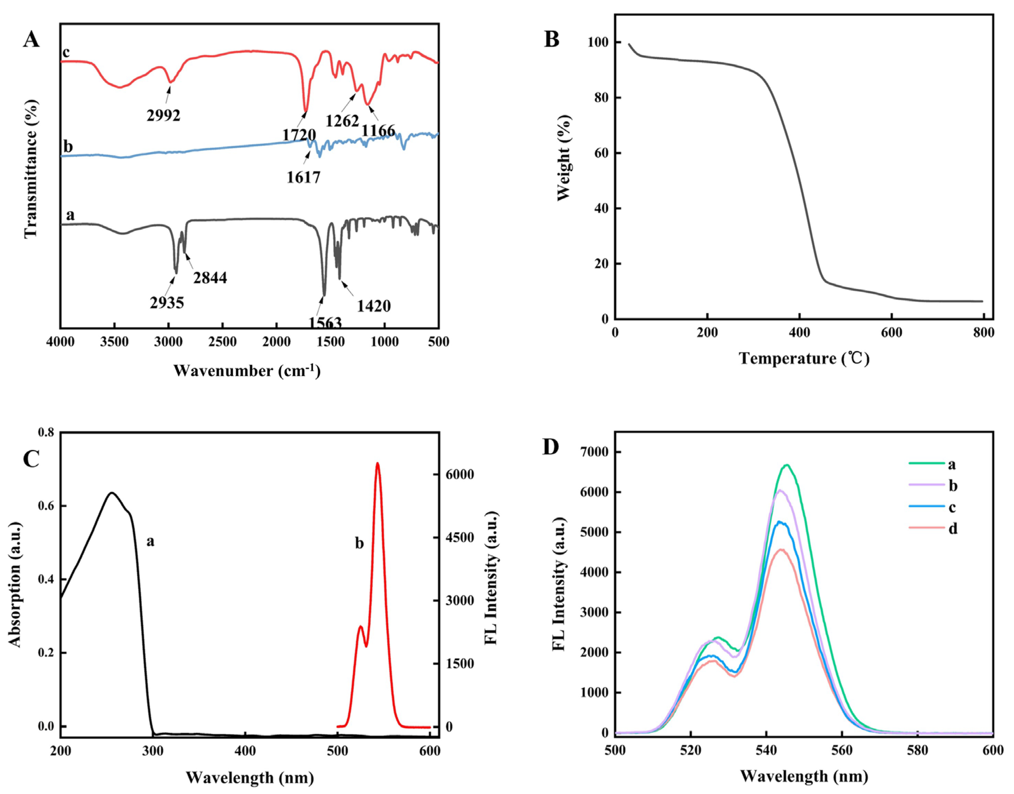
3.2. Characterization of MIPs Based on UCNP-Grafted COFs

The TEM image of UCNPs (Figure 1A) illustrated that UCNPs had a uniform hexagonal structure. The SEM image of COFs is exhibited in Figure 1B. The interlayer π–π force of COFs caused layer accumulation to form a multilayer stacked structure, and the morphology of COFs material was regular. The SEM image presented in Figure 1C clearly reveals that the surface morphology of UCNP-grafted COFs@MIPs was highly uniform spheres with a rough surface. In the process of the formation of MIPs, some molecular cavities were left on the surface for specific identification of target molecules, so the surface morphology was relatively rough. In addition, there was no significant difference in the morphologies of MIPs and NIPs (Figure 1D). The abovementioned results displayed that the polymers were formed.
In order to further confirm the imprinting process, FT-IR was used to identify the corresponding functional groups of the materials obtained in different steps. Figure 2A (a) demonstrates obvious characteristic peaks at 2844 cm−1 and 2935 cm−1, which were caused by the stretching vibration of methylene and vinyl groups, and the characteristic bands at 1420 cm−1 and 1563 cm−1 were attributed to the carboxyl groups of oleic acid, which displayed that UCNPs were successfully synthesized [33]. The FT-IR spectrum of COFs in Figure 2A (b) displays a vibrational band at about 1617 cm−1, proving the existence of the C=N bond. Several absorption bands are presented in Figure 2A (c) containing 2992 cm−1 (C=H), 1262 cm−1 and 1166 cm−1 (O-C-O) and 1720 cm−1 (C=O), which corresponded to the successful reaction between MAA, AM, and EGDMA during the imprinting process. Apparently, the results also indicated the effective synthesis of MIPs [34].
TGA analysis of UCNPs, COFs (Figure S1), and UCNP-grafted COFs@MIPs (Figure 2B) was carried out in nitrogen stream. From room temperature to 800 °C, there was an obvious two-stage decomposition. The primary stage of the decomposition of the components occurred at around 300 °C on account of the expulsion of residual solvents and the decomposition of the imprinted layer and UCNPs, and the weight loss was 83.24% for the UCNP-grafted COFs@MIPs. The decomposition of the moment stage occurred at about 450 °C. Due to the decomposition of the COFs, the weight loss was 10%. This indicated that the MIP layer and COFs were successfully combined on the UCNP surface.
In order to explore the sensing mechanism, we studied the UV-Vis absorption spectrum of MMZ and fluorescence emission spectrum of UCNP-grafted COFs@MIPs (Figure 2C). The UV-Vis absorption spectrum of MMZ was 254 nm, and the fluorescence emission wavelength of MIPs was 544 nm. The possibility of energy transfer and internal filtering effect was ruled out because there was no spectral overlap [35]. Thus, it was inferred that the mechanism of fluorescence quenching was presumably the photo-induced electron transfer. As displayed in Figure S2, when MMZ was adsorbed and combined with MIPs through hydrogen bonding, the lone pair of electrons located on MIPs (the electron donor) were transferred to the acceptor MMZ, causing fluorescence quenching. This result was also consistent with the previously reported studies [36].
The XRD patterns of UCNPs, COFs, and UCNP-grafted COFs@MIPs are displayed in Figure S3. The characteristic diffraction peaks of UCNPs were observed with a cubic structure, which was consistent with the literature [37]. There was a weak diffraction peak in the COFs spectrum, and the characteristic diffraction peaks of UCNPs and COFs were presented in UCNP-grafted COFs@MIPs. These results indicated the successful synthesis of UCNP-grafted COFs@MIPs.
In this study, COFs as matrix-supporting material were grafted onto the fluorescent nanosensor to progress the sensitivity and adsorption capacity of the sensor. As shown in Figure 2D, the fluorescence intensity of UCNP-grafted COFs@MIPs was essentially higher than that of UCNPs@MIPs, which was attributed to the fact that the grafting of COFs on UCNPs changed the surface characteristics of UCNPs, thereby enhancing the fluorescence intensity. In addition, there were a lot of π–π-conjugated structures of COFs, and the formation of the C=N double bond further enhanced the conjugated system and increased the fluorescence intensity [17,38]. As can be seen from Figure 2D, the fluorescence quenching degree of UCNP-grafted COFs@MIPs was about three higher times than that of UCNPs@MIPs when UCNPs@MIPs and UCNP-grafted COFs@MIPs adsorbed the same concentration of MMZ, demonstrating that the introduction of COFs was beneficial to improving the sensitivity of the sensor.

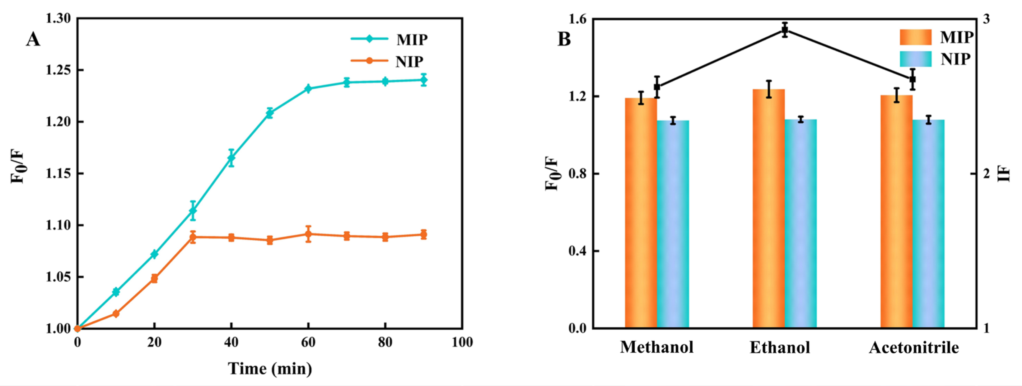
3.3. Optimization of Adsorption Parameters

The dynamic adsorption experiment was investigated by changing the adsorption time of UCNP-grafted COFs@MIPs with a high concentration of MMZ (3 mg L−1). As shown in Figure 3A, fluorescence quenching effect (F0/F) of both UCNP-grafted COFs@MIPs and UCNP-grafted COFs@NIPs gradually increased with extending contact time. The F0/F value remained unchanged over time, which indicated that the combination of the template MMZ and MIPs based on UCNP-grafted COFs had reached equilibrium. Based on the results, 60 min was defined as the ideal response time for the entire experiment.
Adsorption solvent was a pivotal factor in the assessment of adsorption performance, so ethanol, methanol and acetonitrile were chosen for optimization. As shown in Figure 3B and Table S3, when IF was used as a reference index, the ethanol solution had the highest IF, meaning that in ethanol, the prepared sensor MIPs had more significant signal changes relative to NIPs. Therefore, ethanol was chosen as the solution matrix for the sensors in the following experiments.

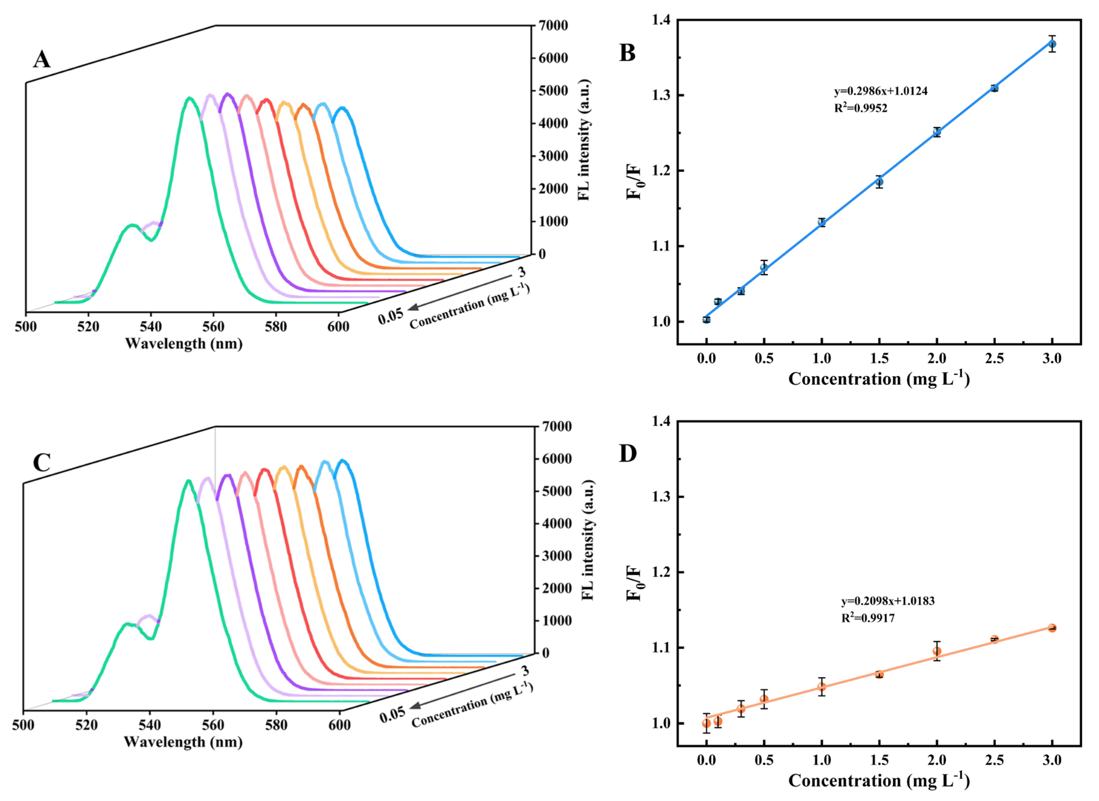
3.4. Establishment of Sensing Methods

The fluorescence spectra were displayed (Figure 4A,C) by recording the fluorescence intensity of UCNP-grafted COFs@MIPs and UCNP-grafted COFs@NIPs in different MMZ concentrations (0.05–3 mg L−1). For both MIPs and NIPs, the fluorescence intensities were quenched linearly. In addition, under identical MMZ concentrations, the fluorescence reduction degree of MIPs was larger than that of NIPs, which was attributed to the formation of recognition sites corresponding to MMZ on the surface of MIPs. The fluorescence quenching degree (F0/F) of MIPs and NIPs and the concentration of MMZ conformed to the Stern–Volmer equation:
F0/F = Ksv [Q] + 1
where F0 indicates the fluorescence intensity of sensor before adsorbing MMZ, F represents the fluorescence intensity of sensor after binding to MMZ, Ksv is the Stern–Volmer constant, and [Q] is the concentration of MMZ. The proportion of the Ksv values of MIPs and NIPs (Ksv,MIPs/Ksv,NIPs) was characterized as the imprinting factor (IF) to determine the selectivity of the sensor.
IF = (F0,MIP/FMIP − 1)/(F0,NIP/FNIP − 1)
Typically, IF is used as a measure for the selection of synthesized materials, with a larger IF representing a greater degree of fluorescence burst for MIPs compared to NIPs, demonstrating better adsorption capacity and selectivity of the synthesized material.
Within the extent of 0.05–3 mg L−1, the linear equation of MIPs (Figure 4B) was F0/F = 0.2986 [Q] + 1.0124 and R2 was 0.9952. The linear equation of NIPs (Figure 4D) was F0/F = 0.2098 [Q] + 1.0183, and R2 was 0.9917. The imprinting factor (IF) of the sensing system was 3.37, which revealed that MIPs had better specificity than NIPs. In addition, when MIPs were taken as a sensor for detection, the LOD for MMZ was evaluated to be 3 μg L−1.

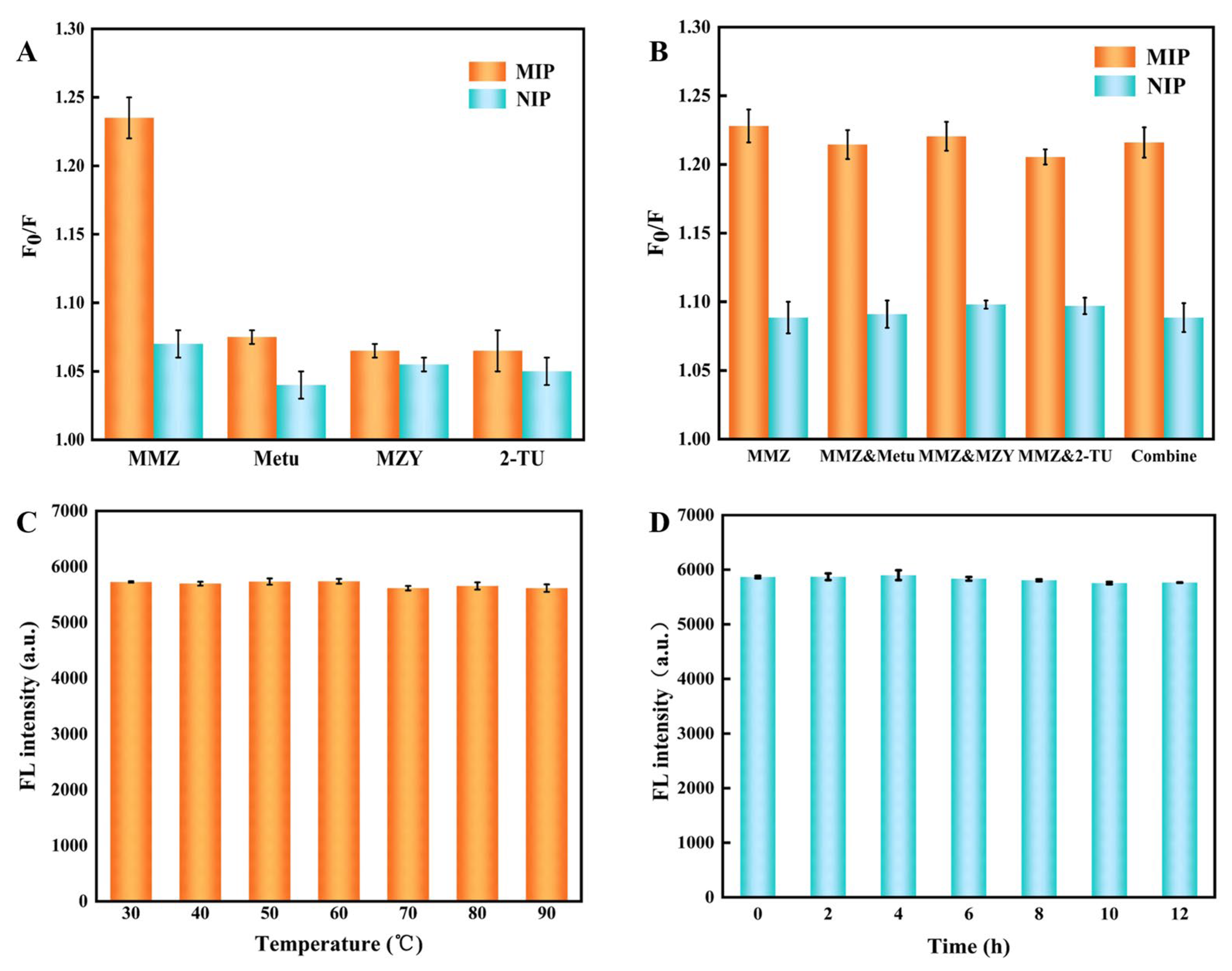
3.5. Selectivity Analysis

Aiming at assessing the specific recognition capability of UCNP-grafted COFs@MIPs for MMZ, the control experiments were carried out via using 2-mercaptoimidazole (MZY), N-methylthiourea (Metu), 2-thiouracil (2-TU). Figure S4 demonstrates the structures of MMZ and its structural analogs. Figure 5A exhibits that the fluorescence quenching degree of UCNP-grafted COFs@MIPs with MMZ was significantly higher than other structural analogs, while MMZ and structural analogs had the similar effects on the fluorescence intensity of UCNP-grafted COFs@NIPs. Additionally, the fluorescence response of UCNP-grafted COFs@MIPs to the MMZ was the most sensitive, and the extent of fluorescence diminishing was additionally the most noteworthy among the analytes. The main reason was that during the imprinting process, MMZ-related sites were shaped on the surface of the MIPs. The MMZ can be bound to the MIPs via hydrogen bond, causing fluorescence to weaken as a result of the photoinduced electron transfer. The fluorescence response of UCNP-grafted COFs@NIPs was insensitive to MMZ and other structural analogues because of the pure physical adsorption among them.
In order to study the anti-interference ability of MIPs to MMZ in complex environments, MMZ was added to other structural analogs in equal quantities. As shown in Figure 5B, the addition of structural analogs had no effect on the recognition of MMZ by MIPs, indicating that UCNP-grafted COFs@MIPs had superior selective recognition performance.

3.6. Stability and Reusability Performance

Stability, an imperative figure for the nanosensor performance, was explored by observing the fluorescence intensity of UCNP-grafted COFs@MIPs every 10 °C and every 2 h. As shown in Figure 5C, the fluorescence intensity remained almost constant with temperature varying from 30 °C to 90 °C, which further revealed that the thermal stability of UCNP-grafted COFs@MIPs was sufficient. In addition, Figure 5D indicates that the fluorescence intensity of UCNP-grafted COFs@MIPs demonstrated only little changes over 12 h, indicating that the sensor possessed a stable emission.
The reusability of the fluorescent sensor was appraised through five adsorption–desorption cycles, and the results are recorded in Figure S5. Briefly, 25 mg MIP nanoparticles were added into 50 mL MMZ solution (3 mg L−1), and the corresponding fluorescence intensity was measured. After equilibrium adsorption, the fluorescence intensity was monitored once more. Imprinted MMZ was removed from the mixed solution by methanol/acetic acid (v:v, 9:1), and the MIPs were collected by centrifugation. In addition, after five tests, the fluorescence intensity was still 96.37% of the initial fluorescence intensity, indicating a great reusability. The abovementioned findings further revealed that the constructed fluorescence sensor can adsorb MMZ at least five times without significant difference.

3.7. Method Performance Comparison

Compared with the existing fluorescence sensor detection technology (Table S4), the detection range and detection limit of the constructed fluorescence sensor detection technology were significantly higher than those of some detection methods, and the proposed sensor did not need complex extraction process in actual application. The developed sensor had a wider linear range and lower detection limit for MMZ detection. The introduction of COFs with large surface area and excellent structural performance granted the sensing system high sensitivity and superior adsorption capacity. Moreover, as a luminescent material, UCNPs had advantages of narrow emission bands, weak self-fluorescence background, and remarkable photochemical stability compared with traditional quantum dots. Finally, the silica-based imprinted layer was introduced as the recognition unit of the sensor, which not only maintains the sensitive signal of the fluorescent element, but also further improves the anti-interference ability of the sensor. In conclusion, due to the introduction of UCNPs, COFs and MIPs, the constructed UCNP-grafted COFs@MIPs sensor exhibited high selectivity, remarkable sensitivity, outstanding stability, and excellent repeatability.

3.8. Sample Analysis

The constructed UCNP-grafted COFs@MIPs fluorescent sensor was utilized to study the measurement of MMZ in pork and milk. MMZ was not identified in the clear tests; thus, the tests implemented the spiked and recovered experiments. Each spiked sample underwent three repeated tests to assess the precision of the method (Table 1). The recovery rate of the proposed method was between 88.24% and 91.89%, and the relative standard deviation was between 2.43% and 3.78%. Compared with the classical HPLC method, the results of HPLC were consistent with that of the fluorescence method with a higher sensitivity. Therefore, UCNP-grafted COFs@MIPs can accurately measure MMZ in food samples, indicating that UCNP-grafted COFs@MIPs can be used to analyze real samples.

4. Conclusions

In this study, UCNP-grafted COFs@MIPs were synthesized successfully using one-pot polymerization and applied to the rapid determination of MMZ. The sensor exhibited a superior stability, outstanding selectivity, excellent accuracy, and high sensitivity. The photosensor had the merits of shortening the experimental analysis time, obtaining better recovery rate and repeatability. The actual samples measurement results showed that the fluorescence method based on UCNP-grafted COFs@MIPs had a favorable practical application prospect in food detection.

Supplementary Materials

The following supporting information can be downloaded at: https://www.mdpi.com/article/10.3390/pr12030626/s1, Figure S1: TGA curves of UCNPs, COFs; Figure S2: The energy transfer principle; Figure S3: XRD curves of UCNPs (a), COFs (b), UCNPs-grafted COFs@MIPs (c); Figure S4: The chemical structures of methimazole (MMZ), 2-mercaptoimidazole (MZY), N-methylthiourea (Metu) and 2-thiouracil (2-TU); Figure S5: Reusability of UCNPs-grafted COFs@MIPs for the determination of MMZ; Table S1: Optimization of addition amount of COFs; Table S2: Optimization of the molar ratio of MMZ to AM to MAA to EGDMAs; Table S3: Optimization of the matrix of tests; Table S4: The comparison of the analytical methods with other reported methods. See Refs. [1,10,39,40,41,42,43].

Author Contributions

Conceptualization, Y.L. and G.F.; methodology, Y.L.; software, Y.L. and T.Z.; validation, Y.L., S.L. and Y.C.; formal analysis, Y.L. and T.Z.; investigation, Y.L. and T.Z.; resources, Y.L. and G.F.; data curation, Y.L.; writing—original draft preparation, Y.L.; writing—review and editing, Y.L., Y.C. and G.F.; visualization, Y.L. and T.Z.; supervision, Y.L. and S.L.; project administration, Y.L.; funding acquisition, G.F. All authors have read and agreed to the published version of the manuscript.

Funding

This research was funded by the National Natural Science Foundation of China, grant number 32372433.

Data Availability Statement

Data are contained within the article.

Conflicts of Interest

The authors declare that they have no known competing financial interests or personal relationships that could have appeared to influence the work reported in this paper.

References

  1. Saleh, T.A.; Al-Shalalfeh, M.M.; Al-Saadi, A.A. Silver nanoparticles for detection of methimazole by surface-enhanced Raman spectroscopy. Mater. Res. Bull. 2017, 91, 173–178. [Google Scholar] [CrossRef]
  2. Tavakkoli, N.; Soltani, N.; Sadeghi, M.; Salavati, H. Electrochemical determination of methimazole using nanoporous gold film electrode modified with MoO2 thin film. Microchem. J. 2019, 150, 104153. [Google Scholar] [CrossRef]
  3. Lega, F.; Contiero, L.; Biancotto, G.; Angeletti, R. Determination of thyreostats in muscle and thyroid tissues by QuEChERS extraction and ultra-performance liquid chromatography tandem mass spectrometry. Food Addit. Contam. Part A Chem. Anal. Control Expo. Risk Assess. 2013, 30, 949–957. [Google Scholar] [CrossRef]
  4. Tatara, M.R.; Golynski, M.; Radzki, R.P.; Bienko, M.; Krupski, W. Effects of long-term oral administration of methimazole on femur and tibia properties in male Wistar rats. Biomed. Pharmacother. 2017, 94, 124–128. [Google Scholar] [CrossRef] [PubMed]
  5. Pérez-Fernández, V.; Marchese, S.; Gentili, A.; García, M.Á.; Curini, R.; Caretti, F.; Perret, D. Analysis of antithyroid drugs in surface water by using liquid chromatography–tandem mass spectrometry. J. Chromatogr. A 2014, 1367, 78–89. [Google Scholar] [CrossRef]
  6. Nezhadali, A.; Mehri, L.; Shadmehri, R. Determination of methimazole based on electropolymerized-molecularly imprinted polypyrrole modified pencil graphite sensor. Mater. Sci. Eng. C Mater. Biol. Appl. 2018, 85, 225–232. [Google Scholar] [CrossRef] [PubMed]
  7. Lai, H.; Li, G.; Zhang, Z. SnS2/AuNPs surface-enhanced Raman scattering sensor for rapid and selective quantification of methimazole in serum and meat samples. Sens. Actuators B Chem. 2023, 380, 133325. [Google Scholar] [CrossRef]
  8. Golparvar, A.; Kim, J.; Boukhayma, A.; Briand, D.; Carrara, S. Highly accurate multimodal monitoring of lactate and urea in sweat by soft epidermal optofluidics with single-band Raman scattering. Sens. Actuators B Chem. 2023, 387, 133814. [Google Scholar] [CrossRef]
  9. Amjadi, M.; Hallaj, T.; Asadollahi, H.; Song, Z.; de Frutos, M.; Hildebrandt, N. Facile synthesis of carbon quantum dot/silver nanocomposite and its application for colorimetric detection of methimazole. Sens. Actuators B Chem. 2017, 244, 425–432. [Google Scholar] [CrossRef]
  10. Yu, C.; Qin, D.; Jiang, X.; Zheng, X.; Deng, B. Facile synthesis of bright yellow fluorescent nitrogen-doped carbon quantum dots and their applications to an off–on probe for highly sensitive detection of methimazole. Microchem. J. 2021, 168, 106480. [Google Scholar] [CrossRef]
  11. Ge, Y.; Ji, R.; Shen, S.; Cao, X.; Li, F. A ratiometric fluorescent probe for sensing Cu2+ based on new imidazo [1,5-a] pyridine fluorescent dye. Sens. Actuators B Chem. 2017, 245, 875–881. [Google Scholar] [CrossRef]
  12. Yao, J.; Huang, C.; Liu, C.; Yang, M. Upconversion luminescence nanomaterials: A versatile platform for imaging, sensing, and therapy. Talanta 2020, 208, 120157. [Google Scholar] [CrossRef]
  13. Tessitore, G.; Mandl, G.A.; Brik, M.G.; Park, W.; Capobianco, J.A. Recent insights into upconverting nanoparticles: Spectroscopy, modeling, and routes to improved luminescence. Nanoscale 2019, 11, 12015–12029. [Google Scholar] [CrossRef] [PubMed]
  14. Fan, D.; Wang, E.; Dong, S. Upconversion-chameleon-driven DNA computing: The DNA-unlocked inner-filter-effect (DU-IFE) for operating a multicolor upconversion luminescent DNA logic library and its biosensing application. Mater. Horiz. 2019, 6, 375–384. [Google Scholar] [CrossRef]
  15. Yin, M.; Jing, C.; Li, H.; Deng, Q.; Wang, S. Surface chemistry modified upconversion nanoparticles as fluorescent sensor array for discrimination of foodborne pathogenic bacteria. J. Nanobiotechnol. 2020, 18, 41. [Google Scholar] [CrossRef]
  16. Zhao, W.; Xia, L.; Liu, X. Covalent organic frameworks (COFs): Perspectives of industrialization. CrystEngComm 2018, 20, 1613–1634. [Google Scholar] [CrossRef]
  17. Zhang, Y.; Zhang, D.; Liu, A.H. Luminescent Molecularly Imprinted Polymers Based on Covalent Organic Frameworks and Quantum Dots with Strong Optical Response to Quinoxaline-2-Carboxylicacid. Polymers 2019, 11, 708. [Google Scholar] [CrossRef] [PubMed]
  18. Bagheri, A.R.; Aramesh, N.; Sher, F.; Bilal, M. Covalent organic frameworks as robust materials for mitigation of environmental pollutants. Chemosphere 2021, 270, 129523. [Google Scholar] [CrossRef] [PubMed]
  19. Solomos, M.A.; Claire, F.J.; Kempa, T.J. 2D molecular crystal lattices: Advances in their synthesis, characterization, and application. J. Mater. Chem. A 2019, 7, 23537–23562. [Google Scholar] [CrossRef]
  20. Peng, Y.; Wong, W.K.; Hu, Z.; Cheng, Y.; Yuan, D.; Khan, S.A.; Zhao, D. Room Temperature Batch and Continuous Flow Synthesis of Water-Stable Covalent Organic Frameworks (COFs). Chem. Mater. 2016, 28, 5095–5101. [Google Scholar] [CrossRef]
  21. Sun, Y.; Waterhouse, G.I.N.; Xu, L.; Qiao, X.; Xu, Z. Three-dimensional electrochemical sensor with covalent organic framework decorated carbon nanotubes signal amplification for the detection of furazolidone. Sens. Actuators B Chem. 2020, 321, 128501. [Google Scholar] [CrossRef]
  22. Skorjanc, T.; Shetty, D.; Valant, M. Covalent Organic Polymers and Frameworks for Fluorescence-Based Sensors. ACS Sens. 2021, 6, 1461–1481. [Google Scholar] [CrossRef] [PubMed]
  23. Chen, L.; Wang, X.; Lu, W.; Wu, X.; Li, J. Molecular imprinting: Perspectives and applications. Chem. Soc. Rev. 2016, 45, 2137–2211. [Google Scholar] [CrossRef] [PubMed]
  24. BelBruno, J.J. Molecularly Imprinted Polymers. Chem. Rev. 2019, 119, 94–119. [Google Scholar] [CrossRef]
  25. Zhang, H. Molecularly Imprinted Nanoparticles for Biomedical Applications. Adv. Mater. 2020, 32, e1806328. [Google Scholar] [CrossRef] [PubMed]
  26. Pandey, H.; Khare, P.; Singh, S.; Singh, S.P. Carbon nanomaterials integrated molecularly imprinted polymers for biological sample analysis: A critical review. Mater. Chem. Phys. 2020, 239, 121966. [Google Scholar] [CrossRef]
  27. Yuan, Y.; Yang, Y.; Zhu, G. Molecularly Imprinted Porous Aromatic Frameworks for Molecular Recognition. ACS Cent. Sci. 2020, 6, 1082–1094. [Google Scholar] [CrossRef] [PubMed]
  28. Zhang, Y.; Yuan, X.; Jiang, W.; Liu, H. Determination of nereistoxin-related insecticide via quantum-dots-doped covalent organic frameworks in a molecularly imprinted network. Microchim. Acta 2020, 187, 1–9. [Google Scholar] [CrossRef] [PubMed]
  29. Hu, X.; Cao, Y.; Tian, Y.; Qi, Y.; Fang, G.; Wang, S. A molecularly imprinted fluorescence nanosensor based on upconversion metal–organic frameworks for alpha-cypermethrin specific recognition. Microchim. Acta 2020, 187, 632. [Google Scholar] [CrossRef]
  30. Guo, J.-X.; Qian, H.-L.; Zhao, X.; Yang, C.; Yan, X.-P. In situ room-temperature fabrication of a covalent organic framework and its bonded fiber for solid-phase microextraction of polychlorinated biphenyls in aquatic products. J. Mater. Chem. A 2019, 7, 13249–13255. [Google Scholar] [CrossRef]
  31. Liu, H.; Zhang, Y.; Zhang, D.; Zheng, F.; Huang, M.; Sun, J.; Sun, X.; Li, H.; Wang, J.; Sun, B. A fluorescent nanoprobe for 4-ethylguaiacol based on the use of a molecularly imprinted polymer doped with a covalent organic framework grafted onto carbon nanodots. Microchim. Acta 2019, 186, 182. [Google Scholar] [CrossRef] [PubMed]
  32. Ni, T.; Zhang, D.; Wang, J.; Wang, S.; Liu, H.; Sun, B. Grafting of quantum dots on covalent organic frameworks via a reverse microemulsion for highly selective and sensitive protein optosensing. Sens. Actuators B Chem. 2018, 269, 340–345. [Google Scholar] [CrossRef]
  33. Cao, Y.; Hu, X.; Zhao, T.; Mao, Y.; Fang, G.; Wang, S. A core-shell molecularly imprinted optical sensor based on the upconversion nanoparticles decorated with Zinc-based metal-organic framework for selective and rapid detection of octopamine. Sens. Actuators B Chem. 2021, 326, 128838. [Google Scholar] [CrossRef]
  34. Yu, Q.; He, C.; Li, Q.; Zhou, Y.; Duan, N.; Wu, S. Fluorometric determination of acetamiprid using molecularly imprinted upconversion nanoparticles. Microchim. Acta 2020, 187, 222. [Google Scholar] [CrossRef] [PubMed]
  35. Yue, X.; Zhou, Z.; Li, M.; Jie, M.; Xu, B.; Bai, Y. Inner-filter effect induced fluorescent sensor based on fusiform Al-MOF nanosheets for sensitive and visual detection of nitrofuran in milk. Food Chem. 2021, 367, 130763. [Google Scholar] [CrossRef] [PubMed]
  36. Dong, F.; Hu, K.; Han, H.; Liang, J. A novel method for methimazole determination using CdSe quantum dots as fluorescence probes. Microchim. Acta 2008, 165, 195–201. [Google Scholar] [CrossRef]
  37. Li, J.; Zhang, C.; Yin, M.; Zhang, Z.; Chen, Y.; Deng, Q.; Wang, S. Surfactant-Sensitized Covalent Organic Frameworks-Functionalized Lanthanide-Doped Nanocrystals: An Ultrasensitive Sensing Platform for Perfluorooctane Sulfonate. ACS Omega 2019, 4, 15947–15955. [Google Scholar] [CrossRef] [PubMed]
  38. Xue, R.; Guo, H.; Wang, T.; Gong, L.; Wang, Y.; Ai, J.; Huang, D.; Chen, H.; Yang, W. Fluorescence properties and analytical applications of covalent organic frameworks. Anal. Methods 2017, 9, 3737–3750. [Google Scholar] [CrossRef]
  39. Hu, X.; Guo, Y.; Zhang, J.; Wang, X.; Fang, G.; Wang, S. A signal-amplified ratiometric fluorescence biomimetic sensor based on the synergistic effect of IFE and AE for the visual smart monitoring of oxytetracycline. Chem. Eng. J. 2022, 433, 134499. [Google Scholar] [CrossRef]
  40. Pan, M.; Fang, G.; Lu, Y.; Kong, L.; Yang, Y.; Wang, S. Molecularly imprinted biomimetic QCM sensor involving a poly(amidoamine) dendrimer as a functional monomer for the highly selective and sensitive determination of methimazole. Sens. Actuators B Chem. 2015, 207, 588–595. [Google Scholar] [CrossRef]
  41. Zakrzewski, R. Determination of methimazole in urine with the iodine-azide detection system following its separation by reversed-phase high-performance liquid chromatography. J. Chromatogr B Analyt. Technol. Biomed. Life Sci. 2008, 869, 67–74. [Google Scholar] [CrossRef] [PubMed]
  42. Xie, M.; Chen, Z.; Zhao, F.; Lin, Y.; Zheng, S.; Han, S. Selection and Application of ssDNA Aptamers for Fluorescence Biosensing Detection of Malachite Green. Foods 2022, 11, 801. [Google Scholar] [CrossRef] [PubMed]
  43. Yin, X.; Li, H.; Wu, S.; Lu, Y.; Yang, Y.; Qin, L.; Li, L.; Xiao, J.; Liang, J.; Si, Y.; et al. A sensitive and specific enzyme-linked immunosorbent assay for the detection of pymetrozine in vegetable, cereal, and meat. Food Chem. 2023, 418, 135949. [Google Scholar] [CrossRef] [PubMed]
Scheme 1. The preparation route of UCNP-grafted COFs@MIPs sensor.
Processes 12 00626 sch001
Figure 1. (A) TEM image of UCNPs. (B) SEM images of the COFs. (C) UCNP-grafted COFs@MIPs. (D) UCNP-grafted COFs@NIPs.
Processes 12 00626 g001
Figure 2. (A) FT-IR spectra of UCNPs (a), TFPB-PA COFs (b) and UCNPs grafted COFs@MIPs (c). (B) TGA curve of UCNP-grafted COFs@MIPs. (C) The UV absorption spectrum of MMZ (a) and fluorescence emission spectrum of UCNP-grafted COFs@MIPs (b). (D) Fluorescent spectra of UCNP-grafted COFs@MIPs (a), UCNPs@MIPs (b), UCNPs@MIPs with MMZ (c), UCNP-grafted COFs@MIPs with MMZ (d).
Processes 12 00626 g002
Figure 3. (A) Effect of adsorption time (0–90 min) on the fluorescence response of UCNP-grafted COFs@MIPs and UCNP-grafted COFs@NIPs. (B) Effect of adsorption solvent (methanol, ethanol, acetonitrile) on the fluorescence response of UCNP-grafted COFs@MIPs and UCNP-grafted COFs@NIPs.
Processes 12 00626 g003
Figure 4. (A) Fluorescence responses of the UCNP-grafted COFs@MIPs toward different concentrations of MMZ. (B) The linear relationship of the UCNP-grafted COFs@MIPs toward different concentrations of MMZ. (C) Fluorescence responses of the UCNP-grafted COFs@NIPs toward different concentrations of MMZ. (D) The linear relationship of the UCNP-grafted COFs@NIPs toward different concentrations of MMZ. (MMZ concentrations: 0.05, 0.1, 0.3, 0.5, 1, 1.5, 2, 2.5, 3 mg L−1).
Processes 12 00626 g004
Figure 5. (A) The changes in fluorescence intensity of UCNP-grafted COFs@MIPs and UCNP-grafted COFs@NIPs by 3 mg L-1 MMZ and its analogs. (B) The effect of interferents on the fluorescence response of MMZ. (C) Effect of temperature on the fluorescence intensity of the UCNP-grafted COFs@MIPs. (D) Effect of irradiation time at 544 nm on the fluorescence intensity of the UCNP-grafted COFs@MIPs.
Processes 12 00626 g005
Table 1. The recovery of MMZ-containing samples by fluorescence analysis and HPLC detection.
SamplesAdded (μg kg−1)Fluorescence DetectionHPLC Detection
Found (μg kg−1)Recovery %
(Mean ± SD)
(n = 3)
RSD%
(n = 3)
Found
(μg kg−1)
Recovery %
(Mean ± SD)
(n = 3)
RSD%
(n = 3)
Pork0ND a--ND a--
10088.5488.24 ± 2.42.7579.5579.53 ± 2.53.14
200179.7289.56 ± 3.84.21166.9583.45 ± 3.03.61
500455.1591.03 ± 3.03.27429.3485.89 ± 3.0 3.53
Milk0ND a--ND a--
10090.3790.34 ± 3.84.1887.5987.50 ± 2.52.86
200183.2591.54 ± 2.62.79170.4985.03 ± 2.02.35
500460.1391.89 ± 3.53.78420.5684.01 ± 4.04.76
a Not Detectable.
Disclaimer/Publisher’s Note: The statements, opinions and data contained in all publications are solely those of the individual author(s) and contributor(s) and not of MDPI and/or the editor(s). MDPI and/or the editor(s) disclaim responsibility for any injury to people or property resulting from any ideas, methods, instructions or products referred to in the content.

