Article

Investigation of the Antimicrobial and Physico-Mechanical Properties of Nature-Friendly Nanosilver-Loaded Pig Lining Leather Prepared Using Exhaustion Method

1
Department of Textile Material and Chemical Processing, School of Textile-Leather and Fashion, Hanoi University of Science and Technology, 1 Dai Co Viet, Hanoi 11615, Vietnam
2
Faculty of Fashion Design Technology, Ho Chi Minh City Industry and Trade College, 20 Tang Nhon Phu, Ho Chi Minh 71210, Vietnam
3
Faculty of Garment Technology—Fashion, Industrial University of Ho Chi Minh City, 12 Nguyen Van Bao, Ho Chi Minh 71408, Vietnam
*
Author to whom correspondence should be addressed.
Processes 2023, 11(7), 1891; https://doi.org/10.3390/pr11071891
Submission received: 17 May 2023 / Revised: 20 June 2023 / Accepted: 21 June 2023 / Published: 23 June 2023

Abstract

:
The natural hydrophilic property of leather makes it a possible growing substrate for microorganisms. Microorganisms such as bacteria, yeasts, and molds frequently grow on shoe lining leather, causing skin diseases, unpleasant odors, and discomfort for the wearers. To address these issues, herein, bio-synthesized silver nanoparticles from Piper betle L. leaf extract were applied as an antimicrobial agent to pig leather via the exhaustion method. The characterization of nanosilver-treated pig leather was performed using colorimetry, scanning electron microscopy (SEM), energy-dispersive X-ray spectroscopy (EDX), atomic absorption spectroscopy (AAS), and FTIR techniques. Evaluation of antimicrobial efficacy of the nanosilver-treated leather was both qualitatively and quantitatively assessed against two bacteria (Escherichia coli and Staphylococcus aureus) and two fungi (a yeast Candida albicans and a mold Aspergillus niger) in accordance with AATCC TM90, AATCC TM30, and ISO 16187:2013 standards. The results indicated that the nanosilver was immobilized on the surface of collagen fibers as well as within the collagen matrix of the pig leather. The treated leather exhibited highly effective antibacterial and antifungal activities against all tested microorganisms, and the inhibition increased with an increase in the initial nanosilver concentration in the treated solution. Furthermore, the exhaustion technique used for the antimicrobial treatment of pig leather had no negative effects on its physico-mechanical properties, and it met the standard requirements of ISO 20882:2007 for shoe upper lining. Therefore, based on the efficient antimicrobial and suitable physico-mechanical properties, nanosilver-treated pig leather adapts the criteria for making hygienic shoe upper lining.

1. Introduction

Footwear materials are chosen typically based on their physico-mechanical and chemical properties, accompanied by well-established criteria such as the construction type of shoe and the stress to which these materials are subjected throughout the production and wearing processes [1,2]. Apart from these critical requirements, the antimicrobial properties of footwear materials play a significant role in the creation of hygienic shoes that meet comfort standards [3,4,5,6,7,8]. The shoe lining layer, which is often made of pig leather, is in close and intimate contact with the foot skin; therefore, it can adsorb moisture and sweat-containing proteins [9,10,11,12,13]. Natural leather is primarily made up of collagen protein, which contains functional groups such as –NH2, –COO–, and –OH [14,15,16]. These functional groups make the leather naturally hydrophilic and create a suitable medium for the growth of microorganisms including bacteria, yeasts, and molds, resulting in skin problems, unpleasant smells, and discomfort for the wearer [17,18,19]. The unpleasant odor emanating from feet and shoes is due to the degradation of amino acids found in sweat and skin by Brevibacterium linens and Staphylococcus epidermidis [20,21]. In detail, the breakdown of methionine by Brevibacterium linens produces methyl mercaptan gas, while Staphylococcus epidermidis breaks down leucine in sweat to isovaleric acid, causing malodor in shoes and feet [17]. Moreover, leather footwear products are often not washed during use, allowing microorganisms to accumulate and grow [19]. Vietnam’s hot and humid climate provides favorable conditions for microbial growth on leather products during storage, transportation, and use, thus making the antimicrobial properties of these products a concern for both consumers and businesses.
To incorporate antimicrobial properties in the shoe lining leather, various antimicrobial agents and treatment methods were utilized during the leather finishing process [6,7,13,17,18,22]. Numerous antibacterial and antifungal agents such as chitosan and its derivatives, quaternary ammonium-containing polymer compounds, zinc oxide nanoparticles, and silver nanoparticles have been examined for their efficacy in leather treatment [23,24,25,26,27]. These agents exert their effects by interacting with microorganisms through contact mechanisms and disrupting their cell membranes [28,29,30]. Although some chemical antimicrobial agents are employed in the tanning process, their primary role is to inhibit leather biodegradation rather than provide antimicrobial properties [6,14,16]. Moreover, several agents and strategies used to impart antimicrobial properties into leather have been constrained by health and environmental concerns [31,32,33,34]. As a result, it is imperative to produce highly effective antimicrobial agents that are efficient against broad-spectrum bacterial and mold strains while still being ecologically safe for leather material [29,35,36]. In view of these ecological and environmental concerns, bio-synthesized silver nanoparticles (AgNPs) treated on leather have attracted considerable attention mainly due to their antimicrobial activities toward a wide variety of microorganisms [28,29,31,37]. The green synthesis of AgNPs involves the use of bioactive reductants extracted from natural resources including plants, algae, and microorganisms [30,37,38,39,40,41]. The AgNPs are then immobilized into the interwoven collagen fibers of leather to enhance its antimicrobial durability and decrease its toxicity to mammalian cells, making it a desirable option for producing hygienic leather goods [6,7,12,19].
We have recently presented a green and facile strategy to fabricate silver nanoparticles by utilizing Piper betle L. leaf extract (PBL) as a bioactive reductant to reduce Ag+ ions into silver metal [42,43]. The spherical shape and narrow size distribution of the obtained silver nanoparticles (AgPBL) showed good synergistic antimicrobial activities against three common bacteria (Escherichia coli, Pseudomonas aeruginosa, and Staphylococcus aureus) and two fungi (Candida albicans and Aspergillus niger). The present work aims to develop high-potent antibacterial and antifungal lining leather, which has been facilely fabricated by applying an exhaustion method to immobilize AgPBL into the tanned pig leather. The exhaustion method was chosen for the antimicrobial treatment of pig leather due to its utilization in the wet finishing stage of leather manufacturing. To assess the presence and dispersion of AgPBL in the leather matrix, several analytical techniques were employed such as colorimetry, scanning electron microscopy (SEM), energy-dispersive X-ray spectroscopy (EDX), atomic absorption spectroscopy (AAS) and Fourier-transform infrared spectroscopy (FTIR). Evaluation of antibacterial and antifungal efficacy of the nanosilver-treated leather was performed both qualitatively and quantitatively using established protocols for antimicrobial testing of textile and leather materials in accordance with AATCC TM90, AATCC TM30, and ISO 16187:2013 against two bacterial strains (Escherichia coli and Staphylococcus aureus) and two fungal strains (Aspergillus niger and Candida albicans). To the best of the author’s knowledge, there has been no published work to date on the utilization of bio-synthesized AgPBL for the antibacterial and antifungal treatment of pig leather specifically for making hygienic shoe linings.

2. Materials and Methods

2.1. Materials

Analytical grade silver nitrate (Ag(NO)3 99.99%, Aladdin Biochemical Technology Co., Ltd., Shanghai, China) and Piper betle L. leaves (PBL, Hai Duong province, Vietnam) were used for the synthesis of silver nanoparticles (AgPBL) under optimal conditions according to our published work [42]. The samples of pristine pig leather (Le) in the wet-blue tanning form were purchased from Hung Thai Brothers Tannery Co., Ltd., Ho Chi Minh, Vietnam. The leather was then prepared in our laboratory using a DS818-420L leather splitter (Wenzhou Dashun Machinery Manufacture Co., Ltd., Wenzhou, Zhejiang, China) to obtain a uniform thickness of 1 ± 0.1 mm, which is proper for use as a shoe lining material. The pig lining leather was further cut into small pieces (100 × 100 mm) and dried in a Mesdan M250-RH conditioning chamber (Mesdan SpA, Brescia, Italy) at 65% RH and 25 °C for 24 h before being stored in a plastic bag for further study. Double distilled water from an EYELA Still Ace SA-2100E (Tokyo Rikakikai Co., Ltd., Tokyo, Japan) was used as the solvent in all experiments. All microbial strains, including two bacterial strains Escherichia coli (E. coli, ATCC 25922) and Staphylococcus aureus (S. aureus, ATCC 29213), a mold strain Aspergillus niger (A. niger, ATCC 16404), and a yeast strain Candida albicans (C. albicans, ATCC 10231) were obtained from the School of Biotechnology—International University, NTT Hi-Tech Institute—Nguyen Tat Thanh University, Institute of Tropical Biology—Vietnamese Academy of Science and Technology.

2.2. Synthesis and Application of AgPBL to Pig Lining Leather

The bio-synthesized silver nanoparticles AgPBL were fabricated using Piper betle L. leaf extract as bio-reductant according to our previous report, with an average diameter of 20 nm, as shown in Supplementary Material [42]. The pig lining leather samples were then treated with the bio-synthesized AgPBL solutions via the exhaustion technique. The leather samples were immersed in AgPBL solutions with various concentrations (160, 80, 40, and 20 μg/mL), namely iLeAg1/1, iLeAg1/2, iLeAg1/4, and iLeAg1/8, respectively, for 30 min at a liquor-to-leather ratio of 5:1 (w/w). The impregnated samples were then dried at 105 ± 3 °C for 3 min using SDL mini-drier 398 laboratory thermo-fixation (SDL Atlas China, Shenzhen, China). The immersing–drying processes of the leather samples were repeated two times. All processed leather samples were conditioned at 65% RH and 25 °C in a Mesdan M250-RH conditioning chamber for 24 h before storage in plastic bags for microbiological analysis. The processes of synthesizing AgPBL and applying it onto pig lining leather are presented in Figure 1.

2.3. Antibacterial and Antifungal Activities of the Treated Pig Leather

The antibacterial and antifungal activities of the blank and AgPBL-treated leather samples against E. coli, S. aureus, C. albicans, and A. niger were evaluated qualitatively and quantitatively using established protocols for testing the antimicrobial activity of textile and leather materials, including AATCC TM90, AATCC TM30, and ISO 16187:2013 test methods [44,45,46].
For qualitative tests (AATCC TM90 and AATCC TM30), the disk diffusion method was used to determine the zone of inhibition. A volume of 0.1 mL of each organism strain (approximately 106 CFU/mL) was spread on Luria-Bertani (LB) agar plates for bacteria and SDA agar plates for fungi. Next, the control and AgPBL-treated leather samples were placed on the surface of the agar plates. The plates were then incubated at 37 °C for 24 h for bacteria and C. albicans, and at 28 °C for 7 and 14 days for A. niger. Zone of inhibition (ZOI) around and on the leather samples were visually examined and measured using Equation (1). The results were expressed as the mean ± standard deviation (SD) of three independent tests.
W = (T − D)/2
where
  • W: width of clear zone of inhibition, mm;
  • T: total diameter of the test specimen and clear zone, mm;
  • D: diameter of the test specimen, mm.
For quantitative tests (ISO 16187:2013), the static challenge protocol was performed to determine the percentage reduction of bacteria. Six control samples (pristine leather, Le) and six treated leather samples (iLeAg) at each AgPBL concentration were prepared with dimensions of 25 × 25 × 1 mm and placed in individual sterile glass flasks. To each flask, 1 mL of bacterial suspension with a concentration of 5.0 × 10⁵ CFU/mL was added. At the initial time (zero contact time), three control samples and three treated leather samples were collected and washed out with 20 mL of dedicated medium (SCDLP). The remaining six flasks were incubated for 24 h at 37 °C (24 h contact time) and then washed out with 20 mL of the SCDLP medium. All flasks were tightly capped and shaken in an incubator shaker at 120 rpm and 37 °C for 30 s. A series of ten-fold dilutions of the bacterial sample solutions were made using NaCl 0.85% aqueous solution, and 100 μL of each diluted bacterial solution was spread over LB agar plates. The plates were incubated at 37 °C for 24 h, and the surviving bacteria were counted. The bacterial reduction percentage was calculated using Equation (2).
R = (Ct − Tt) × 100%/Ct
where:
  • R: the bacterial reduction percentage, %;
  • Ct and Tt: the average number of colonies of three control samples and three test samples after 24 h, respectively, CFU/mL.

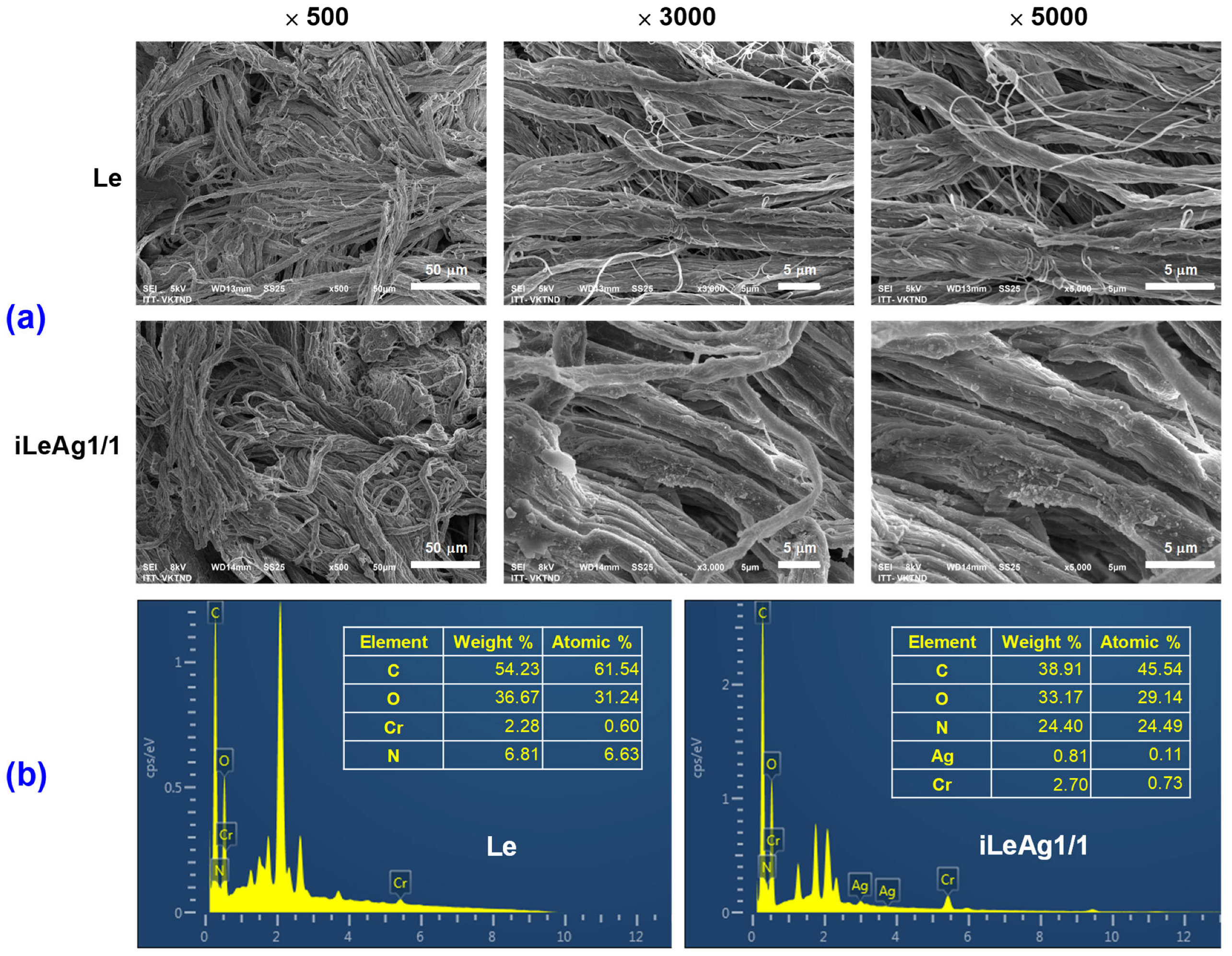
2.4. Characterization of the Treated Pig Leather

A scanning electron microscope (SEM, SM-6510LV JEOL, Tokyo, Japan) coupled with an energy dispersive X-ray spectroscope (EDX, Oxford EDS Microanalysis System, Oxford Instruments NanoAnalysis, High Wycombe, UK) was used to determine the morphologies and elemental compositions of the control and AgPBL-treated leather samples after platinum sputtering and was operated at accelerating voltages ranging from 5 to 8 kV.
Silver contents in the treated leather samples were measured via an Atomic Absorption Spectrometer (AAS, PinAAcle 900T, PerkinElmer, Waltham, MA, USA).
The FTIR analyses of the blank and treated leather samples were carried out using an FTIR spectrometer (Nicolet 6700, Thermo Scientific, Waltham, MA, USA), in transmission mode and the range of 4000–400 cm−1 with a spectral resolution of 4 cm−1.
Color changes of the blank and treated leather samples, in terms of L*, a*, and b* values, and color differences (ΔE*) were determined using a reflectance spectrophotometer (Ci4200, X-rite, Grandville, MI, USA) with D65 illumination and a 10° observer. In the CIELab color space, L* represents lightness, while a* and b* represent chromaticity parameters. The average color parameter values were evaluated at three positions for each sample. The total color difference was calculated based on Equation (3).
E = L 2 + a 2 + b 2
where ΔL*, Δa*, and Δb* represent the changes in L*, a*, and b* between the initial and three values, respectively.

2.5. Physico-Mechanical Properties of the Treated Pig Leather

The AgPBL-treated leather was evaluated for its physico-mechanical properties, which are essential requirements for shoe upper lining materials, in accordance with the standard ISO 20882:2007 [47]. These properties include tear strength (ISO 17696), abrasion resistance (ISO 17704), flex resistance (ISO 17694), lining water vapour permeability and absorption (ISO 17699), and lining water absorption and desorption (ISO 22649). The physico-mechanical tests were performed at the Institute of Footwear Research, Vietnam.

3. Results and Discussion

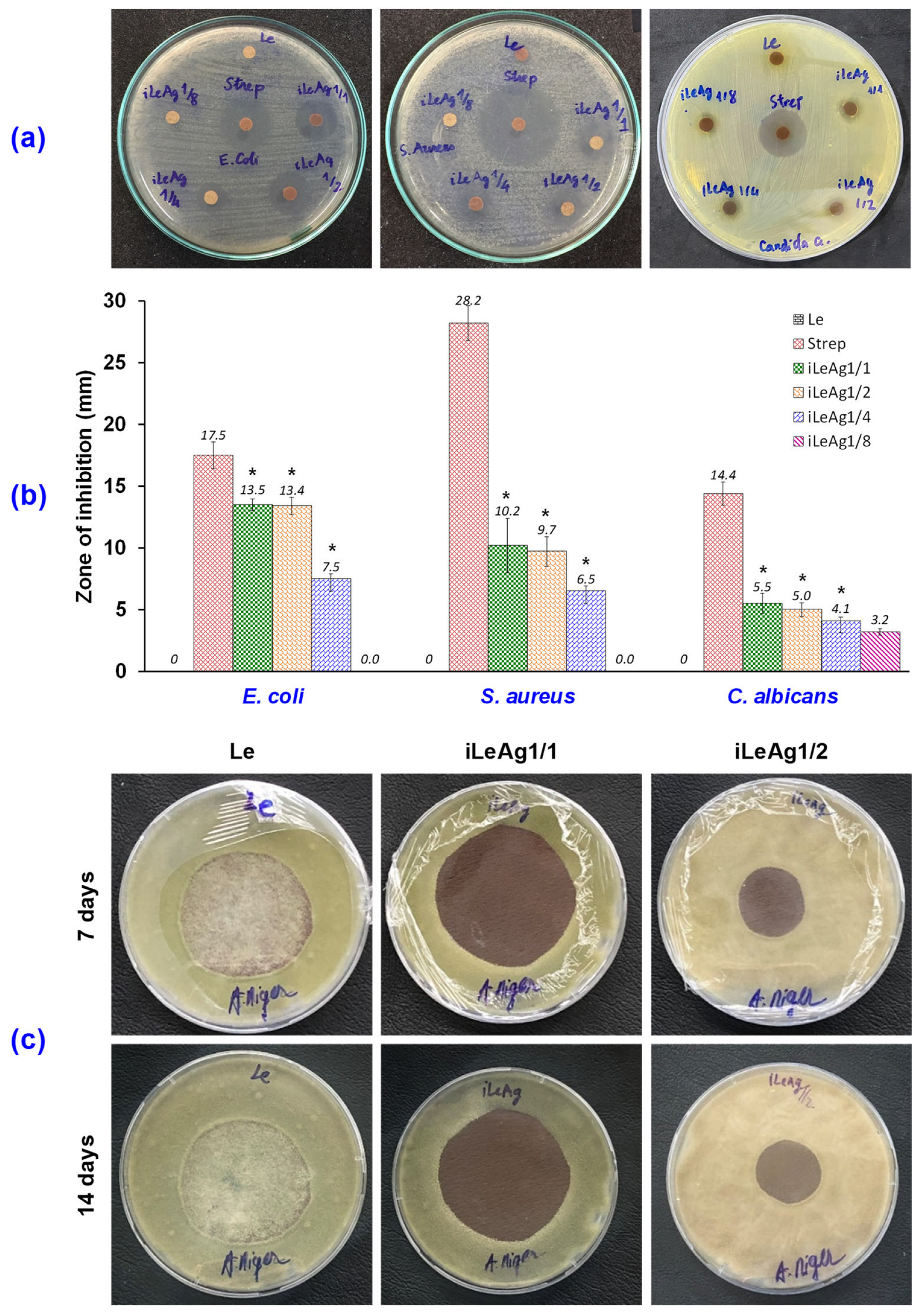
3.1. Antibacterial and Antifungal Efficacy of the AgPBL-Treated Pig Leather

The antimicrobial properties of blank and treated pig leather samples were evaluated against different microorganisms, including two bacteria (E. coli and S. aureus) and two fungi (C. albicans and A. niger). Both qualitative (the disk diffusion method) and quantitative (the static challenge protocol of dynamic contact method) tests were employed to determine the antimicrobial activities of the treated leather. The pig leather samples were treated with 160, 80, 40, and 20 μg/mL AgPBL and were labeled with iLeAg1/1, iLeAg1/2, iLeAg1/4, and iLeAg1/8, respectively.

3.1.1. Antibacterial Efficacy

The AgPBL-treated leather samples obtained via the exhaustion method were subjected to antibacterial tests against a gram-negative bacterium E. coli and a gram-positive bacterium S. aureus. The negative control for the tested bacteria was pristine leather, while the positive one was leather treated with the standard reference antibiotic Streptomycin (Strep, 80 μg/mL). The effect of the AgPBL concentration on the antibacterial efficacy of the treated leather was investigated. As shown in Figure 2a, all the AgPBL-treated leather samples and the Strep-impregnated leather exhibited an obvious ZOI against E. coli and S. aureus; whereas, the pristine leather (Le) showed no activity at all. Furthermore, the leather sample iLeAg1/8 impregnated with AgPBL concentration of 20 μg/mL did not clearly reveal a growth inhibition halo against both tested bacteria. The results of this work indicate that the antibacterial activity of the treated leather was effective when the initial AgPBL concentration in the immersing solution was above 40 μg/mL. The antibacterial results of the immersed leather samples are plotted in Figure 2b, confirming that higher concentrations of AgPBL corresponded to larger zones of inhibition. However, the relation between AgPBL concentrations and inhibition zones was not proportional. Indeed, as compared to the iLeAg1/1 sample, when the AgPBL concentrations were decreased by 50% and 75%, the ZOI of iLeAg1/2 and iLeAg1/4 samples decreased by 0.74% and 44.44% against E. coli, and by 4.90% and 36.27% against S. aureus, respectively. In addition, there was a slight difference in the inhibition zone observed between leather samples treated with AgPBL concentrations of 160 and 80 μg/mL. Furthermore, the findings revealed that the AgPBL-impregnated leather samples had a greater efficacy against gram-negative bacterium than gram-positive ones, which contradicted the results obtained from the antibacterial assessment of the AgPBL solution [42,48]. This disparity may be attributed to the electrostatic interaction between Ag+ ions and the negative charge of RCOO or the lone-pair electrons of N atoms in the amino acids of collagen proteins present in the leather [18,23]. These interactions could hinder the release of AgPBL from the iLeAg sample into the medium containing the tested bacteria. Consequently, the concentration of released AgPBL in the bacterial medium during the static conditions of the qualitative test might not be sufficient to effectively inhibit the growth of S. aureus, in contrast to its impact on E. coli, leading to a smaller ZOI. It is important to emphasize that this hypothesis requires additional evidence to confirm its validity.
The quantitative assessment of the antibacterial activities of AgPBL-treated leather samples was conducted to determine the percentage reduction of bacteria according to the static challenge protocol of the dynamic contact method (ISO 16187:2013). The results depicted in Figure 3 were consistent with those obtained from the disk diffusion method, in which the treated leather exhibited significant antimicrobial activity against the tested bacteria. In Figure 3a, the blank leather sample (Le) did not reveal any antibacterial activity against the tested bacteria, which were observed to form dense bacterial colonies on the LB agar plates. On the contrary, the leather impregnated with AgPBL demonstrated considerable antibacterial activity due to a significant decrease in bacterial colonies after 0 h and 24 h contact times. Furthermore, iLeAg1/1 had the highest antibacterial activity, while iLeAg1/8 had the lowest, as indicated by agar turbidity.
The bacterial reduction percentage (%R) of the treated leather plotted in Figure 3b showed that the iLeAg1/1 samples treated with 160 μg/mL AgPBL exhibited the highest antibacterial efficiency against both E. coli and S. aureus after 0 h and 24 h contact times. After 0 h contact time, the %R values on E. coli and S. aureus were 41.07% and 66.03%, respectively, and both increased to 99.99% after 24 h contact time. The %R values of iLeAg12 samples treated with 80 μg/mL AgPBL against E. coli and S. aureus decreased by 22.72% and 2.46%, respectively, after 24 h contact time compared to iLeAg1/1 samples. Comparatively, iLeAg1/8 samples treated with 20 μg/mL AgPBL showed a significant decline in antibacterial activity against both bacteria. Their bactericidal rates against E. coli and S. aureus decreased by 83.62% and 35.36%, respectively, after 24 h contact time compared to iLeAg1/1 samples. It is worth noting that although zones of inhibition of iLeAg1/8 samples were not detected in the qualitative antibacterial test, the quantitative test revealed that these samples still had slight antibacterial activities. The results indicated that in the dynamic contact method, the iLeAg samples displayed greater antibacterial efficacy against gram-positive bacteria compared to gram-negative bacteria. This observation aligns with the findings from the antibacterial assessment of the pure AgPBL solution [42]. In this quantitative method, the AgPBL was sturdily released from the iLeAg samples into the SCDLP medium and directly attached to the targeted bacteria. Consequently, the antibacterial mechanism observed in the quantitative tests for the iLeAg samples is similar to that of the pure AgPBL solution. Although the precise antibacterial mechanisms of nanosilver remain incompletely understood, the prevailing hypothesis suggests that nanosilver primarily acts by adhering onto the surface of bacterial cell membranes and subsequently infiltrating the interior of the bacteria. Within the bacterial cells, the silver nanoparticles release silver ions, which intensify their bactericidal efficacy by interacting with DNA and compounds rich in phosphorus and sulfur. This interaction ultimately leads to cell death [30,33,37,42,48].

3.1.2. Antifungal Efficacy

The antifungal properties of the AgPBL-treated leather samples were analyzed qualitatively by checking them against one yeast strain (C. albicans) and one mold strain (A. niger) according to AATCC TM30. The results from the disk diffusion tests were reported in Figure 2. It is obvious that the presence of nanosilver on pig leather samples significantly improved the antifungal activities. Indeed, although a very low concentration of AgPBL (20 μg/mL) was employed to treat the pig leather; a well-defined inhibition halo to C. albicans growth was clearly observed around the treated sample. Despite being chrome-tanned leather, the untreated leather samples did not expose any antifungal activities against both tested fungi. Figure 2a,b demonstrate that the antifungal ability of the treated leather reduced with the decrease in AgPBL concentration. This was supported by a decrease in the ZOI values against the C. albicans strain of 9.09% and 25.45% when the AgPBL concentration was reduced by 50% and 75%, respectively. The iLeAg1/1 sample impregnated with 160 μg/mL AgPBL solution exhibited noteworthy antifungal efficacy against A. niger after seven and 14 days, as reported in Figure 2c. This was evidenced by no mold spore germination or growth surrounding the samples. However, mold spore growth was clearly observed on the iLeAg1/2 sample, which was immersed in the 80 μg/mL AgPBL solution, after seven days of incubation, and it became even more pronounced after 14 days. These obtained results prove again the significant influence of the AgPBL concentration on the antimicrobial effectiveness of the treated leather. Notably, both tested fungi showed greater resistance to the AgPBL-treated leather compared to the tested bacteria, as observed in the antimicrobial efficacy of the treated samples against both bacterial and fungal strains. These findings suggest that the AgPBL concentration of 160 μg/mL should be selected for the treatment of pig leather in order to achieve effective antimicrobial activities against both bacteria and fungi. This treated leather (iLeAg1/1) was further characterized by SEM, EDX, AAS, and FTIR to confirm the presence of nanosilver on the leather matrix, which is associated with its antimicrobial efficacy.

3.2. Coloration and Characteristics of the AgPBL-Treated Pig Leather

Due to the surface plasmon absorption of the silver nanoparticles, a brownish color was visually observed after the treatment of the leather with AgPBL solutions using the exhaustion method. The coloration of the treated leather was analyzed by examining the colorimetric data (L*, a*, b*) and color differences (ΔE*) to evaluate the impact of AgPBL concentration in the immersing solutions. The colorimetric data and color differences of the blank and treated leather samples are summarized in Table 1. The results indicated that the blank leather exhibited a bright yellow color with relatively high L*, a*, and b* values of 62.42, 9.77, and 23.86, respectively. In comparison, the treated leather samples showed slightly smaller L*, a*, and b* values when compared to the blank leather.
The decrease in the L* value corresponded to an increase in the AgPBL concentration, resulting in a darker appearance of the treated leather samples. Additionally, the color difference (ΔE*) of the treated samples increased with higher AgPBL concentrations, indicating a greater uptake of nanosilver onto the leather surfaces. This observation suggests that the AgPBL nanoparticles were evenly adhered to the leather surface, resulting in a uniform brownish color across the entire treated leather.
SEM analysis was conducted on both the blank and treated leather samples to visually confirm the presence of AgPBL embedded onto the sample surfaces. Figure 4a shows distinct hierarchically suprafibrillar structures of the collagen fiber bundles in both the blank and treated leather. In the micrographs of the treated leather samples (iLeAg1/1), nanoparticles were observed as small bright spots loosely attached to the collagen fiber surface. It is worth noting that in SEM images, elements with higher atomic numbers exhibit higher contrast, resulting in brighter spots. Therefore, the brighter spots observed in Figure 4a can be attributed to inorganic matter with a high atomic number, such as silver nanoparticles, while the organic polymer (collagen) provides a low-contrast background [36,49]. However, the silver nanoparticles utilized in this study had a size of approximately 20 nm and were capable of penetrating into the collagen fiber bundles. As a result, their detection in SEM images could be challenging due to this technique only offering insights into the surface morphology of the sample. Therefore, the EDX analytical technique was employed to confirm the existence of nanosilver on the treated leather sample.
EDX has proven to be a highly effective technique for the elemental analysis of a given material [18,23]. The EDX spectrum of the blank leather in Figure 4b did not show any signal of silver, but it did exhibit a signal for chromium because chrome-tanned leather was employed in this study. Compared to the blank leather, the EDX spectrum of the iLeAg1/1 sample displayed a strong signal at 3 keV, indicating the presence of AgPBL on the leather surface [17,23,36]. This finding further confirms that the bright spots observed in the SEM images of the iLeAg1/1 sample are indeed AgPBL.
The presence of AgPBL in the treated leather was further validated by AAS characterizations. This facilitated a direct assessment of the amount of AgPBL permeating into the pig leather after the exhaustion treatment. As reported in Table 2, the analysis results demonstrate that the total silver content of the iLeAg1/1 sample was approximately 513.3 mg/kg, while the blank leather sample had no silver content.
FTIR spectra were obtained for the Le, iLeAg1/1, and AgPBL samples to assess the chemical differences between the blank and treated leather. The aim was to investigate alterations in the functional groups of collagen proteins resulting from the antimicrobial finishing process using AgPBL. As depicted in Figure 5, the FTIR spectrum of Le displayed all the functional groups associated with the pristine pig leather. A broad peak observed at 3304.5 cm−1 was assigned to the N–H stretching vibration. The peaks at 1633.5 and 1547.8 cm−1 were attributed to the C=O stretching vibration and N–H bending vibration, respectively, representing the amide bands. The peaks at 2921.1 and 2852.3 cm−1 were associated with the C–H stretching vibrations in the protein chain. The peak observed at 1450.4 cm−1 indicated the O–H bending vibration. Additionally, the peak at 1236.6 cm−1 was attributed to the symmetrical stretching of C=O in the amide III bands, while the peak at 1030.9 cm−1 represented the stretching vibration of C–N in amines [18,27,50]. The FTIR spectrum of AgPBL exhibited characteristic peaks corresponding to various functional groups. These included O-H (3440.0 cm−1), C–H of alkanes (2922.2 cm−1), C=O of amino acids and proteins (1630.9 cm−1), N=O of nitro groups (1384.3 cm−1), C–O of phenolics (1285.2 cm−1), and C–N of aliphatic amine (1088.1 cm−1). These peaks are attributed to the presence of residual phytochemicals on the AgPBL surface after bio-synthesis [42]. When comparing the FTIR spectra of the blank leather, bio-synthesized AgPBL, and iLeAg1/1 samples, it was observed that the iLeAg1/1 spectrum exposed fairly similar characteristic peaks, including 1633.3 cm−1, 1547.7 cm−1, 1451.1 cm−1, 1380.8 cm−1, 1238.1 cm−1, and 1029.5 cm−1, to both the blank leather and AgPBL. A peak appeared at 1762.7 cm−1 in the iLeAg1/1 spectrum corresponding to the C=O stretching vibration of amino acids and proteins. This peak suggests that the immersion of the leather in the aqueous AgPBL solution might cause slight hydrolysis of the collagen protein [2,6].
The collagen proteins of leather contain polar groups such as amide and carboxylate functional groups that have an affinity for binding with metal atoms [18,23]. In the case of AgPBL, the absorption onto the collagen surface is likely facilitated by the electrostatic interaction between Ag+ ions and the negative charge of RCOO or the lone-pair electrons of N atoms in amino acids. Furthermore, hydrogen bonding between the amide and carboxylate groups of collagen proteins and the functional groups on the surface of AgPBL further contributes to these binding interactions. The observed peak shifts in the nanosilver-treated leather could be ascribed to the interaction between the silver atoms and the amide groups present in the collagen proteins. This interaction leads to an increase in the intensity of the FTIR peaks of the treated leather. The results obtained from colorimetric, SEM, EDX, AAS, and FTIR analyses of both the blank and treated leather samples provide strong evidence that AgPBL has been effectively incorporated into the leather matrix following the antibacterial treatment via the exhaustion method.

3.3. The Physico-Mechanical Properties of the AgPBL-Treated Pig Leather

In order to assess the impact of the antimicrobial treatment on the intrinsic properties of pig leather, a series of tests were conducted on the leather samples according to the ISO 20882:2007 standard. These tests aimed to evaluate the physico-mechanical properties of the treated leather in comparison to the requirements of the shoe lining material. According to the data summarized in Table 3, the mechanical properties of the nanosilver-loaded leather iLeAg1/1, such as tear strength, abrasion resistance, and flex resistance, were found to be comparable with those of the pristine leather. This indicates that the antimicrobial treatment employed in this study did not induce any significant alterations in the chemical structure of the pig leather. It also suggests that the interaction of AgPBL to the leather matrix was primarily based on physical bonding. The mechanical performance of the treated leather met all the standard requirements for shoe lining with the tear strength exceeding the standard requirement by more than 210%.
Similarly, the water absorption and water desorption properties of the treated leather were not significantly affected by the antimicrobial treatment with AgPBL. The water absorption of the treated leather remained at approximately 90% of the standard requirement for lining material. Its water desorption, however, exceeded the standard requirement by over 160%. The water vapour permeability and water vapour absorption of the treated leather were excellent, surpassing 150% of the standard requirements for upper lining material. These findings could be ascribed to the swelling effect of collagen fiber bundles after the impregnation process, which leads to increased gaps between the fibers. As the leather absorbs water, it swells, and during the drying process, it subsequently shrinks. However, the shrinkage of collagen fiber bundles is greater than that of the pores, resulting in pore expansion and an increase in water vapour permeability. Above all, the application of AgPBL on pig leather successfully enhanced its antimicrobial effectiveness without significantly impacting other comfort-related properties of this natural material. The AgPBL-loaded pig leather met all the necessary criteria outlined in the ISO 20882:2007 standard for shoe lining. The exhaustion method employed in the wet finishing stage, carried out in rotary drums, proved to be suitable and practical for tanneries.

4. Conclusions

Herein, an eco-friendly approach has been employed to develop a leather-based material with highly effective antimicrobial activity. Bio-synthesized silver nanoparticles were utilized as the antimicrobial agent to treat pig leather via the immersing-drying processes. The loading of AgPBL on the treated leather was confirmed by colorimetric, SEM, EDX, AAS, and FTIR analyses. The treated leather exhibited remarkable antimicrobial properties against a wide range of microbial strains, including both gram-negative and gram-positive strains as well as fungi. Furthermore, it also demonstrated suitable physico-mechanical properties that met the requirements of shoe lining material specified in the ISO 20882:2007 standard. These obtained results suggest that the AgPBL-loaded pig leather has great potential as a hygienic lining material for active footwear production.

Supplementary Materials

The following supporting information can be downloaded at: https://www.mdpi.com/article/10.3390/pr11071891/s1, Figure S1: UV-vis spectrum of the bio-synthesized AgPBL; Figure S2: TEM images of the AgPBL with magnification of (a) ×100 k and (c) ×400 k; (c) The particle size distribution of the AgPBL as measured by Image J software. References [42,51,52,53,54] are cited in the supplementary materials.

Author Contributions

Conceptualization, N.-T.N. and V.-H.B.; methodology, N.-T.N., V.-H.B. and T.-H.V.; formal analysis, T.-H.V.; investigation, N.-T.N., V.-H.B. and T.-H.V.; data curation, N.-T.N. and V.-H.B.; writing—original draft preparation, T.-H.V., D.-N.P., T.-H.N. and T.-M.-L.N.; writing—review and editing, N.-T.N., V.-H.B. and D.-N.P.; visualization, N.-T.N. and T.-H.V.; supervision, N.-T.N. and V.-H.B.; project administration, N.-T.N.; funding acquisition, N.-T.N. All authors have read and agreed to the published version of the manuscript.

Funding

This work was funded by the Hanoi University of Science and Technology under Grant No. T2022-PC-092.

Data Availability Statement

The data presented in this study are available in the article and Supplementary Material.

Acknowledgments

The authors would like to thank their colleagues from the School of Textile—Leather and Fashion, Hanoi University of Science and Technology for their efforts in supporting this work.

Conflicts of Interest

The authors declare no conflict of interest.

References

  1. Healy, A.; Dunning, D.N.; Chockalingam, N. Materials used for footwear orthoses: A review. Footwear Sci. 2010, 2, 93–110. [Google Scholar] [CrossRef]
  2. Nalyanya, K.M.; Rop, R.K.; Onyuka, A.S.; Birech, Z. A review of natural plants as sources of substances for cleaner leather tanning technologies. Text. Leather Rev. 2021, 4, 137–148. [Google Scholar] [CrossRef]
  3. Orlita, A. Microbial biodeterioration of leather and its control: A review. Int. Biodeterior. Biodegrad. 2004, 53, 157–163. [Google Scholar] [CrossRef]
  4. Hossain, M.; Azam, F.A.B.; Chowdhury, M. Quality assessment of shoe leather based on the properties of strength and comfort, collected from different footwear and leather industries in Bangladesh. Text. Leather Rev. 2021, 4, 30–37. [Google Scholar] [CrossRef]
  5. Bai, Z.; Wang, X.; Zheng, M.; Yue, O.; Xie, L.; Zha, S.; Dong, S.; Li, T.; Song, Y.; Huang, M.; et al. Leather for flexible multifunctional bio-based materials: A review. J. Leather Sci. Eng. 2022, 4, 16. [Google Scholar] [CrossRef]
  6. Xia, Q.; Yang, L.; Hu, K.; Li, K.; Xiang, J.; Liu, G.; Wang, Y. Chromium Cross-Linking Based Immobilization of Silver Nanoparticle Coating on Leather Surface with Broad-Spectrum Antimicrobial Activity and Durability. ACS Appl. Mater. Interfaces 2019, 11, 2352–2363. [Google Scholar] [CrossRef] [PubMed]
  7. Wu, X.; Wu, J.; Mu, C.; Wang, C.; Lin, W. Advances in Antimicrobial Polymer Coatings in the Leather Industry: A Comprehensive Review. J. Leather Sci. Eng. 2021, 60, 15004–15018. [Google Scholar] [CrossRef]
  8. Fan, Q.; Ma, J.; Xu, Q. Insights into functional polymer-based organic-inorganic nanocomposites as leather finishes. J. Leather Sci. Eng. 2019, 1, 3. [Google Scholar] [CrossRef] [Green Version]
  9. Ławińska, K.; Serweta, W.; Popovych, N.; Sieczyńska, K.; Decka, S.; Woźnicki, D.; Ogrodowczyk, D.; Rostocki, A.; Sprynskyy, M. Microbiological and chemical analysis of bamboo textile materials and leathers modified with bamboo extract at the tanning stage. Fibres Text. East. Eur. 2021, 29, 33–39. [Google Scholar] [CrossRef]
  10. Liu, G.; Haiqi, G.; Li, K.; Xiang, J.; Lan, T.; Zhang, Z. Fabrication of silver nanoparticle sponge leather with durable antibacterial property. J. Colloid Interface Sci. 2018, 514, 338–348. [Google Scholar] [CrossRef]
  11. Lkhagvajav, N.; Koizhaiganova, M.; Yasa, I.; Çelik, E.; Sari, Ö. Characterization and antimicrobial performance of nano silver coatings on leather materials. Braz. J. Microbiol. 2015, 46, 41–48. [Google Scholar] [CrossRef] [PubMed] [Green Version]
  12. Sportelli, M.C.; Picca, R.A.; Paladini, F.; Mangone, A.; Giannossa, L.C.; Di Franco, C.; Gallo, A.L.; Valentini, A.; Sannino, A.; Pollini, M. Spectroscopic characterization and nanosafety of Ag-modified antibacterial leather and leatherette. Nanomaterials 2017, 7, 203. [Google Scholar] [CrossRef] [PubMed] [Green Version]
  13. Abou Elmaaty, T.; Sayed-Ahmed, K.; Mohamed Ali, R.; El-Khodary, K.; Abdeldayem, S.A. Simultaneous sonochemical coloration and antibacterial functionalization of leather with selenium nanoparticles (SeNPs). Polymers 2022, 14, 74. [Google Scholar] [CrossRef] [PubMed]
  14. Biškauskaitė, R.; Valeika, V. Wet Blue Enzymatic Treatment and Its Effect on Leather Properties and Post-Tanning Processes. Materials 2023, 16, 2301. [Google Scholar] [CrossRef]
  15. Thanikaivelan, P.; Rao, J.R.; Nair, B.U.; Ramasami, T. Progress and recent trends in biotechnological methods for leather processing. Trends Biotechnol. 2004, 22, 181–188. [Google Scholar] [CrossRef]
  16. Parisi, M.; Nanni, A.; Colonna, M. Recycling of Chrome-Tanned Leather and Its Utilization as Polymeric Materials and in Polymer-Based Composites: A Review. Polymers 2021, 13, 429. [Google Scholar] [CrossRef]
  17. Velmurugan, P.; Lee, S.-M.; Cho, M.; Park, J.-H.; Seo, S.-K.; Myung, H.; Bang, K.-S.; Oh, B.-T. Antibacterial activity of silver nanoparticle-coated fabric and leather against odor and skin infection causing bacteria. Appl. Microbiol. Biotechnol. 2014, 98, 8179–8189. [Google Scholar] [CrossRef]
  18. Elsayed, H.; Hasanin, M.; Rehan, M. Enhancement of multifunctional properties of leather surface decorated with silver nanoparticles (Ag NPs). J. Mol. Struct. 2021, 1234, 130130. [Google Scholar] [CrossRef]
  19. Xiang, J.; Ma, L.; Su, H.; Xiong, J.; Li, K.; Xia, Q.; Liu, G. Layer-by-layer assembly of antibacterial composite coating for leather with cross-link enhanced durability against laundry and abrasion. Appl. Surf. Sci. 2018, 458, 978–987. [Google Scholar] [CrossRef]
  20. Ara, K.; Hama, M.; Akiba, S.; Koike, K.; Okisaka, K.; Hagura, T.; Kamiya, T.; Tomita, F. Foot odor due to microbial metabolism and its control. Can. J. Microbiol. 2006, 52, 357–364. [Google Scholar] [CrossRef] [Green Version]
  21. Kanlayavattanakul, M.; Lourith, N. Body malodours and their topical treatment agents. Int. J. Cosmet. Sci. 2011, 33, 298–311. [Google Scholar] [CrossRef]
  22. Lyu, B.; Chang, R.; Gao, D.; Ma, J. Chromium Footprint Reduction: Nanocomposites as Efficient Pretanning Agents for Cowhide Shoe Upper Leather. ACS Sustain. Chem. Eng. 2018, 6, 5413–5423. [Google Scholar] [CrossRef]
  23. Velmurugan, P.; Cho, M.; Lee, S.-M.; Park, J.-H.; Bae, S.; Oh, B.-T. Antimicrobial fabrication of cotton fabric and leather using green-synthesized nanosilver. Carbohydr. Polym. 2014, 106, 319–325. [Google Scholar] [CrossRef]
  24. Velmurugan, P.; Shim, J.; Bang, K.-S.; Oh, B.-T. Gold nanoparticles mediated coloring of fabrics and leather for antibacterial activity. J. Photochem. Photobiol. B 2016, 160, 102–109. [Google Scholar] [CrossRef] [PubMed]
  25. Rohaeti, E.; Kasmudjiastuti, E.; Murti, R.; Dodi, I. Enhancement of antibacterial activity of suede leather through coating silver nanoparticles synthesized using Piper betle. Rasayan J. Chem. 2020, 13, 628–635. [Google Scholar] [CrossRef]
  26. Kate, S.; Sahasrabudhe, M.; Pethe, A. Biogenic Silver Nanoparticle Synthesis, Characterization and its Antibacterial activity against Leather Deteriorates. Jordan J. Biol. Sci. 2020, 13, 493–498. [Google Scholar]
  27. Koizhaiganova, M.; Yaşa, I.; Gülümser, G. Assessment of antibacterial activity of lining leather treated with silver doped hydroxyapatite. Int. Biodeterior. Biodegrad. 2015, 105, 262–267. [Google Scholar] [CrossRef]
  28. Abdelghany, T.M.; Al-Rajhi, A.M.H.; Al Abboud, M.A.; Alawlaqi, M.M.; Ganash Magdah, A.; Helmy, E.A.M.; Mabrouk, A.S. Recent Advances in Green Synthesis of Silver Nanoparticles and Their Applications: About Future Directions. A Review. BioNanoScience 2018, 8, 5–16. [Google Scholar] [CrossRef]
  29. Jadoun, S.; Arif, R.; Jangid, N.K.; Meena, R.K. Green synthesis of nanoparticles using plant extracts: A review. Environ. Chem. Lett. 2021, 19, 355–374. [Google Scholar] [CrossRef]
  30. Thi Lan Huong, V.; Nguyen, N.T. Green synthesis, characterization and antibacterial activity of silver nanoparticles using Sapindus mukorossi fruit pericarp extract. Mater. Today Proc. 2021, 42, 88–93. [Google Scholar] [CrossRef]
  31. Gour, A.; Jain, N.K. Advances in green synthesis of nanoparticles. Artif. Cell. Nanomed. Biotechnol. 2019, 47, 844–851. [Google Scholar] [CrossRef] [Green Version]
  32. Rafique, M.; Sadaf, I.; Rafique, M.S.; Tahir, M.B. A review on green synthesis of silver nanoparticles and their applications. Artif. Cell. Nanomed. Biotechnol. 2017, 45, 1272–1291. [Google Scholar] [CrossRef]
  33. Sharma, V.K.; Yngard, R.A.; Lin, Y. Silver nanoparticles: Green synthesis and their antimicrobial activities. Adv. Colloid Interface Sci. 2009, 145, 83–96. [Google Scholar] [CrossRef]
  34. Nguyen, N.-T.; Liu, J.-H. A green method for in situ synthesis of poly(vinyl alcohol)/chitosan hydrogel thin films with entrapped silver nanoparticles. J. Taiwan Inst. Chem. Eng. 2014, 45, 2827–2833. [Google Scholar] [CrossRef]
  35. Ahmad, S.; Munir, S.; Zeb, N.; Ullah, A.; Khan, B.; Ali, J.; Bilal, M.; Omer, M.; Alamzeb, M.; Salman, S.M.; et al. Green nanotechnology: A review on green synthesis of silver nanoparticles—An ecofriendly approach. Int. J. Nanomed. 2019, 14, 5087–5107. [Google Scholar] [CrossRef] [Green Version]
  36. Nguyen, N.-T.; Vo, T.-L.-H. Fabrication of Silver Nanoparticles Using Cordyline fruticosa L. Leave Extract Endowing Silk Fibroin Modified Viscose Fabric with Durable Antibacterial Property. Polymers 2022, 14, 2409. [Google Scholar] [CrossRef] [PubMed]
  37. Sharma, D.; Kanchi, S.; Bisetty, K. Biogenic synthesis of nanoparticles: A review. Arabian J. Chem. 2019, 12, 3576–3600. [Google Scholar] [CrossRef] [Green Version]
  38. Kharissova, O.V.; Dias, H.V.R.; Kharisov, B.I.; Pérez, B.O.; Pérez, V.M.J. The greener synthesis of nanoparticles. Trends Biotechnol. 2013, 31, 240–248. [Google Scholar] [CrossRef] [PubMed]
  39. Alduraihem, N.S.; Bhat, R.S.; Al-Zahrani, S.A.; Elnagar, D.M.; Alobaid, H.M.; Daghestani, M.H. Anticancer and Antimicrobial Activity of Silver Nanoparticles Synthesized from Pods of Acacia nilotica. Processes 2023, 11, 301. [Google Scholar] [CrossRef]
  40. Rizwana, H.; Alzahrani, T.; Alwahibi, M.S.; Aljowaie, R.M.; Aldehaish, H.A.; Alsaggabi, N.S.; Ramadan, R. Phytofabrication of Silver Nanoparticles and Their Potent Antifungal Activity against Phytopathogenic Fungi. Processes 2022, 10, 2558. [Google Scholar] [CrossRef]
  41. Shashiraj, K.N.; Nayaka, S.; Kumar, R.S.; Kantli, G.B.; Basavarajappa, D.S.; Gunagambhire, P.V.; Almansour, A.I.; Perumal, K. Rotheca serrata Flower Bud Extract Mediated Bio-Friendly Preparation of Silver Nanoparticles: Their Characterizations, Anticancer, and Apoptosis Inducing Ability against Pancreatic Ductal Adenocarcinoma Cell Line. Processes 2023, 11, 893. [Google Scholar] [CrossRef]
  42. Vu, T.H.; Bui, V.H.; Nguyen, N.T. Antibacterial Properties of Silver Nanoparticles Synthesized Using Piper betle L. Leaf Extract. Mater. Sci. Forum 2021, 1020, 236–242. [Google Scholar] [CrossRef]
  43. Nguyen, N.-T.; Vu, T.-H.; Bui, V.-H. Antibacterial and Antifungal Fabrication of Natural Lining Leather Using Bio-Synthesized Silver Nanoparticles from Piper betle L. Leaf Extract. Polymers 2023, 15, 2634. [Google Scholar] [CrossRef]
  44. AATCC TM30; Test Method for Antifungal Activity, Assessment on Textile Materials: Mildew and Rot Resistance of Textile Materials. AATCC: Durham, NC, USA, 2017.
  45. AATCC TM90; Test Method for Antibacterial Activity of Textile Materials: Agar Plate. AATCC: Durham, NC, USA, 2016.
  46. ISO 16187:2013; Footwear and Footwear Components—Test Method to Assess Antibacterial Activity. ISO: Geneva, Switzerland, 2013.
  47. ISO 20882:2007; Footwear—Performance Requirements for Components for Footwear—Lining and Insocks. ISO: Geneva, Switzerland, 2007.
  48. Zhang, X.; Wang, W.; Yu, D. Synthesis of waterborne polyurethane–silver nanoparticle antibacterial coating for synthetic leather. J. Coat. Technol. Res. 2018, 15, 415–423. [Google Scholar] [CrossRef]
  49. Shaheen, T.I.; Abd El Aty, A.A. In-situ green myco-synthesis of silver nanoparticles onto cotton fabrics for broad spectrum antimicrobial activity. Int. J. Biol. Macromol. 2018, 118, 2121–2130. [Google Scholar] [CrossRef]
  50. Carvalho, I.; Lima, M.J.; Nobre, D.; Marques, S.M.; Castro, D.; Leite, T.R.; Henriques, M.; Duarte, F.; Ramalho, A.; Carvalho, S. Silver oxide coatings deposited on leathers to prevent diabetic foot infections. Surf. Coat. Technol. 2022, 442, 128338. [Google Scholar] [CrossRef]
  51. Varghese Alex, K.; Tamil Pavai, P.; Rugmini, R.; Shiva Prasad, M.; Kamakshi, K.; Sekhar, K.C. Green Synthesized Ag Nanoparticles for Bio-Sensing and Photocatalytic Applications. ACS Omega 2020, 5, 13123–13129. [Google Scholar] [CrossRef]
  52. Xia, Y.; Xiong, Y.; Lim, B.; Skrabalak, S.E. Shape-Controlled Synthesis of Metal Nanocrystals: Simple Chemistry Meets Complex Physics? Angew. Chem. Int. Ed. 2009, 48, 60–103. [Google Scholar] [CrossRef] [Green Version]
  53. Almansob, A.; Bahkali, A.H.; Albarrag, A.; Alshomrani, M.; Binjomah, A.; Hailan, W.A.; Ameen, F. Effective treatment of resistant opportunistic fungi associated with immuno-compromised individuals using silver biosynthesized nanoparticles. Appl. Nanosci. 2022, 12, 3871–3882. [Google Scholar] [CrossRef] [PubMed]
  54. He, Q.; Lu, J.; Liu, N.; Lu, W.; Li, Y.; Shang, C.; Li, X.; Hu, L.; Jiang, G. Antiviral Properties of Silver Nanoparticles against SARS-CoV-2: Effects of Surface Coating and Particle Size. Nanomaterials 2022, 12, 990. [Google Scholar] [CrossRef]
Figure 1. Schematic illustration of the bio-synthesis of AgPBL and its immersion into pig lining leather via exhaustion method, namely iLeAg.
Processes 11 01891 g001
Figure 2. (a) The photographs show the zone of inhibition of the Le, iLeAg, and Streptomycin (80 μg/mL) treated leather samples against E. coli, S. aureus, and C. albicans strains with change in the AgPBL concentration (160, 80, 40, and 20 μg/mL); (b) mean zone of inhibition of leather samples against E. coli, S. aureus and C. albicans strains (* p < 0.05); (c) antifungal activities of the Le, iLeAg1/1, and iLeAg1/2 samples against A. niger after seven and 14 days of incubation.
Processes 11 01891 g002
Figure 3. (a) The photographs of bacterial growth in the nutrient agar plates; and (b) the bacterial reduction percentage (%R) of the Le and AgPBL-treated leather samples against E. coli and S. aureus (* p < 0.05).
Processes 11 01891 g003aProcesses 11 01891 g003b
Figure 4. (a) The SEM micrographs at different magnifications of ×500, ×3000 and ×5000; and (b) EDX spectra of the blank leather (Le) and the AgPBL-treated leather sample (iLeAg1/1).
Processes 11 01891 g004
Figure 5. The FTIR spectra of the Le, AgPBL, and iLeAg1/1 samples.
Processes 11 01891 g005
Table 1. Colorimetric data, color differences, and images of the AgPBL-treated leather samples (iLeAg) in comparison with the blank pig leather (Le).
SampleAgPBL (μg/mL)L*a*b*ΔE*Real Images
Le062.429.7723.860Processes 11 01891 i001
iLeAg1/116057.289.622.082.39Processes 11 01891 i002
iLeAg1/28057.729.7822.222.23Processes 11 01891 i003
iLeAg1/44059.289.7322.741.51Processes 11 01891 i004
iLeAg1/82059.759.6523.311.19Processes 11 01891 i005
Table 2. Total silver content of the Le and iLeAg1/1 samples.
SampleAgPBL
(μg/mL)
Total Silver Content
(mg/kg)
Le00
iLeAg1/1160513.3 ± 7.2
Table 3. The physico-mechanical properties of the Le and iLeAg1/1 according to the standard ISO 20882:2007.
NoPropertiesUnitLeiLeAg1/1ISO 20882:2007
Requirements
1Tear strength
(ISO 17696)
N3231.9lining ≥ 15 N
Compared to the pristine leather (Le)%100.099.7
Compared to ISO 20882:2007%213.3212.7
2Abrasion resistance
(ISO 17704)
cyclesWithout hole through the thickness of the material component25,600 cycles dry
12,800 cycles wet
3Flex resistance
(ISO 17694)
cycles15,000 cycles dry without visible damageDry 15,000 cycles without visible damage
4Lining water vapour permeability
(ISO 17699)
mg/cm2·h3.133.56WVP of lining ≥ 2.0 mg/cm2·h
Compared to the pristine leather (Le)%100.0113.7
Compared to ISO 20882:2007%156.0178.0
5Lining water vapour absorption
(ISO 17699)
mg/cm221.020.6WVA of lining ≥ 8.0 mg/cm2
Compared to the pristine leather (Le)%100.098.1
Compared to ISO 20882:2007%263.0257.5
6Lining water absorption
(ISO 22649)
mg/cm254.153.6absorption ≥ 70 mg/cm2
Compared to the pristine leather (Le)%100.099.1
Compared to ISO 20882:2007%90.189.0
7Lining water desorption
(ISO 22649)
%97.196.8desorption ≥ 60%
Compared to the pristine leather (Le)%100.099.7
Compared to ISO 20882:2007%161.8161.3
Disclaimer/Publisher’s Note: The statements, opinions and data contained in all publications are solely those of the individual author(s) and contributor(s) and not of MDPI and/or the editor(s). MDPI and/or the editor(s) disclaim responsibility for any injury to people or property resulting from any ideas, methods, instructions or products referred to in the content.

