Article

In Silico Analysis of Plant Flavonoids as Potential Inhibitors of Newcastle Disease Virus V Protein

1
Centre of Agricultural Biochemistry and Biotechnology, University of Agriculture, Faisalabad 38000, Pakistan
2
Department of Zoology, GC University, Lahore 54000, Pakistan
3
Department of Environmental Sciences and Engineering, GC University, Faisalabad 38000, Pakistan
4
Department of Bioinformatics and Biotechnology, GC University, Faisalabad 38000, Pakistan
5
Department of Zoology, College of Sciences, King Saud University, P.O. Box 2455, Riyadh 11451, Saudi Arabia
6
Department of Infection and Immunity, Research Centre, King Faisal Specialist Hospital and Research Centre, Riyadh 11564, Saudi Arabia
*
Author to whom correspondence should be addressed.
Processes 2022, 10(5), 935; https://doi.org/10.3390/pr10050935
Submission received: 11 February 2022 / Revised: 28 April 2022 / Accepted: 5 May 2022 / Published: 9 May 2022
(This article belongs to the Special Issue Molecular Modeling: Computer-Aided Drug Design)

Abstract

:
Newcastle disease is a viral infection causing serious economic losses to the global poultry industry. The V protein of Newcastle disease virus (NDV) is a pathogenicity determinant having various functions such as the suppression of apoptosis and replication of the NDV. This study was designed to assess the resistance potential of plant flavonoids against the V protein of Newcastle disease virus. Sequence analysis was performed using EXPASY and ProtParam tools. To build the three-dimensional structure of V protein, a homology-modeling method was used. Plant flavonoids with formerly reported therapeutic benefits were collected from different databases to build a library for virtual screening. Docking analysis was performed using the modeled structure of V protein on MOE software. Interaction analysis was also performed by MOE to explain the results of docking. Sequence analysis and physicochemical properties showed that V protein is negatively charged, acidic in nature, and relatively unstable. The 3D structure of the V protein showed eight β-pleated sheets, three helices, and ten coiled regions. Based on docking score, ten flavonoids were selected as potential inhibitors of V protein. Furthermore, a common configuration was obtained among these ten flavonoids. The interaction analysis also identified the atoms involved in every interaction of flavonoid and V protein. Molecular dynamics (MD) simulation confirmed the stability of two compounds, quercetin-7-O-[α-L-rhamnopyranosyl(1→6)-β-D-galactopyranoside] and luteolin 7-O-neohesperidoside, at 100 ns with V protein. The identified compounds through molecular docking and MD simulation could have potential as NDV-V protein inhibitor after further validation. This study could be useful for the designing of anti-NDV drugs.

1. Introduction

Newcastle disease, caused by Newcastle disease virus (NDV), is a major threat to the global poultry industry that causes socio-economic crises [1]. The Office International des Epizooties [OIE] has reported it LISTED due to its serious consequences [2]. This disease is prevalent in different regions of the world, affecting about 250 bird species (IICAB, 2016). NDV is a single-stranded RNA virus surrounded by an envelope with a genome size of ~15 kb [3]. The NDV genome consists of six genes coding for six different proteins: nucleocapsid protein (NP), phosphoprotein (P), hemagglutinin-neuraminidase (HN), matrix protein (M), large protein (L), and fusion protein (F) [4]. Through an RNA-editing mechanism, P gene of NDV codes for three different proteins i.e., W, V, and P [5,6]. NDV infects a great variety of free-living birds and is spread throughout the world. NDV is also called avian paramyxovirus serotype 1 as it belongs to the family Paramyxoviridae [7]. The NDV is included in the Avulavirus class of the Paramyxoviridae family [8]. Even though NDV just has one serotype, there are eighteen distinct genotypes (from genotype I to genotype XVIII), recognized according to the nucleotide sequence of the fusion (F) protein gene [9,10].
The V protein (36 kDa) of NDV is comprised of 239 amino acids [11]. It is produced by the addition of one G residue in the sequence of P protein through RNA editing. It interacts with N protein and modulates viral RNA replication [12]. NDV encoded V protein prevents the interferon α/β reaction through its carboxyl-terminal domain, which is essential for infection. It has been observed that V protein inhibits host interferon reaction by two means. It hinders interferon signaling by steering the signal transducer and activator of transcription 1 (STAT1) for its deprivation by the carboxyl end domain. Secondly, the carboxyl end of the V protein of NDV binds to melanoma differentiation-associated protein 5 (MDA5) and prevents interferon reaction [13]. The addition of two G amino acids at the RNA editing location on the P gene codes for W mRNA. Nevertheless, W mRNA has been identified in NDV-diseased cells, while W protein in an NDV-diseased cell has not ever been described. The replication of NDV needs the evasion of the pro-apoptotic pathway to achieve effective virus production and extension; V protein is involved in the interference of cell apoptotic pathway. That is the reason that this protein is used to treat cancer. The V protein of NDV has a C-terminal domain that is cysteine-rich and attached with two atoms of Zn2+ like other paramyxovirus V proteins [14].
Flavonoids are plant secondary metabolites that are recognized as the typical colored pigments of plant tissues [15]. Flavonoids are chemicals with different phenolic residues and are present in different plant parts [16]. These compounds have been known for their advantageous properties for human health long before these have been isolated as effective compounds. The starting materials of flavonoids are phenylalanine and malonyl-CoA [17]. The enzymes that take part in the biosynthesis of flavonoids are oxoglutarate-dependent dioxygenases, glycosyl transferases, and other flavonoids [18]. Epidemiologic research proposes a defensive part of flavonoids found in the diet to counter coronary artery disease [19]. Another significant effect of flavonoids is the scavenging of oxygen derivative-free radicals. These days, attention to their potential health benefits has increased as flavonoid activities have been detected in vitro and in vivo. These studies exhibited that flavonoids possess anti-carcinogenic, anti-inflammatory, anti-viral, and anti-allergic characteristics [20].
Flavonoids are well-known therapeutic, organic compounds. Their antivirus potential has been established recently [21], but their function as anti-NDV has not yet been explored. In this study, we have analyzed the anti-NDV potential of flavonoids by docking and binding analysis with the V protein of NDV.

2. Results

2.1. Sequence Analysis and Chemical Properties

The EXPASY server was used to obtain and analyze the chemical parameters. From this analysis, the theoretical molecular weight of V protein was obtained as 25,152.62 Da. The isoelectric point was 5.27, indicating the acidic nature of the protein. There were 28 negatively charged amino acids and 22 amino acids with positive charge, giving an overall negative charge to the protein. The value of the extinction coefficient was calculated as 36,355. The instability index was obtained as 41.10, indicating that this protein was unstable.

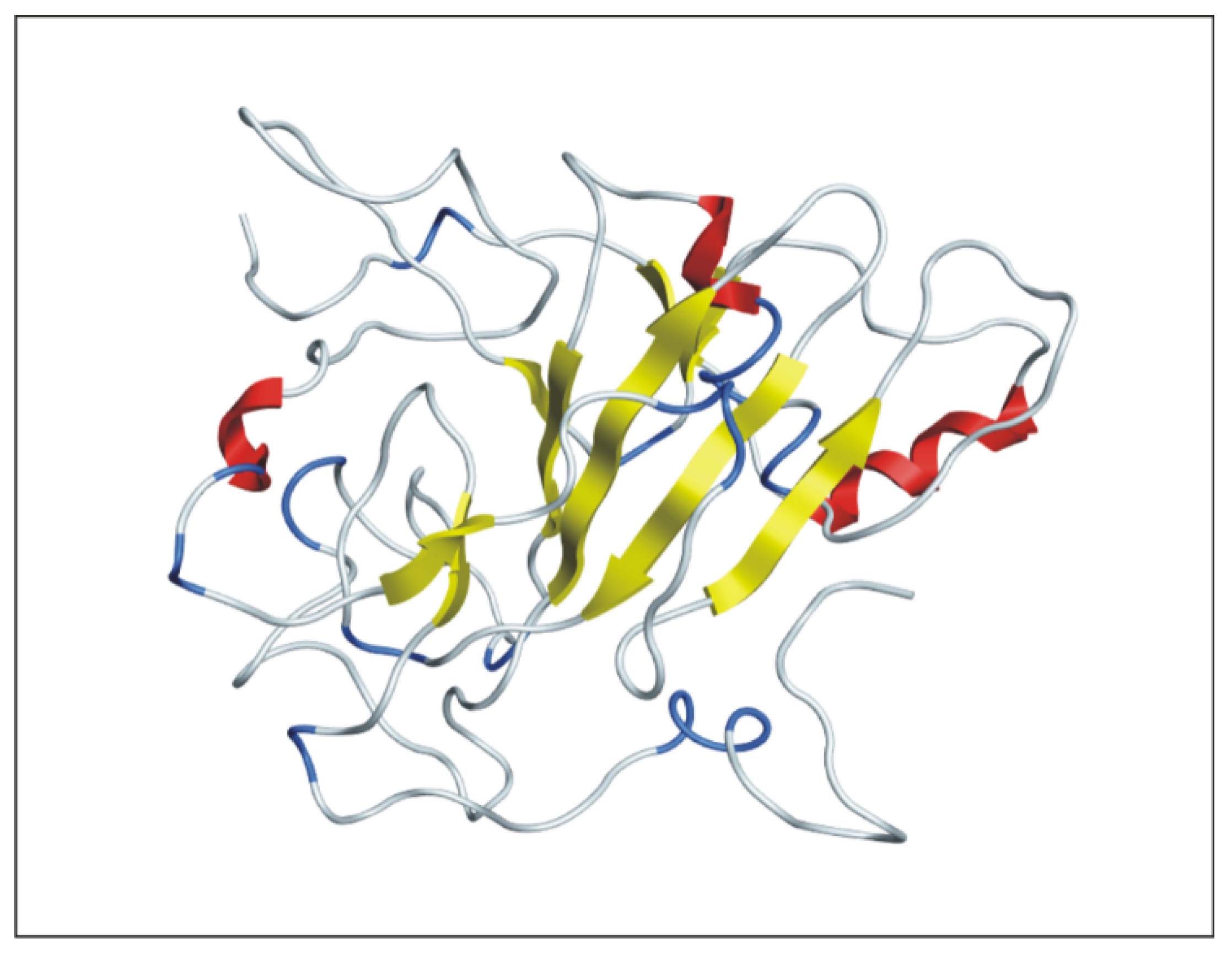
2.2. D Structure Predictions

Five models were obtained using Modeler v9.19 software. Template identification was performed by Swiss model. For homology modeling, PDB ID: 2HYE was used as a template. Based on different model evaluation tools, the best model (Figure 1) was selected. For model evaluation, the Ramachandran plot from RAMPAGE indicated that 92 percent of residues were in the most favored region (Figure 2). The Z-score obtained by Pros-web indicated that the quality of the model is in the X-ray crystallographic region (Figure 3). The superimposition of the template and model was performed by a molecular operating environment (MOE) platform and resulted in a high similarity with an RMSD value of 0.24 Å. In this model, there were three α-helices, eight β-pleated sheets, and four coiled regions as shown in red, yellow, and white colors, respectively, in Figure 1. This structure was used in further docking experiments.

2.3. Docking Experiments

Protein–ligand binding assays for V protein were performed by the MOE compute option. The top model of the V protein of NDV was used as a central molecule in docking experiments based on the results of model evaluation. A library of 250 flavonoids was constructed to perform docking experiments. From these experiments, the docking score for each ligand with each different configuration was obtained. Based on higher docking scores, ten flavonoids were selected. These flavonoids could be potential inhibitors of NDV V protein, and the chemical structures of these flavonoids are presented in Figure 4. The plant sources of each flavonoid along their ChEBI ID and docking scores are shown in Table 1.

2.4. Calculation of Common Scaffold

The flexible alignment of the ten best flavonoids was performed to obtain a common structural configuration by MOE software using the Ligand Alignment feature. By this alignment, a common scaffold was obtained. The structure of this scaffold is shown in Figure 5.

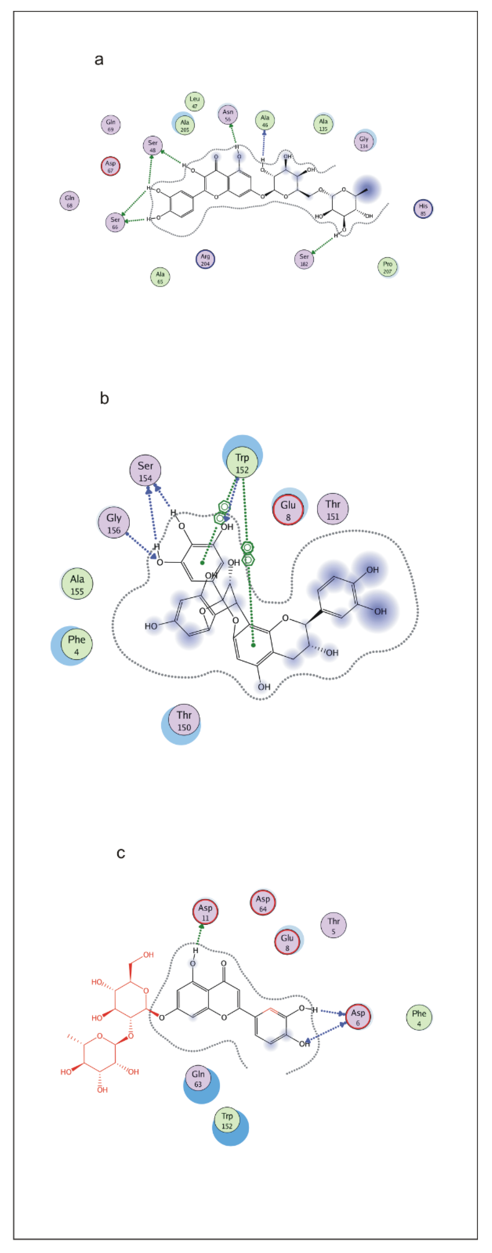
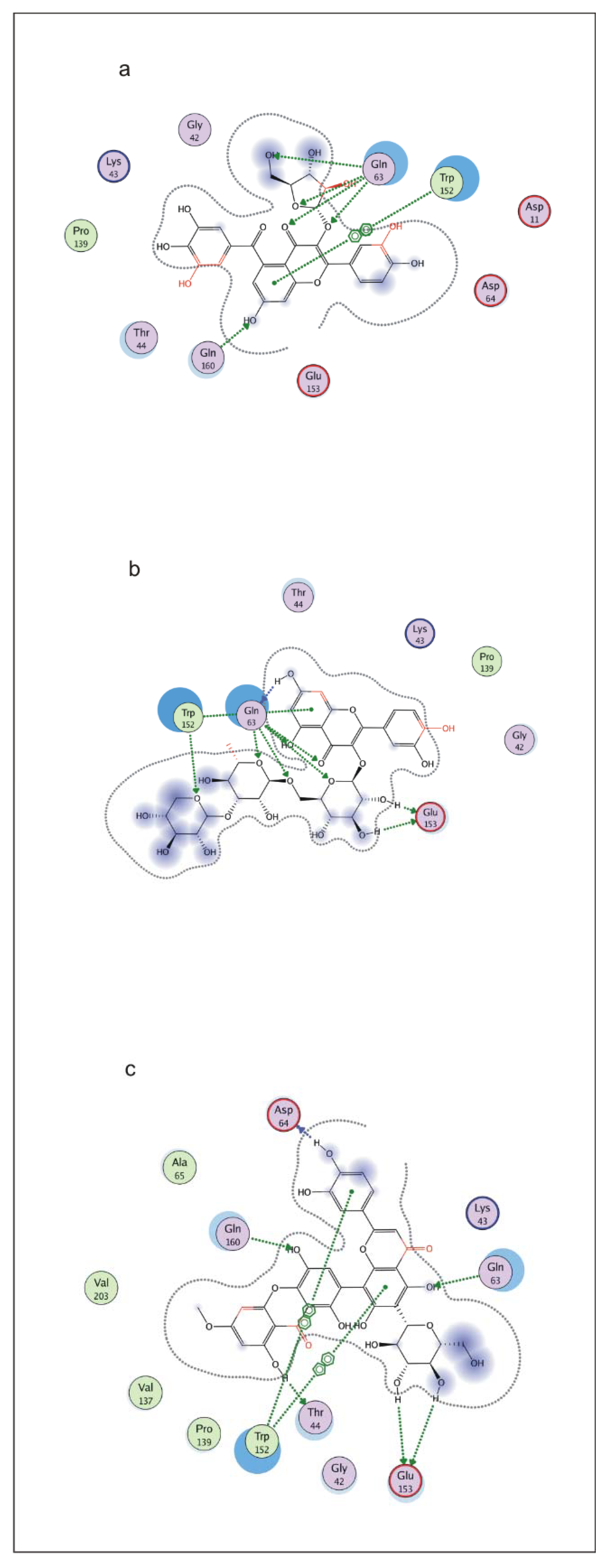
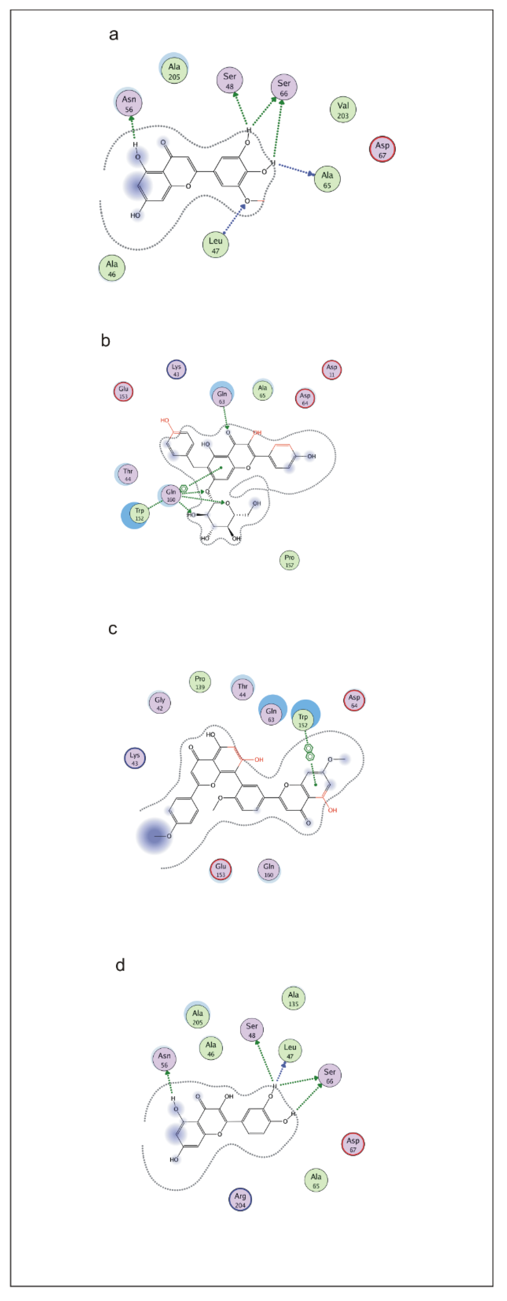
2.5. Ligand Interactions

Interaction studies were performed by MOE software. The results obtained from interaction studies provide the information of atoms involved in the interaction and types of bonds along with distances. The illustrated sight of interactions is presented in Figure 6. Flavonoid 1 showed interactions such as hydrogen bonding with Ser 48, Ser 66, Asn 56, Ala 46, and Ser 182 residues of V protein (Figure 7a). Flavonoid 2 formed interactions with Gly 56 and Ser 154 and an arene–arene link with Trp 152 (Figure 7b). Flavonoid 3 formed links with receptor residues at positions Asp 11 and Asp 6 (Figure 7c). Flavonoid 4 formed a link with Gln 63 and Gln 160 and an arene–arene bond with Trp 152 (Figure 8a). Flavonoid 5 formed an interaction with receptor atoms at the following positions: Gln 63, Trp 152, and Glu 153 (Figure 8b). Flavonoid 6 developed an interaction with V protein at the following residues: Asp 64, Gln 160, Gln 63, Glu 153, and Trp 152 (Figure 8c). Flavonoid 7 developed an interaction with the following V protein residues: Ser 48, Ser 66, Asn 56, Ala 65, and Leu 47 (Figure 9a). Flavonoid 8 showed interactions with Gln 63, Gln 160, and Trp 152 residues of the receptor protein (Figure 9b). Flavonoid 9 interacted with the Trp 152 residue of V protein (Figure 9c). Flavonoid 10 made interactions with Asn 56, Ser 48, Leu 47, and Ser 66 (Figure 9d).

2.6. Molecular Dynamic Simulation

Molecular dynamics (MD) simulation is an in silico method commonly used to study the dynamic behavior and stabilization of the protein–ligand complex during different conditions. To validate the docking results, a molecular dynamics (MD) simulation of the best ligand poses was performed using the Desmond Package (http://www.schrodinger.com/products/desmond (accessed on 3 May 2022)). The MD simulation was conducted at 100 ns of initial confirmation of the protein–ligand complexes obtained after the docking analysis.
The MD simulation of V Protein with quercetin-7-O-[α-L-rhamnopyranosyl(1→6)-β-D-galactopyranoside] and luteolin 7-O-neohesperidoside was executed with the Desmond Simulation Package for 100 ns per complex. The stability of the modeled proteins was verified by plotting the root mean square deviation (RMSD) during the production run, as shown in Figure 10a,b. The RMSD for the complex showed the compact fluctuation falling at an average of 0.5 s and the stable trajectory throughout the production run with a maximum deviation in the range of 1.8 Å and 2.4 Å.

3. Discussion

Newcastle disease (ND) is a widely reported epidemic of domestic poultry and free-living birds. Its morbidity and mortality rate is very high as reported by the World Health Organization for Animals (OIE) [22]. The virus encodes seven nonstructural proteins; V protein is one of them [23,24] involved in viral replication and acts as interferon antagonistic [25]. It is also involved in the circumvention of the cell apoptotic pathway. which is necessary for viral replication [26]. Both live and inactivated vaccination has been used to control Newcastle disease virus (NDV) infection. These vaccines, although eliminating the clinical disease, fail to reduce viral load [27]. Moreover, the virulence of NDV has also been reported in vaccinated chickens [28]. Therefore, it is necessary to develop an anti-NDV drug.
The current study was designed to perform an in silico analysis based on molecular docking using plant flavonoids as ligands to inhibit the V protein of NDV. The predicted structure of the V protein was assessed by the available online tools for its stereochemistry and quality and then subjected to docking experiments. A library of 250 flavonoids was built for docking experiments. Finally, the ten best flavonoid structures were obtained as suitable binding molecules of the V protein of NDV. The ligands that bind to the same protein have usually similar structural and functional features. To find structural similarities among the selected flavonoids in this study, we performed the structural alignment of all ten flavonoids and found a common scaffold in the structures of these flavonoids. This information is significantly important for future drug designing against NDV [21]. In our study, Flavonoid No.1 was quercetin-7-O-[α-L-rhamnopyranosyl(1→6)-β-D-galactopyranoside], which has been reported to possess antimalarial activity [29]. Flavonoid No. 2 was epigallocatechin-(4β→8,2β→O-7)-epicatechin, which was reported earlier as an HIV-1 protease inhibitor [30]. Flavonoid No. 3 was luteolin 7-O-neohesperidoside, which has shown antiarthritic [31] and antibacterial [32] activities. Flavonoid No. 4 was 5-galloylquercetin-3-O-α-L-arabinofuranoside amd was reported for antimalarial activity [33]. Flavonoid No. 5 was quercetin O-glucoside and was reported as an antioxidant [34]. Swertifrancheside (Flavonoid No. 6) was previously reported as an HIV-reverse transcriptase inhibitor [35,36] and human DNA ligase I inhibitor [37]. Flavonoid No. 7, (3′-O-methyltricetin), and 8, (7-O-β-D-glucoside kaempferol), have shown excellent antioxidant activities [38]. Flavonoid No. 9 (7,4′,4‴-trimethyl ether amentoflavone) has shown the potential inhibition of phosphatase in regenerating liver [39] and the suppression of mouse lymphocyte proliferation [40]. The most interesting and well-studied plant flavonoid is quercetin (Flavonoid No. 10), which has shown the reticence of tobacco mosaic virus infection [41], hepatoprotective effect [3], anticancer [42], antitumor [43], and anti-inflammatory activities [44]. Moreover, quercetin-based derivatives have shown excellent anti-viral activities against porcine epidemic diarrhea virus and influenza virus mainly through mediating cellular reactive oxygen species, related genes, and symptoms-based treatments [45,46]. Quercetin 3-glucoside is also effective against the influenza virus [45,46]. Recently quercetin had shown anti-hepatitis B virus (HBV) activity in vitro [47]. It is also well tested against murine coronavirus and dengue virus (DENV) [48]. Rhinovirus is responsible for the common cold, asthma, and chronic obstructive pulmonary disease. Quercetin showed potent activity against rhinovirus infection in vitro and in vivo [49]. To further validate our docking results, the protein–ligand complexes were subjected to molecular dynamics (MD) simulation, which is the common approach used in molecular modeling [50]. Hence, a 100 ns MD simulation was performed using the Desmond Package, and the stability of the docked protein–ligand complexes was analyzed through specific parameters such as RMSD [51]. The results revealed that the compounds quercetin-7-O-[α-L-rhamnopyranosyl(1→6)-β-D-galactopyranoside] and luteolin 7-O-neohesperidoside derived from Grevillea and Daucus (Table 1), respectively, established a good stability with V protein of NDV during the simulation time, confirming their inhibitory effect. The RMSD values were calculated to analyze the structural changes of the protein–ligand complexes [52], and both ligand compounds showed slight variations. However, the complex with quercetin-7-O-[α-L-rhamnopyranosyl(1→6)-β-D-galactopyranoside] appeared more stable. This shows that the compound quercetin-7-O-[α-L-rhamnopyranosyl(1→6)-β-D-galactopyranoside] appears to be the best NDV-V inhibitor candidate.
The ten flavonoids identified in our study possess different activities that have been reported earlier for each flavonoid separately. We conclude that these flavonoids can bind to the V protein of NDV and might inhibit the activity of this protein; so, all these ten flavonoids might show potential anti-NDV activity, but Flavonoid No. 1, quercetin-7-O-[α-L-rhamnopyranosyl (1→6)-β-D-galactopyranoside], was found to be the best potential inhibitor of NDV V protein. This flavonoid can be used as an anti-viral compound directly from the plant extract of Grevillea and can be analyzed for oral and infusion routes for its anti-viral activity. Our molecular dynamics data support our hypothesis. Further in vitro and in vivo studies are required to confirm the anti-viral effects of these flavonoids to develop effective therapeutic solutions for NDV.

4. Materials and Method

4.1. Analysis of Protein Sequence

The V protein sequence of New Castle Disease Virus (NDV) was obtained from the NCBI gene bank (ACL68410). The physical and chemical properties including isoelectric point, the composition of atoms, instability index, the composition of amino acids, molecular mass, aliphatic index, calculated half-life, and extinction coefficient were calculated by the EXPASY website [53].

4.2. 3D Structure Building

The V protein structure was not available on the protein data bank, so a model of V protein was developed by the homology modeling method [54]. The template was selected from the Swiss model [55]. The template and query protein comparison was performed using the Modellerv 9.19 program [56]. A file generated in PIR format was then used to build 3D models of V protein using Modellerv9.19. The energy minimization of the 3D model was performed using MOE software. The quality of the model was assessed using ProSA-web Z-score [57], Errat, and 3D Qmean plot [58]. PROCHECK software was used to produce the Ramachandran plot [59] for model assessment. After that, the RMSD value was achieved by the superimposition of the template structure and query model with UCSF Chimera 1.11.2 [60].

4.3. Docking Analysis

MOE software was used to perform docking analysis [61]. The ligand’s chemical structures were retrieved from ChEBI (http://www.ebi.ac.uk/chebi/ (accessed on 3 May 2022)) in mol format. Using default parameters, energies were minimized, and hydrogen atoms were added for modifications. The best-modeled structure was then subjected to docking analysis. The protein structure was also protonated, and its energy was minimized with default MOE values. This structure was then used as a central molecule for the docking experiment. The docking experiment was executed by default constraints of MOE. The docking score obtained by MOE provided free energies of binding for each ligand.

4.4. Ligand Interaction Studies and Alignment of Flavonoids

The alignment of the best flavonoids was performed for the detection of the common structural scaffold by the Flexible alignment option of the LigX module of the MOE program. The interactions of the viral protein and ligand, i.e., flavonoids, were obtained by the Interaction option of the LigX module of MOE software. Ligand interactions provided the diagrammatic representations of interacting atoms. It calculated the distances among these atoms and provided information on the types of bonding between each contributing atom.

5. Conclusions

Our study provides insights into the structure and interactions of the V protein of Newcastle disease virus with plant flavonoids. We report here plant flavonoids as potential inhibitors of V protein of NDV for the first time. This information would be helpful for developing anti-viral drugs for NDV.

Author Contributions

W.S., N.N. and M.S.N.-u.-R. performed most experiments and took part in drafting the manuscript. M.M., S.A. and A.A.A.-Q. designed the work, analyzed the data, and participated in drafting the manuscript. I.L., T.Y. and M.M. helped to analyze the data and edited the final version of the manuscript. All authors have read and agreed to the published version of the manuscript.

Funding

This work was funded by Research Supporting Project number (RSP-2021/26), King Saud University, Riyadh, Saudi Arabia.

Institutional Review Board Statement

Not applicable.

Informed Consent Statement

Not applicable.

Data Availability Statement

All data generated or analyzed during this study are included in this manuscript.

Acknowledgments

We are thankful to the members of the virology lab for their contributions and help. We are thankful to Waqar for his help in simulation experiments. This work was funded by Research Supporting Project number (RSP-2021/26), King Saud University, Riyadh, Saudi Arabia.

Conflicts of Interest

The authors declare that they have no conflict of interest.

References

  1. Yune, N.; Abdela, N. Update on epidemiology, diagnosis and control technique of Newcastle disease. J. Vet. Sci. Technol. 2017, 8, 429. [Google Scholar] [CrossRef] [Green Version]
  2. Boynukara, B.; Gülhan, T.; Çöven, F.; Kiziroğlu, İ.; Durmuş, A. Determination of Newcastle disease virus among wild bird populations in Lake Van basin, Turkey. Turkish J. Vet. Anim. Sci. 2013, 37, 86–93. [Google Scholar]
  3. Miltonprabu, S.; Tomczyk, M.; Skalicka-Woźniak, K.; Rastrelli, L.; Daglia, M.; Nabavi, S.F.; Alavian, S.M.; Nabavi, S.M. Hepatoprotective effect of quercetin: From chemistry to medicine. Food Chem. Toxicol. 2017, 108, 365–374. [Google Scholar] [CrossRef]
  4. Yusoff, K.; Tan, W.S. Newcastle disease virus: Macromolecules and opportunities. Avian Pathol. 2001, 30, 439–455. [Google Scholar] [CrossRef] [PubMed]
  5. Horvath, C.M. Weapons of STAT destruction. Interferon evasion by paramyxovirus V protein. Eur. J. Biochem. 2004, 271, 4621–4628. [Google Scholar] [CrossRef] [PubMed]
  6. Horvath, C.M. Silencing STATs: Lessons from paramyxovirus interferon evasion. Cytokine Growth Factor Rev. 2004, 15, 117–127. [Google Scholar] [CrossRef]
  7. Alexander, D. Newcastle disease and other paramyxovirus infections. Dis. Poult. 1991, 19, 443–462. [Google Scholar] [CrossRef]
  8. Amarasinghe, G.K.; Bào, Y.; Basler, C.F.; Bavari, S.; Beer, M.; Bejerman, N.; Blasdell, K.R.; Bochnowski, A.; Briese, T.; Bukreyev, A. Taxonomy of the order Mononegavirales: Update 2017. Arch. Virol. 2017, 162, 2493–2504. [Google Scholar] [CrossRef]
  9. Dimitrov, K.M.; Afonso, C.L.; Yu, Q.; Miller, P.J. Newcastle disease vaccines-A solved problem or a continuous challenge? Vet. Microbiol. 2017, 206, 126–136. [Google Scholar] [CrossRef]
  10. Snoeck, C.J.; Owoade, A.A.; Couacy-Hymann, E.; Alkali, B.R.; Okwen, M.P.; Adeyanju, A.T.; Komoyo, G.F.; Nakoune, E.; Le Faou, A.; Muller, C.P. High genetic diversity of Newcastle disease virus in poultry in West and Central Africa: Cocirculation of genotype XIV and newly defined genotypes XVII and XVIII. J. Clin. Microbiol. 2013, 51, 2250–2260. [Google Scholar] [CrossRef] [Green Version]
  11. Lamb, R.A.; Parks, G.D. Paramyxoviridae: The viruses and their replication. In Fields Virology, 5th ed.; Fields, B.N., Knipe, D.N., Howley, P.M., Eds.; Lippincott Williams & Wilkins: Philadelphia, PA, USA, 2001; pp. 1449–1496. [Google Scholar]
  12. Horikami, S.M.; Smallwood, S.; Moyer, S.A. The Sendai virus V protein interacts with the NP protein to regulate viral genome RNA replication. Virology 1996, 222, 383–390. [Google Scholar] [CrossRef] [PubMed]
  13. Park, M.S.; Garcia-Sastre, A.; Cros, J.F.; Basler, C.F.; Palese, P. Newcastle disease virus V protein is a determinant of host range restriction. J. Virol. 2003, 77, 9522–9532. [Google Scholar] [CrossRef] [PubMed] [Green Version]
  14. Steward, M.; Samson, A.; Errington, W.; Emmerson, P. The Newcastle disease virus V protein binds zinc. Arch. Virol. 1995, 140, 1321–1328. [Google Scholar] [CrossRef] [PubMed]
  15. Winkel-Shirley, B. Flavonoid biosynthesis. A colorful model for genetics, biochemistry, cell biology, and biotechnology. Plant Physiol. 2001, 126, 485–493. [Google Scholar] [CrossRef] [PubMed] [Green Version]
  16. Middleton, E. Effect of plant flavonoids on immune and inflammatory cell function. Adv. Exp. Med. Biol. 1998, 439, 175–182. [Google Scholar]
  17. Kumar, S.; Pandey, A.K. Chemistry and biological activities of flavonoids: An overview. Sci. World J. 2013, 2013, 162750. [Google Scholar] [CrossRef] [Green Version]
  18. Tanaka, Y.; Sasaki, N.; Ohmiya, A. Biosynthesis of plant pigments: Anthocyanins, betalains and carotenoids. Plant J. 2008, 54, 733–749. [Google Scholar] [CrossRef]
  19. Knekt, P.; Jarvinen, R.; Reunanen, A.; Maatela, J. Flavonoid intake and coronary mortality in Finland: A cohort study. BMJ 1996, 312, 478–481. [Google Scholar] [CrossRef] [Green Version]
  20. Nijveldt, R.J.; Van Nood, E.; Van Hoorn, D.E.; Boelens, P.G.; Van Norren, K.; Van Leeuwen, P.A. Flavonoids: A review of probable mechanisms of action and potential applications. Am. J. Clin. Nutr. 2001, 74, 418–425. [Google Scholar] [CrossRef]
  21. Sarwar, M.W.; Riaz, A.; Dilshad, S.M.R.; Al-Qahtani, A.; Nawaz-Ul-Rehman, M.S.; Mubin, M. Structure activity relationship (SAR) and quantitative structure activity relationship (QSAR) studies showed plant flavonoids as potential inhibitors of dengue NS2B-NS3 protease. BMC Struct. Biol. 2018, 18, 6. [Google Scholar] [CrossRef] [Green Version]
  22. Samal, S.K. Newcastle disease and related avian paramyxoviruses. Biol. Paramyxoviruses 2011, 1, 69–114. [Google Scholar]
  23. Chambers, P.; Millar, N.S.; Bingham, R.W.; Emmerson, P.T. Molecular cloning of complementary DNA to Newcastle disease virus, and nucleotide sequence analysis of the junction between the genes encoding the haemagglutinin-neuraminidase and the large protein. J. Gen. Virol. 1986, 67, 475–486. [Google Scholar] [CrossRef] [PubMed]
  24. Seal, B.S.; King, D.J.; Sellers, H.S. The avian response to Newcastle disease virus. Dev. Comp. Immunol. 2000, 24, 257–268. [Google Scholar] [CrossRef]
  25. Huang, Z.; Krishnamurthy, S.; Panda, A.; Samal, S.K. Newcastle disease virus V protein is associated with viral pathogenesis and functions as an α interferon antagonist. J. Virol. 2003, 77, 8676–8685. [Google Scholar] [CrossRef] [PubMed] [Green Version]
  26. Ravindra, P.; Tiwari, A.K.; Sharma, B.; Rajawat, Y.S.; Ratta, B.; Palia, S.; Sundaresan, N.; Chaturvedi, U.; Kumar, G.A.; Chindera, K. HN protein of Newcastle disease virus causes apoptosis in chicken embryo fibroblast cells. Arch. Virol. 2008, 153, 749–754. [Google Scholar] [CrossRef]
  27. Kapczynski, D.R.; Afonso, C.L.; Miller, P.J. Immune responses of poultry to Newcastle disease virus. Dev. Comp. Immunol. 2013, 41, 447–453. [Google Scholar] [CrossRef] [Green Version]
  28. Rehmani, S.F.; Wajid, A.; Bibi, T.; Nazir, B.; Mukhtar, N.; Hussain, A.; Lone, N.A.; Yaqub, T.; Afonso, C.L. Presence of Virulent Newcastle Disease Virus in Vaccinated Chickens Farms In Pakistan. J. Clin. Microbiol. 2015, 53, 1715–1718. [Google Scholar] [CrossRef] [Green Version]
  29. Ovenden, S.P.; Cobbe, M.; Kissell, R.; Birrell, G.W.; Chavchich, M.; Edstein, M.D. Phenolic glycosides with antimalarial activity from Grevillea “Poorinda Queen”. J. Nat. Prod. 2010, 74, 74–78. [Google Scholar] [CrossRef]
  30. Ma, C.; Nakamura, N.; Hattori, M.; Kakuda, H.; Qiao, J.C.; Yu, H. Inhibitory Effects on HIV-1 Protease of Constituents from the Wood of Xanthoceras s orbifolia. J. Nat. Prod. 2000, 63, 238–242. [Google Scholar] [CrossRef]
  31. Lee, J.H.; Han, Y. Antiarthritic effect of lonicerin on Candida albicans arthritis in mice. Arch. Pharm. Res. 2011, 34, 853–859. [Google Scholar] [CrossRef]
  32. Tian, Y.; Tang, H.; Wang, X.; Qiu, F.; Xue, G.; Li, J. Studies on antibacterial constituents of Discocleidion rufescens (2). China J. Chin. Mater. Med. 2009, 34, 1377–1380. [Google Scholar]
  33. Torres-Mendoza, D.; González, J.; Ortega-Barría, E.; Heller, M.V.; Capson, T.L.; McPhail, K.; Gerwick, W.H.; Cubilla-Rios, L. Weakly Antimalarial Flavonol Arabinofuranosides from Calycolpus w arszewiczianus. J. Nat. Prod. 2006, 69, 826–828. [Google Scholar] [CrossRef] [PubMed]
  34. Onodera, K.I.; Hanashiro, K.; Yasumoto, T. Camellianoside, a novel antioxidant glycoside from the leaves of Camellia japonica. Biosci. Biotechnol. Biochem. 2006, 70, 1995–1998. [Google Scholar] [CrossRef] [PubMed] [Green Version]
  35. Wang, J.N.; Hou, C.Y.; Liu, Y.L.; Lin, L.Z.; Gil, R.R.; Cordell, G.A. Swertifrancheside, an HIV-reverse transcriptase inhibitor and the first flavone-xanthone dimer, from Swertia franchetiana. J. Nat. Prod. 1994, 57, 211–217. [Google Scholar] [CrossRef]
  36. Pengsuparp, T.; Cai, L.; Constant, H.; Fong, H.H.; Lin, L.Z.; Kinghorn, A.D.; Pezzuto, J.M.; Cordell, G.A.; Ingolfsdöttir, K.; Wagner, H. Mechanistic evaluation of new plant-derived compounds that inhibit HIV-1 reverse transcriptase. J. Nat. Prod. 1995, 58, 1024–1031. [Google Scholar] [CrossRef]
  37. Tan, G.T.; Lee, S.; Lee, I.S.; Chen, J.; Leitner, P.; Besterman, J.M.; Kinghorn, D.A.; Pezzuto, J.M. Natural-product inhibitors of human DNA ligase I. Biochem. J. 1996, 314, 993–1000. [Google Scholar] [CrossRef] [Green Version]
  38. Freire, K.R.; Lins, A.; Dórea, M.C.; Santos, F.A.; Camara, C.A.; Silva, T. Palynological origin, phenolic content, and antioxidant properties of honeybee-collected pollen from Bahia, Brazil. Molecules 2012, 17, 1652–1664. [Google Scholar] [CrossRef] [Green Version]
  39. Choi, S.K.; Oh, H.M.; Lee, S.K.; Jeong, D.G.; Ryu, S.E.; Son, K.H.; Han, D.C.; Sung, N.D.; Baek, N.I.; Kwon, B.M. Biflavonoids inhibited phosphatase of regenerating liver-3 (PRL-3). Nat. Prod. Res. 2006, 20, 341–346. [Google Scholar] [CrossRef]
  40. Lee, S.; Choi, J.; Son, K.; Chang, H.; Kim, H. Suppression of mouse lymphocyte proliferation in vitro by naturally-occurring biflavonoids. Life Sci. 1995, 57, 551–558. [Google Scholar] [CrossRef]
  41. Krcatović, E.; Rusak, G.; Bezić, N.; Krajačić, M. Inhibition of tobacco mosaic virus infection by quercetin and vitexin. Acta Virol. 2008, 52, 119–124. [Google Scholar]
  42. Khan, F.; Niaz, K.; Maqbool, F.; Ismail Hassan, F.; Abdollahi, M.; Nagulapalli Venkata, K.C.; Nabavi, S.M.; Bishayee, A. Molecular targets underlying the anticancer effects of quercetin: An update. Nutrients 2016, 8, 529. [Google Scholar] [CrossRef] [PubMed]
  43. Wang, C.L.; Li, H.Q.; Meng, W.D.; Qing, F.L. Trifluoromethylation of flavonoids and anti-tumor activity of the trifluoromethylated flavonoid derivatives. Bioorg. Med. Chem. Lett. 2005, 15, 4456–4458. [Google Scholar] [CrossRef] [PubMed]
  44. Pan, M.H.; Lai, C.S.; Ho, C.T. Anti-inflammatory activity of natural dietary flavonoids. Food Funct. 2010, 1, 15–31. [Google Scholar] [CrossRef] [PubMed]
  45. Choi, H.J.; Kim, J.H.; Lee, C.H.; Ahn, Y.J.; Song, J.H.; Baek, S.H.; Kwon, D.H. Antiviral activity of quercetin 7-rhamnoside against porcine epidemic diarrhea virus. Antivir. Res. 2009, 81, 77–81. [Google Scholar] [CrossRef]
  46. Choi, H.J.; Song, J.H.; Kwon, D.H. Quercetin 3-rhamnoside exerts antiinfluenza A virus activity in mice. Phytother. Res. 2012, 26, 462–464. [Google Scholar] [CrossRef]
  47. Parvez, M.K.; Al-Dosari, M.S.; Arbab, A.H.; Al-Rehaily, A.J.; Abdelwahid, M.A. Bioassay-guided isolation of anti-hepatitis B virus flavonoid myricetin-3-O-rhamnoside along with quercetin from Guiera senegalensis leaves. Saudi Pharm. J. 2020, 28, 550–559. [Google Scholar] [CrossRef]
  48. Chiow, K.; Phoon, M.; Putti, T.; Tan, B.K.; Chow, V.T. Evaluation of antiviral activities of Houttuynia cordata Thunb. extract, quercetin, quercetrin and cinanserin on murine coronavirus and dengue virus infection. Asian Pac. J. Trop. Med. 2016, 9, 1–7. [Google Scholar] [CrossRef] [Green Version]
  49. Ganesan, S.; Faris, A.N.; Comstock, A.T.; Wang, Q.; Nanua, S.; Hershenson, M.B.; Sajjan, U.S. Quercetin inhibits rhinovirus replication in vitro and in vivo. Antivir. Res. 2012, 94, 258–271. [Google Scholar] [CrossRef]
  50. Feng, X.Y.; Ding, T.T.; Liu, Y.Y.; Xu, W.R.; Cheng, X.C. In-silico identification of peroxisome proliferator-activated receptor (PPAR)a/c agonists from Ligand Expo Components database. J. Biomol. Struct. 2020, 39, 1853–1864. [Google Scholar] [CrossRef]
  51. Rajagopal, K.; Varakumar, P.; Aparna, B.; Byran, G.; Jupudi, S. Identification of some novel oxazine substituted 9-anilinoacridines as SARS-CoV-2 inhibitors for COVID-19 by molecular docking, free energy calculation and molecular dynamics studies. J. Biomol. Struct. Dyn. 2020, 39, 5551–5562. [Google Scholar] [CrossRef]
  52. Mohankumar, T.; Chandramohan, V.; Lalithamba, H.S.; Jayaraj, R.L.; Kumaradhas, P.; Sivanandam, M.; Hunday, G.; Vijayakumar, R.; Balakrishnan, R.; Manimaran, D.; et al. Design and Molecular dynamic Investigations of 7,8-Dihydroxyflavone Derivatives as Potential Neuroprotective Agents Against A-synuclein. Sci. Rep. 2020, 10, 599. [Google Scholar] [CrossRef] [PubMed] [Green Version]
  53. Gasteiger, E.; Gattiker, A.; Hoogland, C.; Ivanyi, I.; Appel, R.D.; Bairoch, A. ExPASy: The proteomics server for in-depth protein knowledge and analysis. Nucleic Acids Res. 2003, 31, 3784–3788. [Google Scholar] [CrossRef] [PubMed] [Green Version]
  54. Sarwar, M.W.; Saleem, I.B.; Ali, A.; Abbas, F. Insilico characterization and homology modeling of Arabitol dehydrogenase (ArDH) from Candida albican. Bioinformation 2013, 9, 952. [Google Scholar] [CrossRef] [PubMed] [Green Version]
  55. Benkert, P.; Biasini, M.; Schwede, T. Toward the estimation of the absolute quality of individual protein structure models. Bioinformatics 2010, 27, 343–350. [Google Scholar] [CrossRef] [PubMed]
  56. Martí-Renom, M.A.; Stuart, A.C.; Fiser, A.; Sánchez, R.; Melo, F.; Šali, A. Comparative protein structure modeling of genes and genomes. Ann. Rev. Biophys. Biomol. Struct. 2000, 29, 291–325. [Google Scholar] [CrossRef] [Green Version]
  57. Wiederstein, M.; Sippl, M.J. ProSA-web: Interactive web service for the recognition of errors in three-dimensional structures of proteins. Nucleic Acids Res. 2007, 35, W407–W410. [Google Scholar] [CrossRef] [Green Version]
  58. Benkert, P.; Künzli, M.; Schwede, T. QMEAN server for protein model quality estimation. Nucleic Acids Res. 2009, 37, W510–W514. [Google Scholar] [CrossRef] [Green Version]
  59. Laskowski, R.A.; Rullmann, J.A.C.; MacArthur, M.W.; Kaptein, R.; Thornton, J.M. AQUA and PROCHECK-NMR: Programs for checking the quality of protein structures solved by NMR. J. Biomol. NMR 1996, 8, 477–486. [Google Scholar] [CrossRef]
  60. Pettersen, E.F.; Goddard, T.D.; Huang, C.C.; Couch, G.S.; Greenblatt, D.M.; Meng, E.C.; Ferrin, T.E. UCSF Chimera—A visualization system for exploratory research and analysis. J. Comput. Chem. 2004, 25, 1605–1612. [Google Scholar] [CrossRef] [Green Version]
  61. Bürli, R.W.; Wei, H.; Ernst, G.; Mariga, A.; Hardern, I.M.; Herlihy, K.; Cross, A.J.; Wesolowski, S.S.; Chen, H.; McKay, R.D. Novel inhibitors of As(III) S-adenosylmethionine methyltransferase (AS3MT) identified by virtual screening. Bioorg. Med. Chem. Lett. 2018, 28, 3231–3235. [Google Scholar] [CrossRef]
Figure 1. V Protein: 3-dimensional model showing three α-helices, eight β-pleated sheets, and ten coiled regions in red, yellow, and white colors respectively.
Processes 10 00935 g001
Figure 2. Ramachandran plot of V protein: 3-dimensional model showing 93% residues in most favored, 1% in allowed region, and 6% residues in outlier region.
Processes 10 00935 g002
Figure 3. Z-score plot showing the model quality in X-ray region that presents a good quality model.
Processes 10 00935 g003
Figure 4. Structures of plant-based flavonoids showing the best results in docking analysis: (1) quercetin-7-O-[α-L-rhamnopyranosyl(1→6)-β-D-galactopyranoside], (2) epigallocatechin-(4 β→8,2 β→O-7)-epicatechin, (3) luteolin 7-O-neohesperidoside, (4) 5-galloylquercetin-3-O-α-L-arabinofuranoside, (5) quercetin O-glucoside, (6) swertifrancheside, (7) 3′-O-methyltricetin, (8) 7-O-β-D-glucoside kaempferol, (9) 7,4′,4‴-trimethyl ether amentoflavone, and (10) quercetin.
Processes 10 00935 g004
Figure 5. Common scaffold structure found through the sequence alignment of all ten flavonoids.
Processes 10 00935 g005
Figure 6. Three-dimensional view of V protein docking with interacting region of quercetin-7-O-[α-L-rhamnopyranosyl(1→6)-β-D-galactopyranoside]. The values of 29% and 48% show the binding strengths of hydrogen atoms of flavonoids.
Processes 10 00935 g006
Figure 7. Two-dimensional interaction view of flavonoid atoms with specific residues in active site of V protein. Here, dots are showing specific interactions with (a) quercetin-7-O-[α-L-rhamnopyranosyl(1→6)-β-D-galactopyranoside], (b) epigallocatechin-(4β→8,2β→O-7)-epicatechin, and (c) luteolin 7-O-neohesperidoside.
Processes 10 00935 g007
Figure 8. Two-dimensional interaction view of flavonoid atoms with specific residues in active site of V protein. Here, dots are showing specific interactions with (a) 5-galloylquercetin-3-O-α-L-arabinofuranoside, (b) quercetin O-glucoside, and (c) swertifrancheside.
Processes 10 00935 g008
Figure 9. Two-dimensional interaction view of flavonoid atoms with specific residues in active site of V protein. Here, dots are showing specific interactions with (a) swertifrancheside, (b) 3′-O-methyltricetin, (c) 7-O-β-D-glucoside kaempferol, and (d) 7,4′,4‴-trimethyl ether amentoflavone.
Processes 10 00935 g009
Figure 10. The RMSD plot of docked complexes during 100 ns MD simulation. (a) quercetin-7-O-[α-L-rhamnopyranosyl(1→6)-β-D-galactopyranoside] and (b) luteolin 7-O-neohesperidoside.
Processes 10 00935 g010
Table 1. Selected flavonoid molecules downloaded from ChEBI (http://www.ebi.ac.uk/chebi/ (accessed on 3 May 2022)) showing maximum docking score.
Flavonoid No.ChEBI IDNamePlant SourcesDocking Score (Kcal/mol)
170147quercetin-7-O-[α-L-rhamnopyranosyl(1→6)-β-D-galactopyranoside]Grevillea−18.817
265854epigallocatechin-(4β→8,2β→O-7)-epicatechinXanthoceras sorbifolium−18.520
331788luteolin 7-O-neohesperidosideDaucus−17.981
4659425-galloylquercetin-3-O-α-L-arabinofuranosideCalycolpus warscewiczianus−17.953
565569quercetin O-glucosideCamellia japonica−17.895
666537SwertifranchesideSwertia franchetiana−17.745
7599763′-O-methyltricetinCrossostephium chinense
Medicago sativa
−17.598
8656877-O-β-D-glucoside kaempferolCudrania tricuspidata−17.594
990507,4′,4‴-trimethyl ether amentoflavoneGinkgo biloba
Cephalotaxus koreana
−17.278
1016243quercetinVitis vinifera−17.115
Publisher’s Note: MDPI stays neutral with regard to jurisdictional claims in published maps and institutional affiliations.

