Article

Hinokitiol Dysregulates Metabolism of Carcinoma Cell Lines and Induces Downregulation of HPV16E6 and E7 Oncogenes and p21 Upregulation in HPV Positive Cell Lines

by
Erika Sääskilahti
1,
Stina Syrjänen
1,2,*,
Vuokko Loimaranta
1 and
Karolina Louvanto
1,3,4
1
Department of Oral Pathology, Faculty of Medicine, Institute of Dentistry, University of Turku, Lemminkäisenkatu 2, 20520 Turku, Finland
2
Department of Pathology, Turku University Hospital, 20520 Turku, Finland
3
Department of Obstetrics and Gynecology, Faculty of Medicine and Health Technology, Tampere University, 33014 Tampere, Finland
4
Department of Obstetrics and Gynecology, Tampere University Hospital, 33520 Tampere, Finland
*
Author to whom correspondence should be addressed.
Processes 2022, 10(4), 736; https://doi.org/10.3390/pr10040736
Submission received: 16 February 2022 / Revised: 5 April 2022 / Accepted: 7 April 2022 / Published: 11 April 2022

Abstract

:
Background: Hinokitiol (β-thujaplicin), isolated from the wood of Chamaecyparis taiwanensis, has a wide variety of biological properties including anti-inflammatory, anti-microbial, and anti-tumor effects. Therefore, hinokitiol has become a frequent additive in oral and other healthcare products. Objectives: Our goal was to determine the anti-tumor activity of hinokitiol on human papillomavirus (HPV) positive (n = 3) and negative (n = 2) cell lines derived from cervical or head and neck squamous cell carcinoma (HNSCC) and keratinocyte cell lines (n = 3) transformed spontaneously or with HPV16E6 and E7 oncogenes. Methods: The cell-lines were exposed to hinokitiol at different concentrations (0–200 µM) for 24 h. Cell metabolism, proliferation, and the cell cycle distribution were assessed by MTT- and 3H-thymidine incorporation and flow cytometry. Expressions of p21 and on HPV16E6 and E7 oncogenes were assessed by qPCR. Results: In all carcinoma cell lines, hinokitiol treatment declined the metabolic activity irrespective of the HPV status. This decline was statistically significant, however, only in HPV-positive cell lines CaSki and UD-SCC-2 when exposed to hinokitiol concentrations at 100 and 200 µM, respectively (p < 0.05). Immortalized cell lines, HMK and HPV-positive IHGK, were more sensitive as a similar metabolic effect was achieved at lower hinokitiol concentrations of 3.1, 6.25, and 50 µM, respectively. Hinokitiol blocked DNA synthesis of all carcinoma cell lines without evident association with HPV status. G1 cell cycle arrest and p21 upregulation was found in all cell lines after hinokitiol treatment at higher concentration. However, when the p21 results of all HPV-positive cells were pooled together, the increase in p21 expression was statistically significantly higher in HPV-positive than in HPV-negative cell lines (p = 0.03), but only at the highest hinokitiol concentration (200 µM). In HPV-positive cell lines hinokitiol declined the expression of HPV16E7 and E6 along the increase of p21 expression. The dose-dependent inverse correlation between p21 and E7 was statistically significant in SiHa cells (r = −0.975, p-value = 0.03) and borderline in UD-SCC-2 cells (r = −0.944, p-value = 0.06), in which p21 and E6 were also inversely correlated (r = −0.989). Conclusions: Our results indicate that hinokitiol might have potential in preventing the progress of immortalized cells toward malignancy and the growth of malignant lesions. Hinokitiol can also influence on the progression of HPV-associated lesions by downregulating the E6 and E7 expression.

1. Introduction

In 1936, Japanese Professor Tetsuo Nozoe discovered a novel aromatic compound, tropolone monoterpenoid (hinokitiol), extracted from the wood of the Taiwanese ninoki tree (Chamaecyparis taiwanensis) [1]. Later, hinokitiol (2-Hydroxy-4-isopropyl-2,4,6-cycloheptatrien-1-one; C10H12O2), also known as beta-thujaplicin, was identified in several other cupressaceous plants [1,2,3]. Hinokitiol has been recognized as a multi-potential pharmaceutical agent, with anti-inflammatory, anti-bacterial, and anti-fungal activities [2,4,5,6,7,8].
Hinokitiol has also antiviral effects against both HIV and hepatitis B virus, inhibiting viral replication by blocking their RNAseH activity [9,10]. It also exerts antiviral effects against the influenza virus by inhibiting its replication and viral release, but also by inducing apoptosis of the infected cells [11]. Hinokitiol might also be a potential anti-cancer drug as it has been shown to suppress the growth and viability of several cancer cell lines in a dose-dependent manner [7,12,13,14,15,16,17].
Today human papillomaviruses (HPV) account for about 8% of all human cancers. High-risk HPVs (hr-HPV) have a significant role in the carcinogenesis of the uterine cervix and head and neck squamous cell carcinomas (HNSCC) [18,19]. Interestingly, the main molecular mechanisms of hinokitiol seems to be totally opposite to those found with hr-HPVs; hinokitiol induces apoptosis and blocks cell cycling while hr-HPV infection blocks apoptosis and dysregulates cell cycling. There are no previous studies designed specifically to address the antiviral effect of hinokitiol on HPV infection, as such. This might be because HPV cannot be cultured. However, we were able to identify two studies where HPV-positive HeLa cell lines were used, but without any specific note on HPV presence [16,20].
Thus, it was intriguing to study whether hinokitiol could exert HPV-specific anti-tumor effects. Accordingly, we analyzed the effect of hinokitiol on cell metabolism, proliferation, and cell cycling in both HPV-positive and HPV-negative carcinoma cell lines, as well as in transformed cell lines (immortalized either by HPV16 oncogenes E6/E7 or spontaneously). In addition, the expressions of p21 and HPV16E7 and E6 were analyzed because p21 is an important cell cycle inhibitor and anti-proliferative effector, and E6 and E7 are the main oncogenes resulting in cell cycle dysregulation in HPV-infected cells.

2. Materials and Methods

2.1. Cell Lines and Reagents

The effect of hinokitiol on cell metabolism and proliferation was tested with nine cell lines as defined in Table 1 [21,22,23,24]. Of these cell lines, three and two were established from HPV16-positive and HPV-negative squamous cell carcinomas (SCC), respectively. HPV16-positive CaSki and SiHa cell lines originate from cervix uteri, while HPV16-positive UD-SCC-2, and HPV-negative UT-SCC-37 and UT SCC-54 were established from head and neck carcinomas (HNSCC) (Table 1). In addition, three non-tumorigenic cell lines, transformed either spontaneously (HMK and HaCaT) or with HPV16 E6/E7 (IHGK), were tested. Oral fibroblasts established from healthy gingiva were used as a control.
IHGK and HMK cell lines were grown in keratinocyte serum-free media (K-SFM) with low calcium (Thermo Fischer Scientific, Waltham, NJ, USA), supplemented with epidermal growth factor, EGF (0.2 ng/L), and bovine pituitary extract, BPE (20–30 µg/L). All other cell lines were grown in Dulbecco’s modified Eagle’s medium (D-MEM), supplemented with 10% heat-inactivated fetal calf serum, 2 mM L-glutamine, 100 U/mL penicillin, and 100 μg/mL streptomycin (all from Thermo Fischer Scientific, Waltham, NJ, USA). All cell lines have been regularly tested for mycoplasma infection (Mycoplasma Plus PCR primer set, Stratagene, Merck KGaA, Darmstadt, Germany). We cultured different cell lines on separate occasions and did not process the cell lines at the same time to avoid cross-contamination. The cell lines used here also have a typical growth rate and some have phenotypic characteristics that were retained during the experiments. No additional authentication tests were done.
Hinokitiol (β-Thujaplicin, 99% purity) was purchased from Sigma-Aldrich, Merck Life Science, Finland, and dissolved in dimethyl sulfoxide (DMSO, Merck Life Science, Finland) at 0.2 mg/mL, and stored dark at −20 °C until use.

2.2. MTT-Assay for Cell Metabolic Activity and Cell Viability

The metabolic activity was assessed with a colorimetric MTT assay. MTT assay is generally referred to as cell viability assay, but it actually measures metabolic activity, not the viability of the cells. Thus, this is why we use the term “metabolic activity” throughout the manuscript. In total, 40,000–50,000 cells were seeded on 48-well plates (Nunc, Thermo Fischer Scientific, Waltham, NJ, USA) and grown as monolayers until confluence. Each culture well was washed once with phosphate buffered saline solution, PBS (NaCl 0.8%, KCl 0.02%, KH2PO4 0.02%, Na2HPO4 0.12%, pH 7.3), and then exposed to hinokitiol at concentrations of 0, 3.1, 6.25, 12.5, 25, 50, 100, and 200 µM in cell culture media for 24 h. We selected the 24 h exposure because it has been used in the majority of the previous published studies [7,14,16,25]. In the same experiments, additional cell growths were exposed either to the growth media only or to DMSO at the same final concentrations as present in hinokitiol exposures (varying from 0.0025 to 0.05%). All analyses were repeated twice and in case there was a discrepancy between the two experiment rounds the triplicate was then further done for the third time. After hinokitiol exposure, the cells were carefully washed with PBS to remove any residual activities of hinokitiol, followed by a 4 h incubation in a culture medium containing 0.5 mg mL−1 MTT (Acros Organics BVBA, Janssen Pharmaceutical, Geel, Belgium). After incubation, the growth medium was replaced by 250 µL (DMSO) for 1 h to lyse the cells and solubilize the colored formazan crystals. The inhibitory activity of hinokitiol was measured spectrophotometrically at wavelength 569 and 620 nm using a microplate reader (Multiscan FC, Thermo Fisher Scientific instruments Co., Ltd., Shanghai, China). The results of MTT-incorporation assay were given as % ±SD from control.

2.3. Thymidine Incorporation Assay for Cell Proliferation

The cell cultures were grown and exposed to different hinokitiol concentrations (0, 3.1, 6.125, 12.5, 25, 50, 100, and 200 µM) in 96-well plates (Corning Incorporated Costar, Kennebunk, NY, USA) (15,000 cells/well), as described above. After 24 h exposure with hinokitiol, the cell proliferation was measured by the incorporation of a radiolabeled DNA precursor, 3H-thymidine, into the replication strands of DNA produced during cell division. Shortly, 200 μL medium containing 0.075 × 4 μCi of 3H-thymidine (specific activity 3167.2 TBq/mmol ≅ 85.6 Ci/mmol, Amersham, UK) was added to each well 4 h before termination of the culture. The cells were then harvested using a multichannel automated harvester (FilterMate, Perkin Elmer, Downers Grove, IL, USA) and the filters were transferred to filter plates (96UniFilter-96 GF/C, Perkin Elmer, Waltham, IL, USA) and the counts per minute (cpm) were evaluated in a scintillation counter (Wallac 1450 MicroBeta, PerkinElmer, Turku, Finland). Each experiment was performed in triplicate.

2.4. Cell Cycle Assay

For cell cycling we used flow cytometry, using fluorescent DNA-binding dyes to determine the distribution of cells in a population based on distinct nuclear phases. After the cells were exposed to hinokitiol (0, 2.5, 5, 50, and 200 µM) for 24 h, the medium was removed and cells were washed with PBS and detached by trypsinization. Detached cells were washed once with PBS and fixed with 66% ice-cold ethanol at 4 °C. Fixed cells were stained with FxCycle™ PI/RNase Staining Solution (Thermo Fisher Scientific, Waltham, NJ, USA) according to the manufacturer’s instructions. Cell cycle data were collected by flow cytometry (BD LSR Fortessa, Becton, Dickinson Biosciences, Becton Drive, Franklin Lakes, NJ, USA) and analyzed with the FlowJo analysis program v10 (Becton, Dickinson Biosciences, Becton Drive, Franklin Lakes, NJ, USA).

2.5. RNA Extraction and qPCR

Based on the results from MTT and cell proliferation assays we elected to use 0, 2.5, 50, and 200 µM hinokitiol concentrations for qPCR studies. The total RNA was extracted from the hinokitiol-exposed cells by TRIzol ™ reagent (Invitrogen/Thermo Fisher Scientific, Waltham, NJ, USA). Extracted RNA was stored at −70 °C until used. An aliquot of 1 µg was used for cDNA synthesis by a Transcriptor First Strand cDNA Synthesis Kit (Roche, Basel, Switzerland). PCR reactions contained 50 ng of the RT products and were performed by fast-start DNA Green master kit (Roche, Basel, Switzerland) according to the manufacturer’s instructions. Duplicate reactions were performed from triplicate samples. The reaction conditions contained 10 min initial denaturation at 95 °C, and 40 cycles of amplification (95 °C for 10 s, 60 °C 20 s, 72 °C 10 s). PCR was performed by LightCycler® 96 (Roche, Basel, Switzerland). Primers used were as follows: HPV16E7: forward 5′-CAGCTCAGAGGAGGAGGATGAA-3′, reverse 5′-CACACTTGCAACAAAAGGTTACAATATT-3′, and HPV16E6: forward 5′-GAGAACTGCAATGTTTCAGGACC-3′ and reverse 5′-TGTATAGTTGTTTGCAGCTCTGTGC-3′ [26].
p21 and GAPDH primer pairs, HP101023 and HP100003, were acquired from Nordic BioSite, Täby, Sweden. The relative expression of target genes HPV16 E6, HPV16 E7, and p21 were normalized using GAPDH as a reference gene and calculated as dCt = Ct target gene—Ct ref gene. Fold change expression was further determined by 2 ^-(ddCt) where ddCt = dCt of the sample with hinokitiol—dCt of the sample without treatment [27].

2.6. Statistical Analyses

In MTT assay, all analyses were repeated twice and in case there was a discrepancy between the two experiment rounds the triplicate was then further done for a third time. The results were shown as % ±SD from control. The statistical analyses of the results between MTT incorporation in hinokitiol-treated and non-treated cells were done with a two-tail t-test.
Mean values of the triplicate-measured gene expression of p21 (all cell lines) and HPV16E7 (for HPV-positive cell lines) at baseline and the following three exposures of hinokitiol (2.5 µM, 50 µM, and 200 µM) by each cell line was calculated. The trend of gene expression for p21 and HPV16E7 was evaluated for each cell line separately. Correlation coefficients were calculated between the gene expression levels of p21, HPV16E7, and HPV16E7 among the HPV-positive cell lines. Comparisons on the gene expression levels of p21 in combined HPV-positive and HPV-negative cell lines was assessed. All statistical analyses were performed using Stata version 16.1 software (StataCorp, Lakeway Drive, College Station, TX, USA).

3. Results

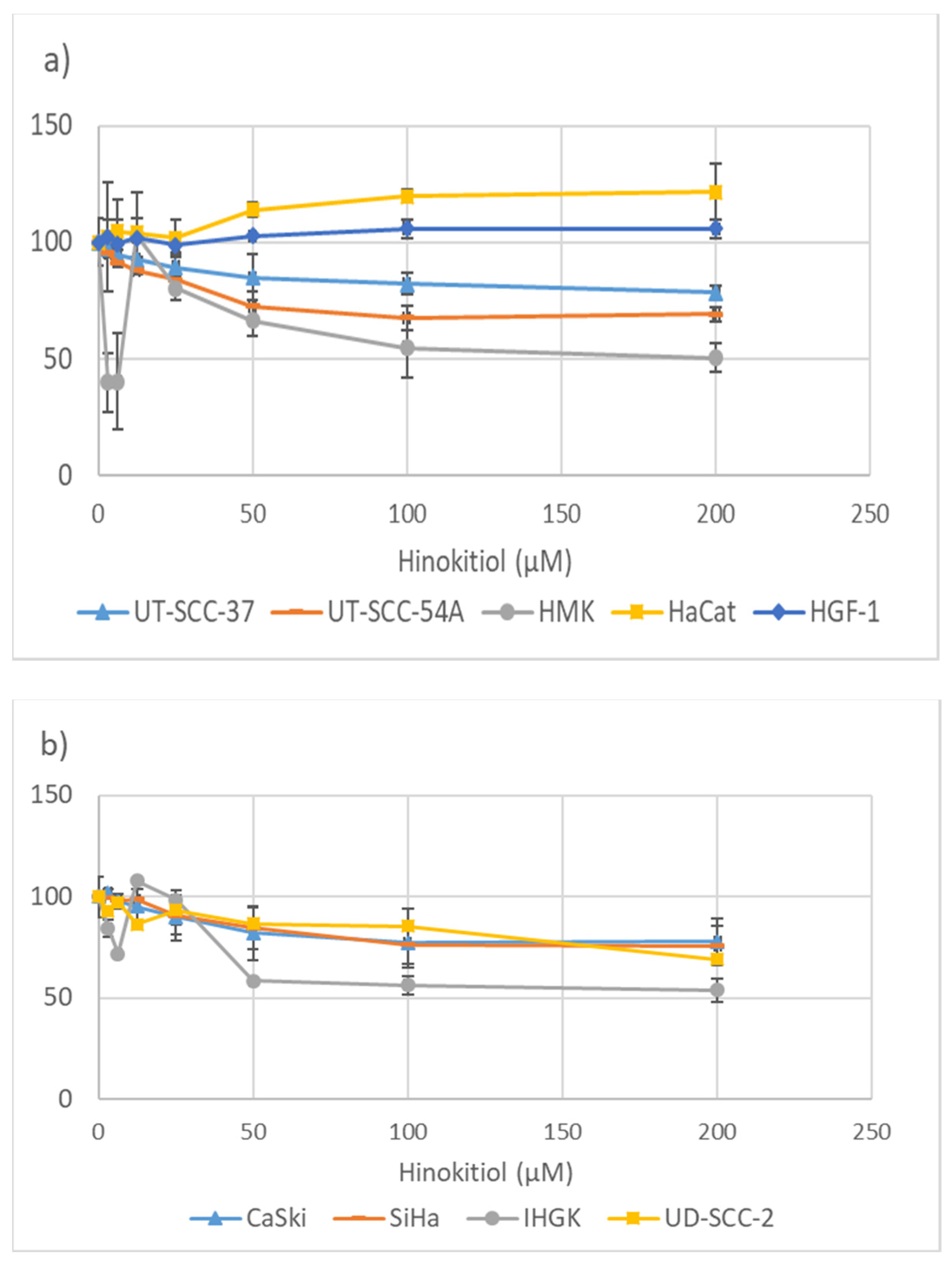
3.1. Hinokitiol and the Metabolic Activity of the Cells

The effect of hinokitiol on cell metabolic activity was assessed with colorimetric MTT-assay and the results of HPV16 negative and HPV16 positive cell lines expressed as % ±SD from control are given in Figure 1a,b, respectively. As shown in the Figure 1a, hinokitiol did not alter the metabolic activity of the normal fibroblasts (HGF-1), even at the higher concentrations. In HaCat cells, hinokitiol at concentrations from 50 µM to 200 µM increased the metabolic activity, being totally different from that found in any other cell lines tested. In all carcinoma cell lines, hinokitiol treatment declined the metabolic activity irrespective of the HPV status. However, the difference in MTT incorporation between the treated and non-treated cells was significant in both HPV-positive CaSki cells at the concentrations of 100 µM, 200 µM, and UD-SCC-2 at 200 µM (p < 0.05, two-tail t-test). In HPV-negative carcinoma cell line UT-SCC-54A, hinokitiol at the concentrations of 100 µM and 200 µM declined the metabolic activity at the borderline statistical significance (p = 0.052 and 0.055, respectively), but not in UT-SCC-37 (p = 0.088 and 0.140, respectively). The effect of hinokitiol on transformed cell lines differed from that found in carcinoma cell lines (Figure 1, HMK and HPV16 + IHGK). Hinokitiol, already at 6.25 µM concentration, suppressed the cell viability of IHGK and HMK by 35% and 80–50%, respectively. Interestingly, hinokitiol at a concentration of 25 µM had no effect on metabolic activity of either cell line, while the treatment of HMK and IHGK with 100 µM hinokitiol inhibited once again the cell viability by 50%. MTT incorporation after hinokitiol treatments of HMK at 3.1, 6.25, and 200 µM and of IHGK at 50, 100, and 200 µM was significantly different from color corporation in non-treated cells (p < 0.05, two-tail t-test).
This indicates that immortalized cell lines (except HaCat, which is closest to normal cells) are more vulnerable to hinokitiol exposure than carcinoma cell lines. Furthermore, the HMK cell line is more sensitive to lower hinokitiol concentrations than HPV16-positive IHGK. We also controlled that the final concentrations (0.0025–0.05%) of DMSO, which were used to dissolve hinokitiol, had no effect on the cell viability of any tested cells (data not shown). Thus, we can conclude that hinokitiol reduces the metabolic activity of immortalized and carcinoma cell lines irrespective of their HPV status, but immortalized cells might be more sensitive to hinokitiol than the carcinoma cell lines, especially the HPV-negative ones.

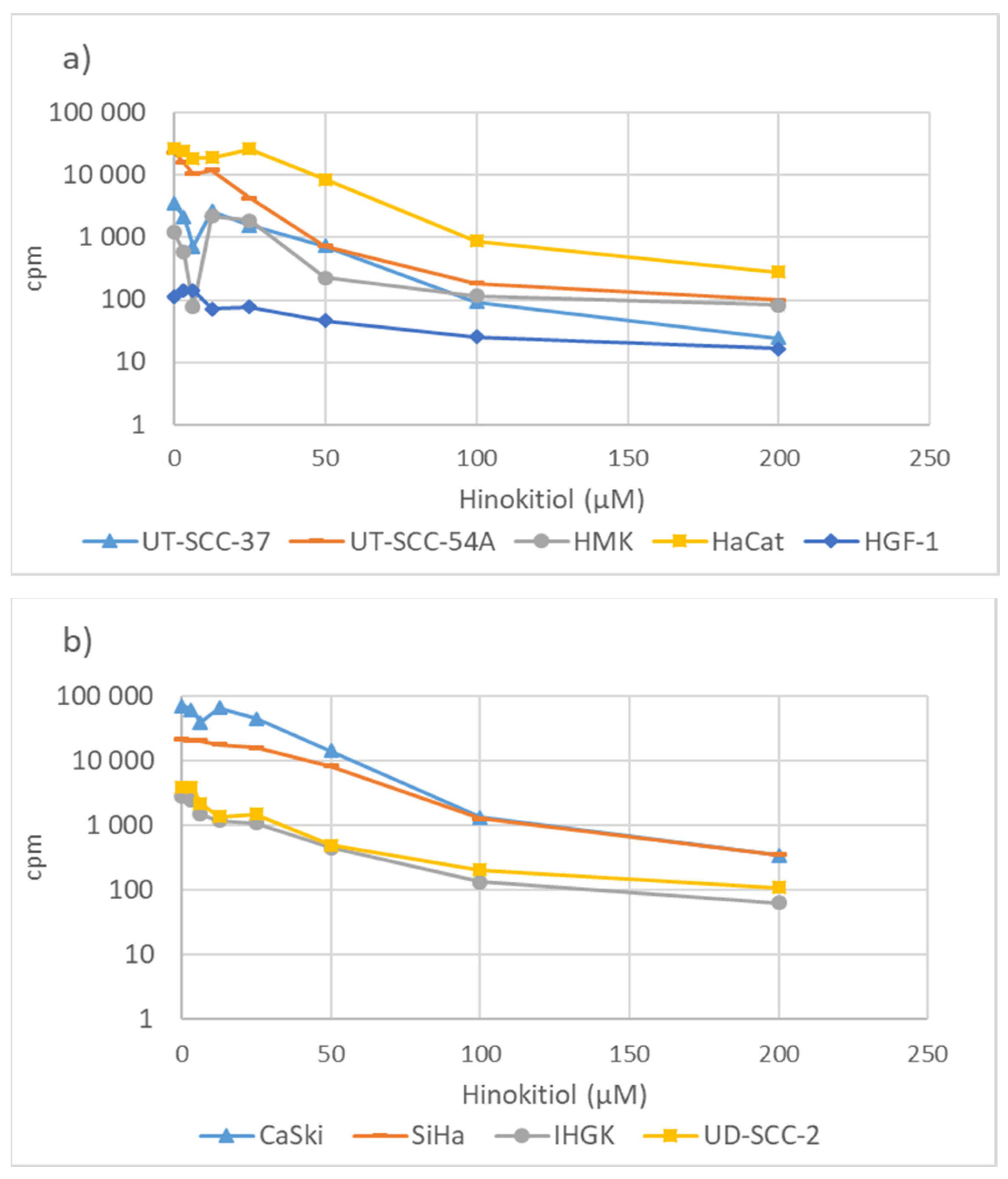
3.2. Hinokitiol and DNA Synthesis

To assess the effect of hinokitiol on DNA synthesis, the cells were pulse-labeled with [3H] thymidine incorporation. In all cell lines, hinokitiol suppressed DNA synthesis in a dose-dependent manner up to 100 µM, after which the effect on DNA synthesis becomes relatively stable (Figure 2a, HPV16 negative cell lines and Figure 2b HPV16 positive cell lines). Interestingly, in spontaneously immortalized HMK cells, 6.25 µM hinokitiol was already able to nearly stop the DNA synthesis, while none of the higher hinokitiol concentrations reached the same level of suppression in DNA synthesis. The pattern was similar to that found with the MTT metabolic activity assay. The suppressive patterns of DNA synthesis after hinokitiol exposure were similar in both HPV-positive and -negative cell lines. Among the HPV-positive cell lines, the suppressive pattern of DNA synthesis was similar in both the cervical carcinoma cell lines (CaSki and SiHa), similarly as found in the MTT assay (Figure 2b). The pattern of DNA synthesis in HPV-positive UD-SCC-2 head and neck carcinoma cell lines was identical to that found in the HPV-positive immortalized cell line IHGK, also originating from the mouth. However, IHGK was more sensitive to hinokitiol when assessed by MTT assay. In fibroblasts, hinokitiol has only a minor effect even at higher concentrations. To conclude, hinokitiol suppressed DNA synthesis in all cell lines and more at concentrations of under 100 µM than at 100–200 µM.

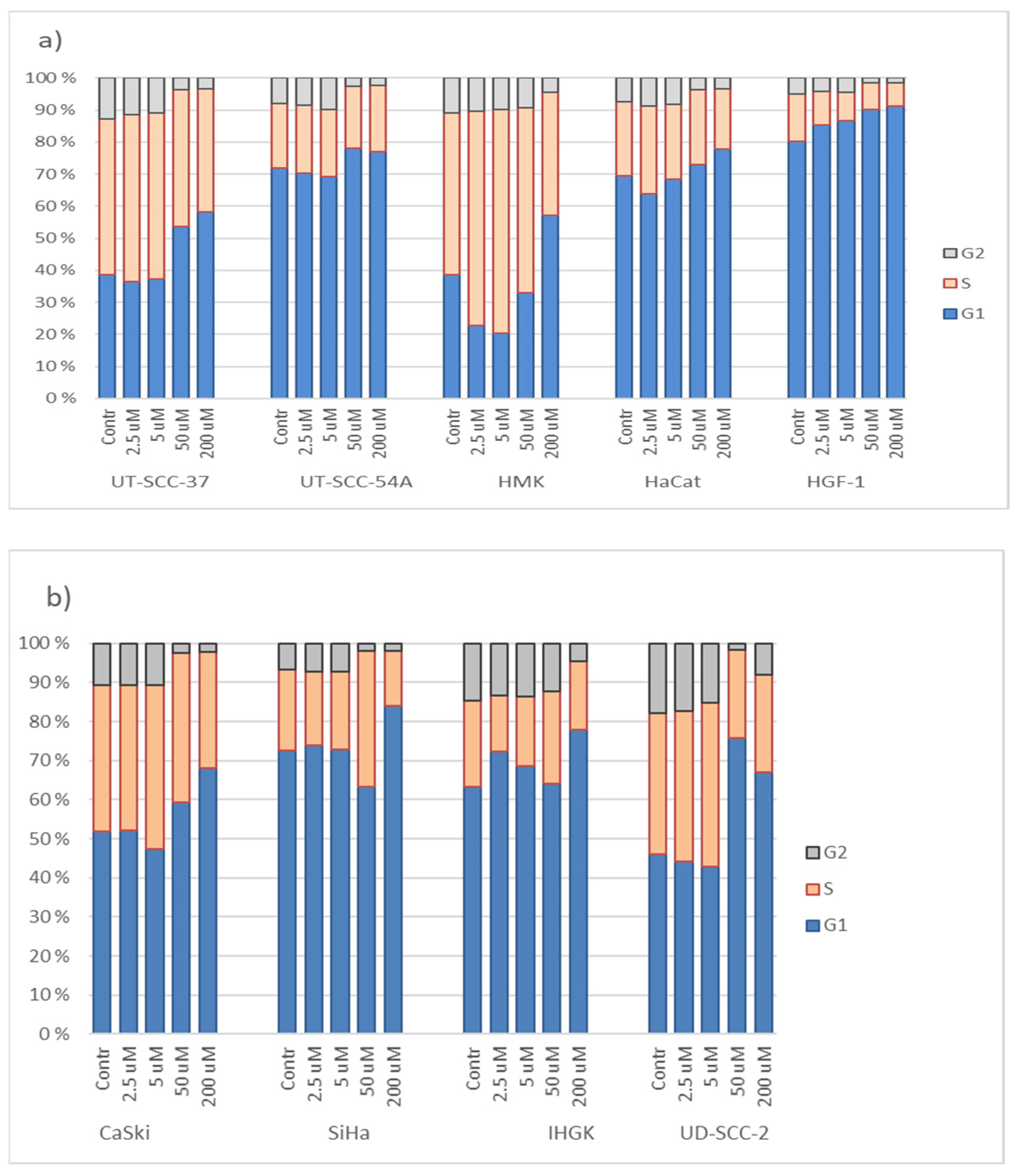
3.3. Hinokitiol and Cell Cycling

We further studied the effect of hinokitiol on cell cycle progression by flow cytometry analysis. The results of the cell cycle distribution in HPV16-negative and HPV16-positive cell lines after hinokitiol exposure at various concentrations are given in Figure 3a,b, respectively). In the normal fibroblasts, hinokitiol treatment increases the percentage of cell population at G1 phase from 80 to 91% in a dose-dependent manner. In HaCaT cell-lines, a similar pattern of increase in cells at the G1 phase was found as in fibroblasts, but only after exposure with higher hinokitiol concentrations (50 µM and 200 µM). The cell cycle distribution of HMK after hinokitiol exposure differs from that found in all other cell lines, as hinokitiol increased the proportion of cells at the S-phase. Only at the highest concentration was hinokitiol able to induce cell cycle arrest at the G1 phase. In the HPV-negative HNSCC cell lines, hinokitiol exposure did not result in nearly any effect at the lower concentration (2.5–5 µM), but concentrations of 50 µM and 200 µM increased the number of cells in the G1 phase at the expense of the cells in the G2 phase. Among the HPV-positive cell lines, hinokitiol treatment resulted in similar cell cycling distribution in CaSki and UD-SCC-2 cell lines as found in both the HPV-negative carcinoma cell lines. For the other two HPV-positive cell lines, an increased arrest of cell population at the G1 phase was found only after hinokitiol exposure at 200 µM. To conclude, the increase in the G1 cell population was mostly at the expense of the G2/M after exposure with the two highest concentrations of hinokitiol. No clear difference in the effect of hinokitiol on cell cycle distribution according to HPV status could be found.

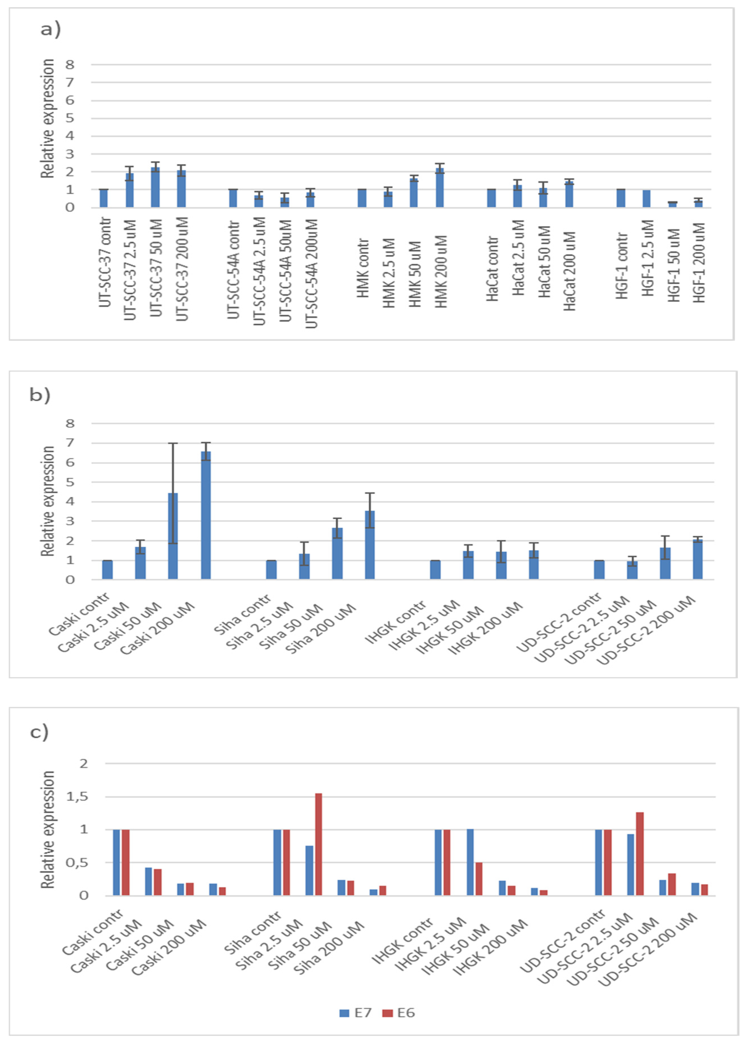
3.4. Hinokitiol, p21 and HPV16E6 and E7

To determine whether p21 is involved in the G1 arrest because of hinokitiol exposure, we quantitated p21 expression by qPCR. The results of HPV16 negative and HPV-positive cell lines are summarized in Figure 4a,b, respectively. In all HPV-positive carcinoma cell lines, p21 expression increased, most clearly in CaSki and SiHa cells-lines and less in HPV16E6 and E7 immortalized IHGK. However, the dose-dependent increase of p21 expression was not significant in any of the individual HPV-positive cell lines as compared with the p21 levels in the non-treated cells. When the results of all HPV-positive cells were pooled together, a statistically significant increase in p21 expression was found, but only at the highest hinokitiol concentration of 200 µM (p = 0.03) (data not shown). Hinokitiol exposure on HPV-negative carcinoma cell lines resulted in two different effects on p21 expression, either a twofold increase (UT-SSC-37) or decline (UT-SCC 54A). In the HaCat cell line, hinokitiol did not have any effect on p21 expression, while p21 expression declined in fibroblast with the two highest concentrations.
Because of the changes found in p21 expression, we analyzed HPV16E6 and E7 gene expression with respect to hinokitiol exposure. As evident from Figure 4c, the expression of HPV16E7 declined nearly dose-dependent in CaSki-, SiHa-, and IHGK-cell lines but the change between hinokitiol-treated and -untreated cell lines remained statistically nonsignificant. The expression of HPV16E6 also declined after hinokitiol treatment, similarly as found with HPV16E7 expression except that hinokitiol resulted in upregulation of HPV16E6 in the SiHa and UD-SCC-2 cell lines, but only at the lowest concentration of 2.5 µM. We also found that the HPVE7 gene was inversely related to the p21 expression in SiHa and CaSki, but not in IHGK. However, only in SiHa cell lines was this inverse dose-dependent correlation statistically significant (r = −0.975, p-value = 0.03) (CaSki: r = −0.788, p-value = 0.21; IHGK: r = −0.596, p-value = 0.40). In the UD-SCC-2 cell line, there was a borderline and significant inverse dose-dependent correlation between p21 and E7 (r = −0.944, p-value = 0.06) and between p21 and E6 (r = −0.989), respectively. Only, in the CaSki cell line, the HPV16E6 and E7 mRNA levels correlated with each other in a dose-dependent matter after hinokitiol treatment (r = 0.9964, p = 0.04). We also analyzed the sensitivity of hinokitiol to decline HPVE6 and E7 mRNA expression by pooling all results from HPV-positive cell lines. Hinokitiol at lower concentration was more sensitive to induce a significant downregulation of E7 (25 µM p = 0.013; 50 µM and 200 µM p = 0.000) than of E6 (50 µM p = 0.002, 200 µM p = 0.001).

4. Discussion

Several in vitro studies exist on the anti-tumor effects of hinokitiol [7,12,13,14,15,16,17,28], but only one of these studies has used oral carcinoma cell lines [7]. Two previous studies have used HeLa cells but without making any note on the presence of HPV18 in this cell line [16,20]. This is the first study to our knowledge to assess the effects of hinokitiol on both HPV-negative and HPV16-positive cell lines, both immortalized or carcinoma cell lines, to identify HPV-specific effects, if any.
At the molecular level, viral oncoproteins E6 and E7 of hr-HPVs are the key players in cancer progression dysregulating cell cycling, inhibiting apoptosis and interfering with several other host proteins leading to accumulation of DNA damage [29,30]. Thus, one important anti-viral and anti-tumor effect of hinokitiol could be the downregulation of HPVE6 and E7 oncogene expression in HPV-positive cell lines. Our experiments showed that hinokitiol was able to upregulate p21 expression significantly more effectively in HPV-positive than in HPV-negative cell lines when pooled results were compared. Furthermore, p21 expression was inversely related to downregulation of HPV16E6 and E7 expression. Hinokitiol at lower concentration was more sensitive to induce a significant downregulation of E7 than of E6. However, we also found differences among the cell lines exposed to hinokitiol, whether HPV-positive or -negative ones.
Fotopoulous and coworkers [20] found that hinokitiol at the concentration of 28 µM was able to achieve 50% growth inhibition (Gl50 value) of HeLa cells, while in our study, the growth inhibition in HPV16-positive carcinoma cell lines varied between 20 and 30% at maximum, which was achieved at the concentration of 50 µM. However, we exposed these cells only for 24 h while the other study used a 48 h exposure. We selected the 24 h exposure because it has been used in a majority of the previous published studies [7,14,16,25].
Interestingly, although hinokitiol was growth-inhibitory at low concentrations (6.25 µM) in both of our immortalized cell lines (IHGK and HMK), it exhibits a paradoxical effect (reversal of growth inhibition) at higher concentrations, as also reported earlier [7]. The mechanism remains unknown but one possible explanation, at least from our study, could be that IHGK and HMK cell lines were grown in keratinocyte serum-free media supplemented with epidermal growth factor (EGF 0.2 ng/L). It has been shown before that the stimulatory effect of EGF on cell growth and cell migration can be blocked by hinokitiol [16], possibly by proteasome-mediated degradation of EGFR [31]. However, in the HPV-positive immortalized IHGK cell line, we found this paradoxical effect only on cell viability, but not on DNA synthesis as found in the HPV-negative HMK cell line. In flow cytometry analyses, we found that hinokitiol treatment at lower concentrations (2.5–50 µM) increased the proportion of HMK cells at the S phase.
Wang and coworkers in 2020 [16] reported that hinokitiol treatment inhibited HeLa cell proliferation by inducing cell cycle arrest at the G1 phase, senescence, and autophagy. The latter is a p53-independent DNA damage response, while mitochondrial-dependent apoptosis might be the hinokitiol-induced pathway in HPV-negative carcinoma cell lines [17,32,33]. In our experiments, we found that hinokitiol at the two highest concentrations declined cell metabolism statistically significantly only in HPV-positive cell lines CaSki and UD-SCC-2. Importantly, we also found that immortalized cell lines might be more sensitive, irrespective of the HPV status. The effect of hinokitiol on the HPV-positive and HPV-negative carcinoma cell lines was rather similar, as revealed by the 3H-thymidine incorporation assay. Using flow cytometry, we could confirm that hinokitiol (at the highest concentration of 200 µM) was able to induce G1 arrest in all cell lines. Wang et al., 2020 [16] reported that the treatment of HeLa cells with 40 µM hinokitiol increased the proportion of cells at the G1 phase by 13% (from 56 to 69%). In our experiments, hinokitiol treatment with 50 µM increased the proportion of cells at the G1 phase from 52 to 59%, and from 46 to 76% in Caski and UD-SCC-2 cell lines, respectively. In Siha cells, the proportion of cells at the G1 phase (over 70%) remained stable after treatment with the two lowest hinokitiol concentrations, while the concentration of 50 µM decreased the number of cells at the G1 phase to 63% and the highest hinokitiol concentration (200 µM) most effectively blocked the cells at the G1 phase (from 72 to 84%). As will be discussed later, hinokitiol resulted in dose-dependent inverse correlation between p21 and E7, which would explain the effective blocking of the SiHa cells at the G1 phase. Our results also indicate that the efficacy of hinokitiol to arrest the cells at the G1 phase is cell-line-specific, which precludes direct comparisons between different studies.
Li and coworkers [32] found that hinokitiol treatment of malignant melanoma cells was associated with G1 arrest, increased the levels of p53 and p21, and concomitantly reduced the expression of cell cycle regulatory proteins, including cyclin D and cyclin E. This mechanism could explain why HPV-positive cells were not more resistant to hinokitiol exposure than the HPV-negative cell lines. Generally, the overexpression of the E6 and E7 oncoproteins in HPV-infected cells will lead to a DNA-synthesis-competent state [18,29,30,34]. The binding of E6 to p53 in the presence of the cellular E6-associated protein (E6AP) will result in p53 degradation. E7 results in cell proliferation by binding to RB protein, which releases E2F transcription factor, leading to transition of the cell cycle from the G1 to S phase. However, when the expression of p21 is high and E7 expression is low, the cell cycle progression is arrested as a result of E7 binding with p21 and cyclin E/Cdk [18,34]. In our experiments, p21 expression increased after hinokitiol treatment, while E7 expression was downregulated. This inverse correlation was significant in a dose-dependent manner only seen with SiHa cells. Downregulation of E7 expression with concomitant upregulation of p21 was more prominent in CaSki than in SiHa cells, which might partly explain that the increase of cells at the G1 phase was found in CaSki cells already after lower hinokitiol treatment (50 µM) than in SiHa cells (200 µM). Furthermore, we found that in the CaSki cells, the dose-dependent downregulated E6 and E7 levels correlated significantly with each other (r = 0.996, p = 0.04).
CaSki and UD-SCC-2 cells have approximately 600 viral copies, while SiHa cells have only two HPV copies. Interestingly, Wang-Johanning et al., 2002 [35] were able to show that even if there is such a great difference in HPV copy numbers between these two cell lines, RNA copy numbers of E6 or E7 in both cell lines were not appreciably different from each other. The RNA copy number per ng RNA in SiHa cells was 1.5 times greater (for E6) and 2.15 times greater (for E7) than these copy numbers in CaSki cells. We only made a relative quantification of HPV16 E6 and E7 RNA expressions, but not the absolute measurements. Thus, we are not able the analyze the changes in E6 and E7 RNA copy numbers caused by hinokitiol at different concentrations, which would be the optimal method to assess the anti-viral effect. Although we found that hinokitiol can upregulate p21 in all cell lines (except in UD-SSC-54A and fibroblasts), the difference between non-treated and treated cells was statistically significant only in HPV-positive cells (pooled results). The p21 is a well-known tumor suppressor; it can also be an oncogenic protein and its induction has been shown to be crucial for promoting cancer cell motility and tumorigenesis [36,37,38]. Because p53 is usually wild-type in HPV-positive carcinoma cell lines, one could argue that hinokitiol is beneficial in HPV-infected cells by increasing the expression of p21 and downregulating HPV16 E6 and E7 expression. With the wild-type p53, p21 acts as a guardian of the genome, whereas in the absence of p53 or with the deficient p53, p21 activity causes genomic instability [36,37,38].
In the present study, we were also interested in analyzing HPV-positive and -negative cell lines from the head and neck region. Even though HPV is the main etiological agent of cervical carcinoma, it also has a role in a subgroup of head and neck carcinomas. Our earlier studies based on cDNA arrays have shown some interesting similarities between HPV-positive cervical and head and neck carcinoma cell lines (CaSki, UT-DEC-1, IHGK, and UD-SCC-2), but also with the HPV-negative cell-line UT-SCC-54A derived originally from an oral lichen planus lesion. In all these cell lines, 13 genes were identified with similar downregulation [22,39]. Our data also indicated that different etiological backgrounds of head and neck carcinoma cell lines might be distinguished by their different global gene expression patterns. Interestingly, we also found in the present study that the effect of hinokitiol on the cell cycle profile was similar in Caski cell lines as found in the HPV-negative UT-SCC-54A and HPV-positive D-SCC-2.
Importantly, we also identified that the hinokitiol declined metabolic activity more effectively in transformed than carcinoma cell lines, which might be important to prevent the immortalized cells from progressing toward malignancy. Finally, we were able to show that hinokitiol has only slight effects on oral fibroblasts, which is in alignment with the earlier reports showing that hinokitiol did not induce DNA damage, apoptosis, or significant levels of autophagy in human stromal fibroblasts [32,33].
Even the highest concentration of hinokitiol (200 µM) was not able to cause any significant effects in any of the parameters that we analyzed. Thus, in further studies, the use of higher hinokitiol concentrations might be more informative than the low doses tested in our experiments. Furthermore, the higher hinokitiol concentrations are more effective for blocking HPV16E6 and E7 oncogene expressions. The use of higher concentrations is also warranted by the fact that several studies have suggested the use of hinokitiol as a therapeutic agent and, thereby, basically the safety (LD50 mouse, 85 mg/kg) and solubility of hinokitiol are the limitations. Hinokitiol has been used, for example, in gels for mouth cleaning in concentration of 2% (appr 12 mM) [40]. Thus, concentrations (µM scale) used in our study fit in these limitations and allow also the use of higher concentrations in further studies. In addition, the use of only one hinokitiol exposure time (24 h) is a noted limitation of the current study.
One limitation of our study was that we have not analyzed the protein expression data of cell cycle-related proteins—neither the proteins nor RNAs that can interact with HPV16E6 and E7 proteins. However, our main aim in this study was to identify virus-specific effects of hinokitiol on HPV, similarly as found earlier on hepatitis B, HIV, and influenza viruses. Here we found that hinokitiol downregulates HPVE6 and E7 expression, which warrants more detailed studies on the mechanisms as these HPV oncogenes are able to exert their effects in the cell by affecting multiple cellular pathways. E6 and E7 proteins are able to do so, aside from the well-known targets p53 and pRb. Thus, the deep understanding of the cellular pathways induced by HPV after hinokitiol treatment needs further studies on several genes and proteins targeted by E6 and E7.

5. Conclusions

Hinokitiol is more toxic to immortalized cell lines than to carcinoma cell lines, and concentrations up to 200 µM induce only minor effects on normal fibroblasts and HaCat cell lines. Hinokitiol was able to induce cell cycle arrest at the G1 phase by upregulating p21 expression in all cell lines expect UD-SCC54 and fibroblasts. The increase in p21 expression was significantly higher in HPV-positive than in HPV-negative cell lines (comparison between pooled results according to HPV status). Importantly, we found that hinokitiol exerted an inverse effect on p21 and HPV16E6 and E7 expression, which was dose dependent in SiHa cells and UD-SCC-2. These results warrant further studies to investigate the effects of hinokitiol, even at higher concentrations, particularly on those key proteins interacting with HPV oncoproteins in inducing malignant progression and permitting tumor growth.

Author Contributions

V.L., S.S. and K.L. designed the study; S.S. and V.L. wrote the initial draft of the manuscript; and E.S. and V.L. contributed to the laboratory work and data analysis. All authors contributed to data interpretation and critically reviewed the manuscript. All authors have read and agreed to the published version of the manuscript.

Funding

This study was supported by the Institute of Dentistry, Faculty of Medicine, University of Turku, the Finnish Medical Foundation (KL), and the Sigrid Juselius Foundation (KL).

Informed Consent Statement

Not applicable.

Data Availability Statement

All data generated and analyzed during current study are included in this published article.

Acknowledgments

We thank Katja Sampalahti and Tatjana Peskova, Institute of Dentistry, University of Turku, for technical assistance in the in vitro experiments. Flow cytometry was performed at the Cell Imaging and Cytometry Core, Turku Bioscience Centre, Turku, Finland, with the support of Biocenter Finland.

Conflicts of Interest

The authors have no conflict of interest to declare.

Statement on Ethics

No ethical permission was needed, as the present study is an in vitro study with cell lines.

References

  1. Komatsu, K. Professor Tetsuo Nozoe and my tropylium ion chemistry. Chem. Rec. 2015, 15, 160–174. [Google Scholar] [CrossRef] [PubMed]
  2. Komaki, N.; Watanabe, T.; Ogasawara, A.; Sato, N.; Mikami, T.; Matsumoto, T. Antifungal mechanism of hinokitiol against Candida albicans. Biol. Pharm. Bull. 2008, 31, 735–737. [Google Scholar] [CrossRef] [PubMed] [Green Version]
  3. National Library of Medicine. National Center for Biotechnology Information. PubChem. Available online: https://pubchem.ncbi.nlm.nih.gov/compound/Hinokitiol (accessed on 15 February 2022).
  4. Domon, H.; Hiyoshi, T.; Maekawa, T.; Yonezawa, D.; Tamura, H.; Kawabata, S.; Yanagihara, K.; Kimura, O.; Kunitomo, E.; Terao, Y. Antibacterial activity of hinokitiol against both antibiotic-resistant and -susceptible pathogenic bacteria that predominate in the oral cavity and upper airways. Microbiol. Immunol. 2019, 63, 213–222. [Google Scholar] [CrossRef] [PubMed]
  5. Kim, D.J.; Lee, M.W.; Choi, J.S.; Lee, S.G.; Park, J.Y.; Kim, S.W. Inhibitory activity of hinokitiol against biofilm formation in fluconazole-resistant Candida species. PLoS ONE 2017, 12, e0171244. [Google Scholar] [CrossRef]
  6. Krenn, B.M.; Gaudernak, E.; Holzer, B.; Lanke, K.; Kuppeveld, F.J.M.V.; Seipelt, J. Antiviral activity of the zinc ionophores pyrithione and hinokitiol against picornavirus infections. J. Virol. 2009, 83, 58–64. [Google Scholar] [CrossRef] [Green Version]
  7. Shih, Y.H.; Chang, K.W.; Hsia, S.M.; Yu, C.C.; Fuh, L.J.; Chi, T.Y.; Shieh, T.M. In vitro antimicrobial and anticancer potential of hinokitiol against oral pathogens and oral cancer cell lines. Microbial. Res. 2013, 168, 254–262. [Google Scholar] [CrossRef]
  8. Ye, J.; Xu, Y.F.; Lou, L.X.; Jin, K.; Miao, Q.; Ye, X. Anti-inflammatory effects of hinokitiol on human corneal epithelial cells: An in vitro study. Eye 2015, 29, 964–971. [Google Scholar] [CrossRef] [Green Version]
  9. Budihas, S.R.; Gorshkova, I.; Gaidamakov, S.; Wamiru, A.; Bona, M.K.; Parniak, M.A.; Crouch, R.J.; McMahon, J.B.; Beutler, J.A.; Grice, S.F.J.L. Selective inhibition of HIV-1 reverse transcriptase-associated ribonuclease H activity by hydroxylated tropolones. Nucleic Acids Res. 2005, 33, 1249–1256. [Google Scholar] [CrossRef] [Green Version]
  10. Hu, Y.; Cheng, X.; Cao, F.; Huang, A.; Tavis, J.E. β-Thujaplicinol inhibits hepatitis B virus replication by blocking the viral ribonuclease H activity. Antiviral Res. 1998, 99, 221–229. [Google Scholar] [CrossRef]
  11. Miyamoto, D.; Kusagaya, Y.; Endo, N.; Sometani, A.; Takeo, S.; Suzuki, T.; Arima, Y.; Nakajima, K.; Suzuki, Y. β-Thujaplicin-copper chelates inhibit replication of human influenza viruses. Antiviral Res. 1998, 39, 89–100. [Google Scholar] [CrossRef]
  12. Hachlafi, N.E.; Lakdar, F.; Khouchlaa, A.; Bakrim, S.; Omari, N.E.; Balahbib, A.; Shariati, M.A.; Zengin, G.; Fikti-Benbrahim, K.; Orlando, G.; et al. Health Benefits and Pharmacological Properties of Hinokitiol. Processes 2021, 9, 1680. [Google Scholar] [CrossRef]
  13. Ko, J.; Bao, C.; Park, H.; Kim, M.; Choi, H.Y.; Kim, Y. β-Thujaplicin modulates estrogen receptor signaling and inhibits proliferation of human breast cancer cells. Biosci. Biotechnol. Biochem. 2015, 79, 1011–1017. [Google Scholar] [CrossRef] [Green Version]
  14. Liu, S.; Yamauchi, H. Hinokitiol, a metal chelator derived from natural plants, suppresses cell growth and disrupts androgen receptor signaling in prostate carcinoma cell lines. Biochem. Biophys. Res. Commun. 2006, 351, 26–32. [Google Scholar] [CrossRef] [PubMed]
  15. Ouyang, W.C.; Liao, Y.W.; Chen, P.N.; Lu, K.H.; Yu, C.C.; Hsieh, P.L. Hinokitiol suppresses cancer stemness and oncogenicity in glioma stem cells by Nrf2 regulation. Cancer Chemother. Pharmacol. 2017, 80, 411–419. [Google Scholar] [CrossRef] [PubMed]
  16. Wang, C.C.; Chen, B.K.; Chen, P.H.; Chen, L.C. Hinokitiol induces cell death and inhibits epidermal growth factor-induced cell migration and signaling pathways in human cervical adenocarcinoma. Taiwan. J. Obstet. Gynecol. 2020, 59, 698–705. [Google Scholar] [CrossRef] [PubMed]
  17. Zhang, G.; He, J.; Ye, X.; Zhu, J.; Hu, X.; Shen, M. β-Thujaplicin induces autophagic cell death, apoptosis, and cell cycle arrest through ROS-mediated Akt and p38/ERK MAPK signaling in human hepatocellular carcinoma. Cell Death Dis. 2019, 10, 255. [Google Scholar] [CrossRef] [Green Version]
  18. Doorbar, J. Molecular biology of human papillomavirus infection and cervical cancer. Clin. Sci. 2006, 110, 525–541. [Google Scholar] [CrossRef] [Green Version]
  19. Syrjänen, S.; Syrjänen, K. HPV in Head and Neck Carcinomas: Different HPV Profiles in Oropharyngeal Carcinomas—Why? Acta Cytol. 2019, 63, 124–142. [Google Scholar] [CrossRef]
  20. Fotopoulou, T.; Ćirić, A.; Kritsi, E.; Calhelha, R.C.; Ferreira, I.C.F.R.; Soković, M.; Zoumpoulakis, P.; Koufaki, M. Antimicrobial/Antibiofilm Activity and Cytotoxic Studies of β-Thujaplicin Derivatives. Arch. Pharm. 2016, 349, 698–709. [Google Scholar] [CrossRef]
  21. Balló, H.; Koldovsky, P.; Hoffmann, T.; Balz, V.; Hildebrandt, B.; Gerharz, C.D.; Bier, H. Establishment and characterization of four cell lines derived from human head and neck squamous cell carcinomas for an autologous tumor-fibroblast in vitro model. Anticancer Res. 1999, 19, 3827–3836. [Google Scholar]
  22. Ruutu, M.; Johansson, B.; Grenman, R.; Syrjänen, S. Two different global gene expression profiles in cancer cell lines established from etiologically different oral carcinomas. Oncol. Rep. 2005, 14, 1511–1517. [Google Scholar] [CrossRef] [PubMed]
  23. Oda, D.; Bigler, L.; Lee, P.; Blanton, R. HPV immortalization of human oral epithelial cells: A model for carcinogenesis. Exp. Cell Res. 1996, 226, 164–169. [Google Scholar] [CrossRef] [PubMed]
  24. Mäkelä, M.; Larjava, H.; Pirilä, E.; Maisi, P.; Salo, T.; Sorsa, T.; Uitto, V.J. Matrix metalloproteinase 2 (gelatinase A) is related to migration of keratinocytes. Exp. Cell Res. 1999, 251, 67–78. [Google Scholar] [CrossRef] [PubMed]
  25. Liu, S.; Yamauchi, H. p27-Associated G1 arrest induced by hinokitiol in human malignant melanoma cells is mediated via down-regulation of pRb, Skp2 ubiquitin ligase, and impairment of Cdk2 function. Cancer Lett. 2009, 286, 240–249. [Google Scholar] [CrossRef] [PubMed]
  26. Turunen, A.; Syrjänen, S. Extracellular calcium regulates keratinocyte proliferation and HPV 16 E6 RNA expression in vitro. Apmis 2014, 122, 781–789. [Google Scholar] [CrossRef] [Green Version]
  27. Livak, K.J.; Schmittgen, T.D. Analysis of relative gene expression data using real-time quantitative PCR and the 2ΔΔCt method. Methods 2016, 25, 402–408. [Google Scholar] [CrossRef] [PubMed]
  28. Viale, A.; Franco, F.D.; Orleth, A.; Cambiaghi, V.; Giuliani, V.; Bossi, D.; Ronchini, C.; Ronzoni, S.; Muradore, I.; Monestiroli, S.; et al. Cell-cycle restriction limits DNA damage and maintains self-renewal of leukaemia stem cells. Nature 2009, 457, 51–56. [Google Scholar] [CrossRef]
  29. Basukala, O.; Banks, L. The Not-So-Good, the Bad and the Ugly: HPV E5, E6 and E7 Oncoproteins in the Orchestration of Carcinogenesis. Viruses 2021, 13, 1892. [Google Scholar] [CrossRef]
  30. Vats, A.; Trejo-Cerro, O.; Thomas, M.; Banks, L. Human papillomavirus E6 and E7: What remains? Tumour Virus Res. 2021, 11, 200213. [Google Scholar] [CrossRef]
  31. Tu, D.G.; Yu, Y.; Lee, C.H.; Kuo, Y.L.; Lu, Y.C.; Tu, C.W.; Chang, W.W. Hinokitiol inhibits vasculogenic mimicry activity of breast cancer stem/progenitor cells through proteasome-mediated degradation of epidermal growth factor receptor. Oncol. Lett. 2016, 11, 2934–2940. [Google Scholar] [CrossRef] [Green Version]
  32. Li, J.Y.; Liu, C.P.; Shiao, W.C.; Jayakumar, T.; Li, Y.S.; Chang, N.C.; Huang, S.Y.; Hsieh, C.-Y. Inhibitory effect of PDGF-BB and serum-stimulated responses in vascular smooth muscle cell proliferation by hinokitiol via up-regulation of p21 and p53. Arch. Med. Sci. 2018, 14, 579–587. [Google Scholar] [CrossRef] [PubMed] [Green Version]
  33. Li, L.H.; Wu, P.; Lee, J.Y.; Li, P.R.; Hsieh, W.Y.; Ho, C.C.; Ho, C.L.; Chen, W.J.; Wang, C.C.; Yen, M.-Y.; et al. Hinokitiol induces DNA damage and autophagy followed by cell cycle arrest and senescence in gefitinib-resistant lung adenocarcinoma cells. PLoS ONE 2019, 9, e104203. [Google Scholar] [CrossRef] [PubMed]
  34. Doorbar, J.; Egawa, N.; Griffin, H.; Kranjec, C.; Murakami, I. Human papillomavirus molecular biology and disease association. Rev. Med. Virol. 2015, 25, 2–23. [Google Scholar] [CrossRef] [Green Version]
  35. Wang-Johanning, F.; Lu, D.W.; Wang, Y.; Johnson, M.R.; Johanning, G.L. Quantitation of human papillomavirus 16 E6 and E7 DNA and RNA in residual material from ThinPrep Papanicolaou tests using real-time polymerase chain reaction analysis. Cancer 2002, 94, 2199–2210. [Google Scholar] [CrossRef]
  36. Shamloo, B.; Usluer, S. p21 in cancer research. Cancers 2019, 11, 1178. [Google Scholar] [CrossRef] [Green Version]
  37. Marhenke, S.; Buitrago-Molina, L.B.; Endig, J.; Orlik, J.; Schweitzer, N.; Klett, S.; Longerich, T.; Geffers, R.; Muñoz, A.S.S.; Dorrell, C.; et al. p21 promotes sustained liver regeneration and hepatocarcinogenesis in chronic cholestatic liver injury. Gut 2014, 63, 1501–1512. [Google Scholar] [CrossRef] [PubMed]
  38. Wei, K.C.; Chen, R.F.; Chen, Y.F.; Lin, C.H. Hinokitiol suppresses growth of B16 melanoma by activating ERK/MKP3/proteosome pathway to downregulate survivin expression. Toxicol. Appl. Pharmacol. 2019, 366, 35–45. [Google Scholar] [CrossRef] [PubMed]
  39. Ruutu, M.; Peitsaro, P.; Johansson, B.; Syrjänen, S. Transcriptional profiling of a human papillomavirus 33-positive squamous epithelial cell line which acquired a selective growth advantage after viral integration. Int. J. Cancer 2002, 100, 318–326. [Google Scholar] [CrossRef] [PubMed]
  40. Iha, K.; Suzuki, N.; Yoneda, M.; Takeshita, T.; Hirofuji, T. Effect of mouth cleaning with hinokitiol-containing gel on oral malodor: A randomized, open-label pilot study. Oral Surg. Oral Med. Oral Pathol. Oral Radiol. 2013, 116, 433–439. [Google Scholar] [CrossRef]
Figure 1. MTT incorporation assay. The effect of hinokitiol on cell metabolic activity was assessed with colorimetric MTT-assay. (a) HPV16 negative and (b) HPV16 positive cell lines. Cells were exposed to hinokitiol at various concentrations (0, 3.1, 6.25, 12.5, 25, 50, 100, and 200 µM) for 24 h. After exposure, the cells were washed and incubated with MTT. The precipitated MTT was dissolved in DMSO, and the amount measured spectrophotometrically. All analyses were repeated twice and in case there was a discrepancy between the two experiment rounds the triplicate was then further done for the third time. The results are shown as % ±SD from control. MTT incorporation after hinokitiol treatments of HMK at 3.1, 6.25, and 200 µM, of CaSki at 100 and 200 µM, of IHGK at 50, 100, and 200 µM and of UD-SCC-2 at 200 µM were significantly different from color corporation in non-treated cells (p < 0.05, two-tail t-test).
Processes 10 00736 g001
Figure 2. Cell proliferation. The cells were exposed to different hinokitiol concentrations (3.1, 6.125, 12.5, 25, 50, 100, and 200 µM) in 96-well plates for 24 h and the cell proliferation was measured by the incorporation of a radiolabeled DNA precursor, 3H-thymidine. Counts per minute (cpm) were evaluated in a scintillation counter. (a) HPV16 negative and (b) HPV16 positive cell lines. Each experiment was performed in triplicate. The results are expressed in logarithmic scale on the x-axis.
Processes 10 00736 g002
Figure 3. The effect of hinokitiol on the cell cycle of (a) HPV-negative and (b) HPV-positive cell lines. The cells were exposed to different hinokitiol concentrations (0, 2.5, 5, 50, and 200 µM) for 24 h and stained with propidium iodide for cell cycle analysis by flow cytometry. The data represents percentages (mean of three independent experiments) of cells in each stage of the cell cycle.
Processes 10 00736 g003
Figure 4. Relative gene expression of p21 and HPV16E6 and HPV16E7. The cells were exposed to different hinokitiol concentrations (0, 2.5, 50, and 200 µM) for 24 h and the p21 expression in (a) HPV16 negative and (b) HPV16 positive cell lines and (c) HPV16 E6, and HPV16E7 expressions were measured by qPCR. The relative expression of target genes HPV16 E6, HPV16 E7, and p21 were normalized using GAPDH as a reference gene.
Processes 10 00736 g004
Table 1. The human papillomavirus positive (CaSki, SiHa and UD-SCC-2) and negative (UT-SCC-37 and UT-SCC-54A) carcinoma and immortalized cell lines (IHGK, HMK, and HaCat) were used in the present study. Normal fibroblasts established from gingiva (HGF-1) served as the controls.
Cell LineOriginOrganHPV Status HPV
Copies/Cell
Acquired from or Reference
CaSkiCervical carcinomauteriHPV16 600 copiesATCC (CRL-1550)
SiHaSquamous cell carcinomacervix uteriHPV16 2 copiesATCC (HTB-35)
UD-SCC-2Squamous cell carcinomahypopharynxHPV16 600 copiesBallo et al., 1999 [21]
UT-SCC-37Squamous cell carcinomagingivaHPV negativeGift from Prof. R. Grenman. Turku University Hospital, Turku, Finland Ruutu et al., 2005 [22]
UT-SCC-54ASquamous cell carcinomabuccal mucosaHPV negativeGift from Prof. R. Grenman. Turku University Hospital, Finland Ruutu et al., 2005 [22]
IHGKKeratinocytegingivatransformed with HPV16 E6/E7Oda 1996 [23]
HMKSpontaneously immortalized keratinocytegingivaHPV negativeMäkelä 1999 [24]
HaCatSpontaneously immortalized keratinocyteskinHPV negativeATCC
HGF-1FibroblastgingivaHPV negativeInstitute of Dentistry, University of Turku, Finland
Publisher’s Note: MDPI stays neutral with regard to jurisdictional claims in published maps and institutional affiliations.

