Article

High-Fat Diets Modify the Proteolytic Activities of Dipeptidyl-Peptidase IV and the Regulatory Enzymes of the Renin–Angiotensin System in Cardiovascular Tissues of Adult Wistar Rats

by
Germán Domínguez-Vías
1,2,*,
Ana Belén Segarra
1,
Manuel Ramírez-Sánchez
1 and
Isabel Prieto
1,*
1
Unit of Physiology, Department of Health Sciences, University of Jaén, Las Lagunillas, 23071 Jaén, Spain
2
Department of Physiology, Faculty of Health Sciences, Ceuta, University of Granada, 18071 Granada, Spain
*
Authors to whom correspondence should be addressed.
Biomedicines 2021, 9(9), 1149; https://doi.org/10.3390/biomedicines9091149
Submission received: 24 July 2021 / Revised: 17 August 2021 / Accepted: 1 September 2021 / Published: 3 September 2021
(This article belongs to the Section Molecular and Translational Medicine)

Abstract

:
(1) Background: The replacement of diets high in saturated fat (SAFA) with monounsaturated fatty acids (MUFA) is associated with better cardiovascular function and is related to the modulation of the activity of the local renin–angiotensin system (RAS) and the collagenase activity of dipeptidyl peptidase IV (DPP-IV). The objective of the work was to verify the capacity of different types of dietary fat on the regulatory activities of RAS and DPP-IV. (2) Methods: Male Wistar rats were fed for 24 weeks with three different diets: the standard diet (S), the standard diet supplemented with virgin olive oil (20%) (VOO), or with butter (20%) plus cholesterol (0.1%) (Bch). The proteolytic activities were determined by fluorometric methods in the soluble (sol) and membrane-bound (mb) fractions of the left ventricle and atrium, aorta, and plasma samples. (3) Results: With the VOO diet, angiotensinase values were significantly lower than with the Bch diet in the aorta (GluAP and ArgAP (mb)), ventricle (ArgAP (mb)) and atrium (CysAP (sol)). Significant decreases in DPP-IV (mb) activity occurred with the Bch diet in the atrium and aorta. The VOO diet significantly reduced the activity of the cardiac damage marker LeuAP (mb) in the ventricle and aorta, except for LeuAP (sol) in the ventricle, which was reduced with the Bch diet. (4) Conclusions: The introduction into the diet of a source rich in MUFA would have a beneficial cardiovascular effect on RAS homeostasis and cardiovascular functional stability.

1. Introduction

The high consumption of a high-fat diet (HFD) has been associated with most of the epidemiological evidence during the last decades, especially the sources of saturated fatty acids (SAFA) and cholesterol have been associated with an increased risk of suffering a series of medical conditions such as obesity, diabetes, metabolic syndrome (MetS), and cardiovascular diseases (CVD); however, these results do not extend to other types of lipid sources [1]. HFD is a cardiac stressor that alters the expression of specific cardiac factors due to glycolipotoxicity [2] and modifies cardiac structure and function. However, trials involving the Mediterranean diet indicate that monounsaturated fatty acids (MUFAs) are more effective than low-fat and low-cholesterol diets in preventing cardiovascular mortality and coronary artery disease [3]. The reduction in SAFA in substitution by MUFA attenuates the increase in blood pressure (BP) [4]. These alterations, depending on the degree of fatty acid saturation, are related to changes in the systemic or local renin–angiotensin systems (RAS) [5,6,7,8,9,10]. The activation of the RAS participates in the development of MetS, heart failure [11], and pathophysiology of hypertension [4,7,12,13,14,15,16,17,18,19,20,21,22,23,24,25]. Within these local RAS, the type of fatty acids consumed with the diet allows modifying various enzymes of the aminopeptidase (AP) family, also called angiotensinases, responsible for metabolizing angiotensin peptides (Ang) [5,9,26]. The APs are relevant in the control of BP and cardiac function [6,27], participating in the regulation of systemic and local RAS, but also as predictive biomarkers of functionality in different organs, including the heart [28,29,30,31,32,33,34,35]. It is suggested that several of the activities of APs involved in the metabolism of angiotensin in the cardiovascular tissues of the rat can be modified according to the amount and type of fat in the diet [6]. Among them, the activity of aspartyl-AP (AspAP; EC 3.4.11.21) is responsible for the metabolism of Ang I to Ang 2–10; glutamyl-AP (GluAP or aminopeptidase A, APA; EC 3.4.11.7) metabolizes Ang II into Ang III; alanyl-AP (AlaAP or aminopeptidase M, APM; EC 3.4.11.2) and/or arginyl-AP (ArgAP or aminopeptidase B, APB; EC 3.4.11.6) are responsible for the metabolism of Ang III to Ang IV and Ang 4–8; and insulin-regulated AP (IRAP; 3.4.11.3)—also called AP-placental leucyl (LAP), cystinyl-AP (CysAP), oxytokinase, or vasopressinase—was identified as the binding site for the Ang IV receptor (AT4) [12,29,36,37,38,39].
Ang II and aldosterone are produced de novo within cardiac tissue [40,41]. RAS activation by diets high in SAFA induces CVD through the manifestation of oxidative stress, inflammation, interstitial fibrosis at sites of vascular injury, ultrastructural abnormalities, and diastolic dysfunction [42,43]. Hemodynamic factors regulate the growth of cardiomyocytes, whereas reactive fibrosis is independent of blood pressure [40]. Under physiological conditions the fibroblasts of the cardiac interstitium explain the gradual turnover of collagen for a correct remodeling of the heart, but an alteration of the RAS would modify the balance between the restructuring behavior [43,44] and collagen degradation [45,46]. Certain APs with broad specificity affect collagen production in cardiac fibroblasts [47]. Dipeptidyl-peptidase IV (DPP-IV; EC 3.4.14.5), also known as collagenase-like peptidase, is a ubiquitously expressed serine-type aminopeptidase enzyme, with subsequent cleavage at the N-terminal amino acids proline and alanine (X-Pro and X-Ala), and also metabolizes the insulinotropic hormone glucagon-like peptide-1 (GLP-1) [48,49,50]. DPP-IV manifests itself on the surface of vascular endothelial cells and in renovascular organs, suggesting a role for DPP-IV in renal and cardiovascular function [51,52], in addition to being associated with ECV [53,54]. The cardiac fibroblast migration and expressions of genes related to fibrosis, such as collagen-I and transforming growth factor-β1 (TGF-β1), are suppressed with a DPP-IV inhibitor [42,55], which, in turn, inhibits MR-dependent oxidative stress and collagen induction mediated by aldosterone.
Thyrotropin-releasing hormone (TRH) has also been identified in the heart and aorta [56], where it can modulate cardiovascular function, either autocrine or paracrine. The enzyme pyroglutamyl-AP (pGluAP; EC 3.4.11.8) is known for its thyrotropin-releasing hormone (TRH) degradation activity [18]. Previous results suggest that the type of fat used in the diet can influence the local activity of pGluAP and modify its biological functions in energy metabolism [4,9,57]. However, its effect on cardiovascular tissues is not entirely clear.
Along with angiotensinases, there are other APs such as leucyl-AP (LeuAP; EC 3.4.11.1) and gamma-glutamyl transferase (GGT; EC 2.3.2.2) involved in the control of BP that act as functional markers of cardiovascular risk [7,9,58] and that are associated with different cardiovascular and kidney diseases [34,54]. GGT is an enzyme of glutathione and cysteine (Cys) metabolism with pro-oxidant activity and a modulating influence on endothelial dysfunction [59]. Elevation of GGT levels is associated with an increased risk of cardiovascular and metabolic diseases [59,60,61,62]. GGT is associated with an increase in mortality from chronic heart disease events such as hypertension and congestive heart failure [28,31,63,64].
The main objectives of the current research were to understand the role of two types of diets with higher MUFA or SAFA fat content on the angiotensinase activities that regulate the classic and non-classic RAS pathways, to identify the implications for DPP-IV collagenase activity, and to verify in each dietary group if there is an inter-relationship between DPP-IV activities and angiotensinases in cardiovascular tissues. Measuring the activities of GGT and LeuAP biomarkers and the degrading activity of TRH (pGluAP) would make it possible to know the functional and metabolic state of cardiovascular tissues. We hypothesized that the inclusion of MUFA in the diet could be a preventive dietary factor for reducing the activation of the RAS pathway through angiotensinases and maintaining the functional integrity of cardiovascular structures.

2. Materials and Methods

2.1. Animals and Diets

Harlan Interfauna Ibérica SA (Barcelona, Spain) supplied adult male Wistar rats for experimentation. The experimental procedures for the use and care of animals were carried out in accordance with Directive 2010/63/EU of the Council of the European Communities and Spanish Regulation RD 53/2013, being previously approved by the Institutional Committee for Animal Use and Care of the University of Jaén with project code number PIUJA_2005_acción 14 (1 January 2006). The rats had the experimental diets and water ad libitum for 24 weeks and were kept under a controlled environment of temperature (20–25 °C) and humidity (50 ± 5%) in a 12 h:12 h light/dark cycle. At the start of the research, the animals were 6 months old and had an average body weight of ± 495 g. The rats were randomly assigned into three experimental groups: (1) A standard diet (S, n = 6), where the rats were fed with commercial food for laboratory rodents (Panlab, Barcelona, Spain) whose nutritional composition was 16.5% protein, 3% total fat, 60% carbohydrates (nitrogen-free extract (NFE)), 5% minerals, and 4% fiber. The other two diets were high in fat (HFD), but with different fatty acid composition. (2) A group of rats was fed diet S supplemented with 20% virgin olive oil (Cooperativa de los Villares, Jaén, Spain) (VOO, n = 5), composed of a total content (%) of 75.5% ω-9-monounsaturated fatty acid (oleic acid, C18:1), 11.5% saturated fatty acid (palmitic acid, C16:0), and 7.5% ω-6-polyunsaturated fatty acid (linoleic acid, C18:2). (3) The second group of rats was fed diet S supplemented with 20% butter (Hacendado, Valencia, Spain) plus cholesterol (0.1%) (Bch, n = 5), composed of a total content (%) of 29% monounsaturated fatty acid (C18:1), 62% saturated fatty acid (C16:0 and C18:0), 4% polyunsaturated fatty acid (C16:0), and short and medium chain fatty acids (C4–C14). The Bch diet was supplemented with cholesterol (0.1%) in order to reach the average cholesterol content of the Western diet. The HFD diets were isocaloric among themselves (VOO diet, 1848 KJ/100 g; diet Bch, 1827 KJ/100 g) and hypercaloric compared with the S diet (1392 KJ/100 g).
At the end of the experimental period, the animals were perfused with a saline solution (0.9% NaCl) through the left heart ventricle under Equithensin anesthesia (2 mL/kg of body weight). A blood sample dissolved in heparin was previously extracted and centrifuged for 10 min at 2000× g to obtain plasma. The heart and aorta were dissected, immediately deposited in liquid nitrogen to separate the samples into different parts as previously described [21], and frozen at −80 °C until use. The atrium and left ventricle were dissected from the heart. The aorta was obtained by cutting from the aortic arch to its abdominal portion.

2.2. Assay of Aminopeptidase Activities

Tissue samples were homogenized in 10 volumes of 10 mM Tris-HCl buffer (pH 7.4) and ultracentrifuged at 100,000× g for 30 min at 4 °C. Analysis of enzyme activities and protein content of the fraction soluble were performed with the supernatant and analyzed in triplicate. The pellets were rehomogenized in 10 mM Tris-HCl buffer (pH 7.4) with 1% Triton-X-100 to solubilize the membrane proteins and ultracentrifuged at 100,000× g for 30 min at 4 °C. The supernatants were kept for at least 4 h at 4 °C and shaken with SM-2 biobeads (100 mg/mL) (Bio-Rad, Richmond, VA, USA) to remove the detergent. Analyses of enzyme activities and protein content of the membrane-bound fraction were conducted with the resulting samples, analyzed also in triplicate.
The enzymatic activities of the soluble (sol) and membrane-bound (mb) fractions of AlaAP, ArgAP, AspAP, CysAP/IRAP, DPP-IV, GGT, GluAP, LeuAP, and pGluAP were measured by a fluorometric analysis using as aminoacyl-β-naphthylamides (aa-β-NA) substrates: L-Ala-β-NA, L-Arg-β-NA, L-Asp-β-NA, L-Cys-β-NA, L-Gly-Pro-β-NA, ɣ-Glu-β-NA, L-Glu-β-NA, L-Leu-β-NA, and L-pGlu-β-NA, respectively, according to the methods of different authors [65,66,67] modified by Prieto and Ramírez [15,68]. An amount of 10 µL/well of the supernatant sample was pipetted in 96-well black plates and was incubated for 30 min at 37 °C in 100 µL of substrate solutions. The enzymatic reactions were then stopped by adding 100 µL of 0.1 M acetate buffer (pH 4.2). The β-NA released as a result of the enzymatic activity was quantified fluorometrically at an emission of 412 nm with an excitation of 345 nm. The activities of each tissue fraction were expressed as pmoles of L-Ala-β-NA, L-Arg-β-NA, L-Asp-β-NA, L-Cys-β-NA, L-Gly-Pro-β-NA, ɣ-Glu-β-NA, L-Glu-β-NA, L-Leu-β-NA, and L-pGlu-β-NA hydrolyzed per minute and per mg of protein (pmol aa-β-NA/min/mg prot). For plasma, it was expressed as picomole per minute and per milliliter (pmol aa-β-NA/min/mL). All chemical products were supplied by Sigma-Aldrich (St. Louis, MO, USA).

2.3. Protein Measurement

For protein quantification, the Bradford method [69] was used, using as standard a bovine serum albumin (BSA; Sigma-Aldrich, St. Louis, MO, USA) standard line.

2.4. Statistical Analysis

Statistical analysis was performed using one-way ANOVA, followed by Tukey’s post hoc test for multiple comparisons. When the normality test failed, a Kruskal–Wallis unidirectional range analysis of variance was performed. Pearson’s correlation coefficient was used to establish the relationship between the APs activities of cardiovascular tissues and plasma. Significant differences were estimated with Sigmaplot v11.0 software (Systat Software, Inc., San José, CA, USA), and p-values less than 0.05 (p < 0.05) were considered statistically significant. All data are presented as mean ± standard error of the mean (SEM).

3. Results

3.1. Angiotensinase Activities

To know how the type of fat in the diet affected the angiotensinase activities that were derived from the classic RAS route, the activities that were studied are represented in the following scheme in green, following the order of action of each angiotensinase based on its precursors and metabolic products (Figure 1).
The values of all angiotensinase activities can be consulted in Supplementary Table S1. None of the angiotensinase activities were significant in plasma. No significant differences were observed in any of the tissues analyzed for AspAP and AlaAP activities. Furthermore, the AspAP activity in the soluble fractions of the atrium and aorta was undetectable. The VOO diet had significantly lower values than the Bch diet for the activities GluAP (mb) and ArgAP (mb) in the aorta (GluAP (mb), VOO: 2024.36 ± 420.28 vs. Bch: 3986.00 ± 582.40; ArgAP (mb), VOO: 1548.75 ± 129.86 vs. Bch: 2087.72 ± 139.98), ArgAP (mb) in the ventricle (VOO: 763.06 ± 15.88 vs. Bch: 1013.07 ± 56.57), and CysAP (sol) in the atrium (VOO: 195.50 ± 23.11 vs. Bch: 310.75 ± 21.51) (Figure 2A,C–E). However, in the ventricle, the ArgAP (sol) activity had a higher value with the VOO diet with respect to the Bch diet (VOO: 255.20 ± 52.95 vs. 127.80 ± 17.60; Figure 2B). Furthermore, the Bch diet significantly increased CysAP (sol) activity of the atrium compared with the S diet (S: 212.18 ± 32.74 vs. Bch: 310.75 ± 21.51; Figure 2E).
Supplementary Figure S1 shows the detection of grouped data correlations for GluAP (sol/mb) and ArgAP (mb) activities between the ventricle and aorta, for CysAP (mb) activity between the aorta and atrium, and for AlaAP (sol) between the plasma and ventricle. However, when the dietary groups were analyzed independently with a stratified analysis, it was observed that the association was lost.

3.2. Dipeptidyl Peptidase IV Activity

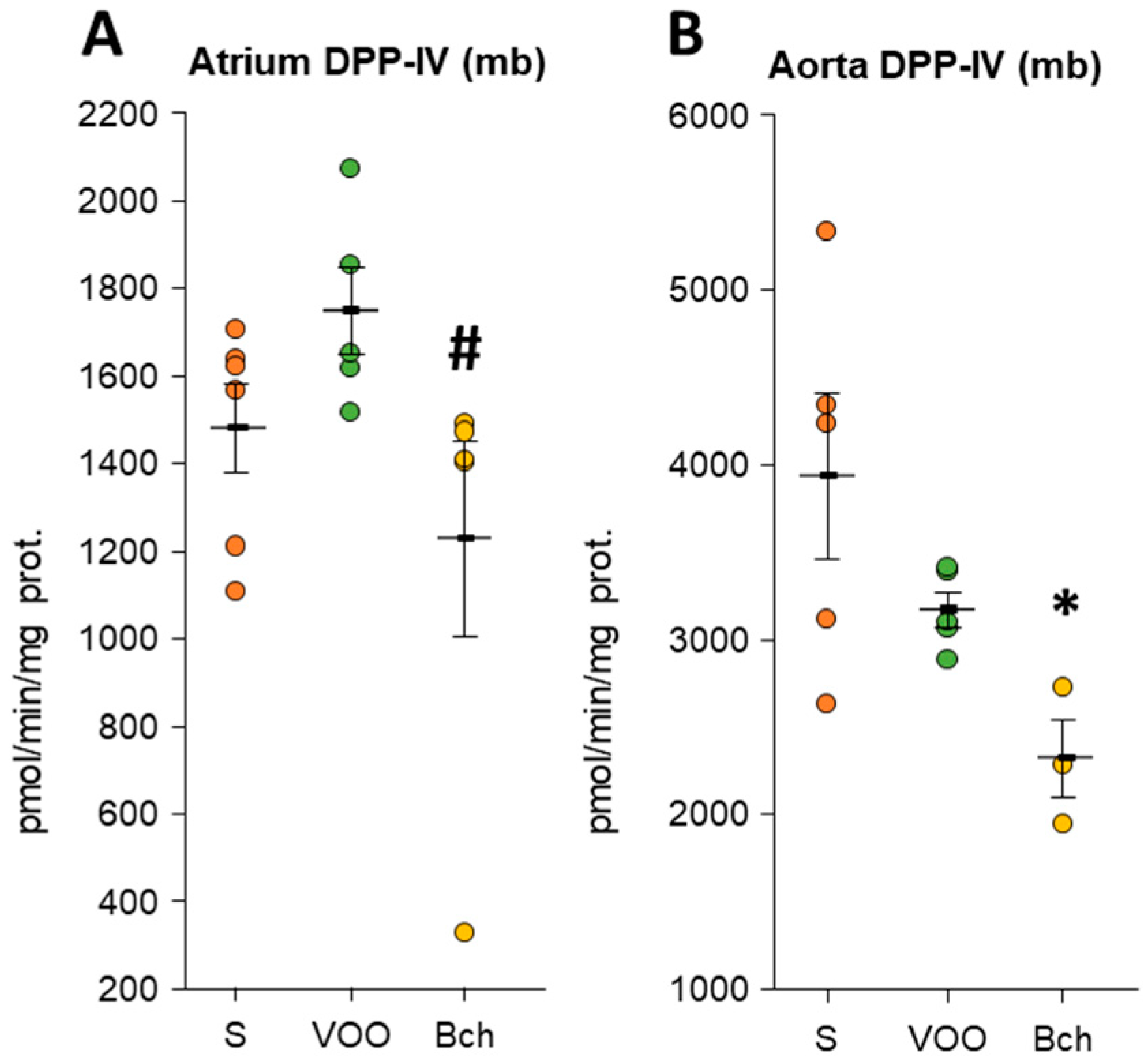
All values of DPP-IV activity can be found in Supplementary Table S2. Collagenase activity of DPP-IV showed significant changes with HFDs in the atrium and aorta (Figure 3). The atrium had higher values of DPP-IV activity (mb) in the VOO diet than in the Bch diet (VOO: 1743.86 ± 99.46 vs. Bch: 1222.50 ± 223.93; Figure 3A). On the other hand, in the aorta, the Bch diet reduced the DPP-IV activity (mb) with respect to the S diet (S: 3937.62 ± 478.42 vs. Bch: 2321.89 ± 224.54; Figure 3B). Despite a positive group correlation with all the data for DPP-IV (sol) activity between aorta and plasma, the stratified correlations did not show significance for independent groups (Supplementary Figure S2).
The DPP-IV activity showed significant grouped correlations of all the point data with the angiotensinase activities in the atrium, ventricle, and aorta (Table 1). However, a stratified analysis for dietary group comparisons showed that only the atrium and ventricle had strong correlations between activities (Table 1: intra-group correlations). DPP-IV and angiotensinase activities seem to be associated with diets, interestingly highlighting the association of the VOO diet in the soluble fractions of the atrium and ventricle.

3.3. Leucyl Aminopeptidase, Gamma-Glutamyl Transferase, and Pyroglutamyl Aminopeptidase Activities

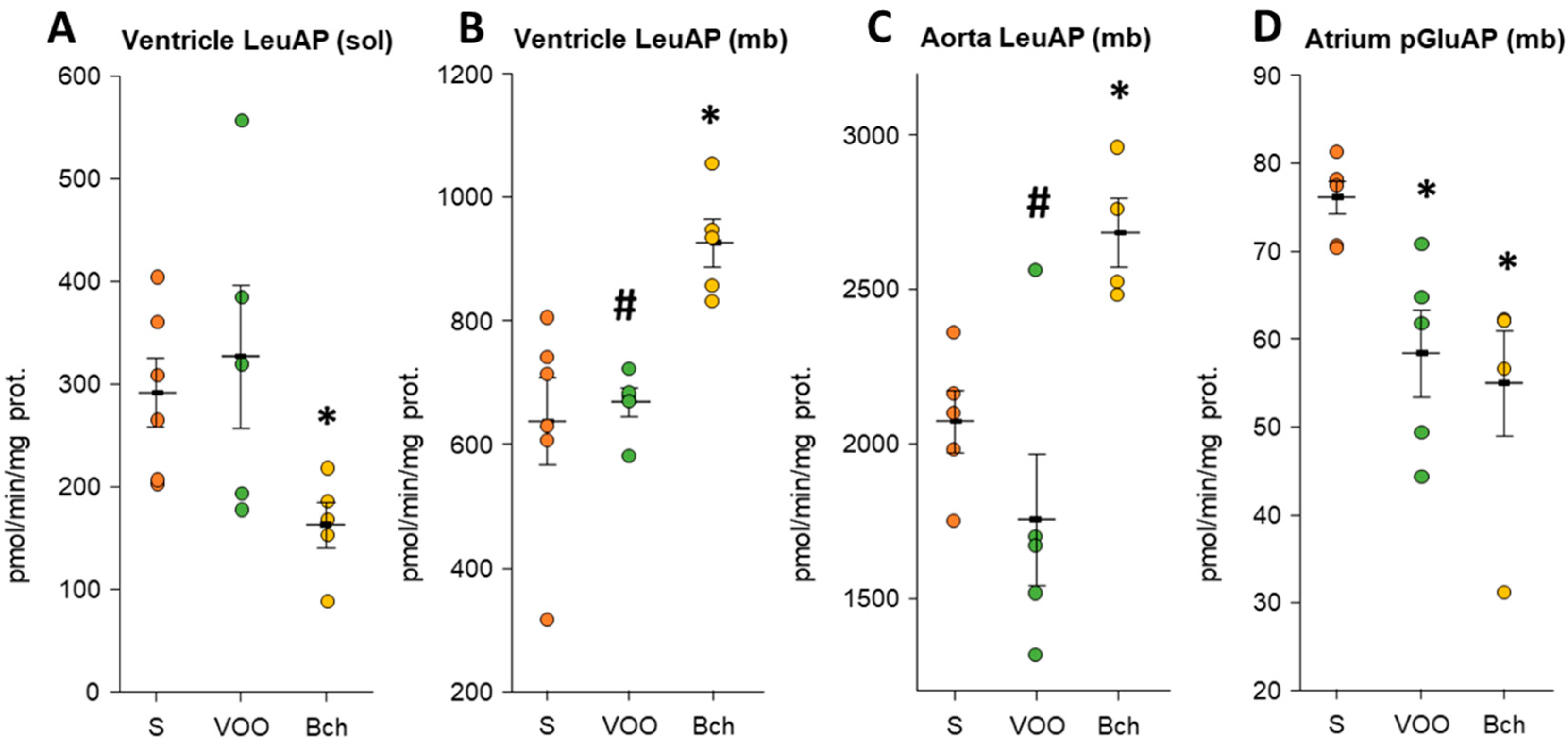
All values of LeuAP, GGT, and pGluAP activities can be found in Supplementary Table S3. Activity levels in the atrium were undetectable for LeuAP (sol/mb), GGT (sol/mb), and pGluAP (mb) activities. The GGT activity did not show that they were regulated by the different HFDs in the different samples examined. The VOO diet presented significantly lower LeuAP (mb) activity values than the Bch diet in the ventricle (VOO: 667.32 ± 23.08 vs. Bch: 924.99 ± 39.31; Figure 4B) and aorta (VOO: 1753.36 ± 213.53 vs. Bch: 2681.70 ± 111.30; Figure 4C). In turn, the Bch diet showed a significant increase in LeuAP (mb) activity compared with the S diet (Figure 4B, S: 636.06 ± 70.51 vs. Bch: 924.99 ± 39.31; Figure 4C, S: 2071.56 ± 100.69 vs. Bch: 2681.70 ± 111.30). On the contrary, LeuAP (sol) activity was significantly reduced with the Bch diet in the ventricle (S: 291.37 ± 33.40 vs. Bch: 162.87 ± 21.53; Figure 4A). With all the point data, a significantly positive grouped correlation was found between the LeuAP activity (mb) of the aorta and the ventricle, but a stratified analysis by dietary groups (Supplementary Figure S3: intra-group correlations) showed that LeuAP was not associated with diet. Furthermore, both HFDs showed a reduction in pGluAP (mb) activity only in the atrium (S: 75.99 ± 1.82; VOO: 58.22 ± 4.92; Bch: 54.83 ± 6.00; Figure 4D).

4. Discussion

Adequate nutritional status is essential for maintaining cardiovascular structure and functionality in the face of metabolic challenges [2,70]. During the last decade, there has been intensive debate about the advice to reduce SAFA and increase MUFA or polyunsaturated (PUFA) to reduce the risk of CVD [71,72]. Virgin olive oil has a preventive effect on the development of atherosclerosis, indicating that endothelial damage triggered by oxidation can be diminished or reversed by the compounds of olive oil [73,74]. In recent work in our laboratory using the same HFDs (VOO and Bch), we confirmed the beneficial effects in animals fed the VOO diet vs. the pernicious results with the Bch diet. With the VOO diet, they do not increase their body weight, they do not increase the values of systolic blood pressure (SBP), and they maintain normal values of triglycerides and the fraction of VLDL cholesterol, reduce total cholesterol and its LDL fraction, and reduce oxidative stress and lipid peroxidation against the pernicious effects that the Bch diet manifests. [4,7,9]. Elevated plasma cholesterol levels are another important risk factor widely recognized for its relationship with angiotensin metabolism [4,5,7,10,12,72], with Mediterranean countries showing lower rates of heart disease than other countries due to the usual diet rich in olive oil [75].
HFDs alter the baroreceptor reflex [76], and the type of fatty acid that makes up the diet is capable of modulating the central [10,26,77] and local RAS regulatory APs [4,5,7,9,57]. Our results show that the VOO diet favored the stabilization of the RAS with respect to the Bch diet, by lowering the values of GluAP (mb) activities in the aorta, ArgAP (mb) in the ventricle and aorta, and CysAP (mb) in the atrium. In turn, it was observed in the ventricle that CysAP (mb) activity was significantly elevated with diet S in addition to VOO. These data may have repercussions with previously published data, where they identified that the local and serum mRNA and/or the activities of AspAP and GluAP progressively increase with the degree of saturation of fatty acids in the diet [4,7,10,72]. GluAP activity is modified by the composition of fatty acids in the diet and by the cholesterol content [4,5,6,9,57], directly or indirectly, and with an important role in the development of cardiovascular disorders and the pathophysiology of hypertension. An analysis of grouped data showed significant correlations of angiotensinase activities in various cardiovascular tissues and plasma (Supplementary Figure S1); however, a stratified analysis isolating dietary groups showed that angiotensinase activities were not associated with each group of diets and that they were not associated with the type of fat used. This absence of correlations does not agree with other studies in which they showed the impact of the fatty acid profile of the diet on central tissues and its correlation with APs activities [26,77]. It is demonstrated that the profile of fatty acids and the levels of APs activities are modified depending on the type of fat used [77], and their activities correlate with dietary fat composition [7,26].
The cardiac interstitium is made up of non-myocytic cells embedded in a highly organized extracellular matrix that contains a three-dimensional collagen network that serves to maintain the architecture of the myocardium and determine its rigidity [78]. It is known that circulating and myocardial cells RAS are directly involved in the regulation of cardiac interstitial remodeling. Oleic acid (n-9), a component of olive oil, prevents Ang II-induced cardiac remodeling (fibrosis and hypertrophy) by suppressing the expression of collagen and fibroblast growth factor 23 (FGF23) in mice [79]. GluAP inhibitors and Ang IV analogs prevent cardiac dysfunction by normalizing central/local GluAP hyperactivity and attenuating cardiac hypertrophy and fibrosis [27,80,81]. Our results show that together with the marked decrease in the RAS activity of the VOO diet compared with the Bch diet, in addition, the VOO diet presented normal values of DPP-IV activity (mb) in the atrium and aorta, but they were significantly lower with the diet Bch. In summary, our data showed higher values of RAS regulatory activities together with lower values of DPP-IV with the Bch diet; these data coincide with investigations that reveal that Ang II stimulates collagen synthesis and inhibits collagenase activity in cardiac fibroblasts [78,82,83]. In a contradictory way, other researchers confirm that HFD with a high content of SAFA increases the expression and activity of DPP-IV in the aorta and atrium, playing a role in the development of aortic stiffness, vascular oxidative stress, endothelial dysfunction, and vascular remodeling by promoting increased deposition of collagen fibers [84,85,86]. The grouped correlation between DPP-IV and angiotensinases activities in cardiovascular tissues (Table 1) manifested a strong association with collagenase DPP-IV activity when the classical and non-classical pathways of RAS were activated. A stratified analysis between independent groups confirmed the association of activities with the variable diet. These results are interesting because it was determined that olive oil has an important role in the association of regulatory activities of RAS (AlaAP, ArgAP, AspAP, CysAP, GluAP) and DPP-IV activity in the atrium and ventricle (Table 1). Second, as we have commented previously, the VOO diet detected in the atrium the lowest values of CysAP activity and normal values of DPP-IV activity compared with the Bch diet. However, the atrium showed a marked increase in CysAP activity with diet Bch compared with diet S and lower values of DPP-IV activity with diet Bch versus diet VOO. Without going further, strong correlations of grouped data were detected between DPP-IV and CysAP activities in all tissues (Table 1), where a stratified analysis showed that the VOO diet was a variable that affected this association in the atrium and ventricle. These results make sense since it is known that there is an interaction of CysAP with DPP-IV that acts in the pathway of the kinin system (metabolism of bradykinin) as a counter-regulation of an overactivation of the RAS [87]. In non-cardiovascular tissues with fibrosis, they are also represented by inflammation and a deposit of cellular matrix, where DPP-IV has a profibrotic role of binding to fibronectin, influencing cell adhesion and migration [88,89].
With the VOO diet vs. the Bch diet, in the ventricular and aortic membrane-bound fractions, the lower values of GluAP and ArgAP angiotensinase activities allowed less arterial damage, as reflected by the activity of the functional marker LeuAP. The LeuAP (mb) activity was elevated in aorta with the Bch diet compared with the S diet; however, the VOO diet showed its benefit by maintaining the LeuAP activity at normal values. Similar results were found in another vascularized organ model using the same type of experimental diets [9]. Our experimental diets did not show significant GGT activity in the cardiovascular tissue fractions; therefore, it does not indicate increased oxidative stress or insufficient oxidation. These results are totally contradictory to the values found in different vascularized organs of animals treated with the same experimental diets, where the VOO diet had lower values than the Bch diet, even behaving as its control [7,9].
The pGluAP (mb) activity was significantly reduced in the atrium with both HFDs with respect to diet S. These data do not agree with other results, where a significant increase in pGluAP (sol/mb) activity of the atrium has been described in renovascular hypertension; however, in the aorta, there are no differences, and in the ventricle, the activity is reduced without being significant with respect to its healthy controls [17]. These differences are probably due to the differences between the animal models.
Treatment of hypertension with AP inhibitors [4,5] has not been shown to totally prevent CVD outcomes [90,91]. Angiotensinases and DPP-IV are objects of study to find new therapeutic approaches targeting RAS and associated peptides in hypertension and heart failure [87]. However, prevention with diets enriched with olive oil has an important influence on the reduction in the central and local RAS pathway [6,7,9,10]. Our results also showed lower angiotensinase activity values with the VOO diet compared with the Bch diet. A main limitation of our study is that the actions of HFDs can be different depending on many variables, such as the species and animal model used, as well as depending on the multiple components of the diet itself; therefore, these experiments cannot be transferred to humans. Adding to that accentuation of the effects of RAS, the experiments were started with adult animals (6 months of age), and the experimental diets were continued with 6-month follow-up. The older age among the animals used in the experiment and the small size of the samples (n = 6–5/group) can increase the variability in the parameters studied and contribute to reducing the differences observed between the groups. Additional research on components of olive oil should be conducted in new preclinical models associated with CDV. In addition, it should be determined in more detail if the cardiovascular benefits of olive oil derive from saponifiable lipids or non-saponifiable lipids, which are minor components, such as phenolic compounds with antioxidant character. For this, it is proposed to try a pure diet in MUFA (refined virgin olive oil) or extracts of olive leaves rich in polyphenols.

5. Conclusions

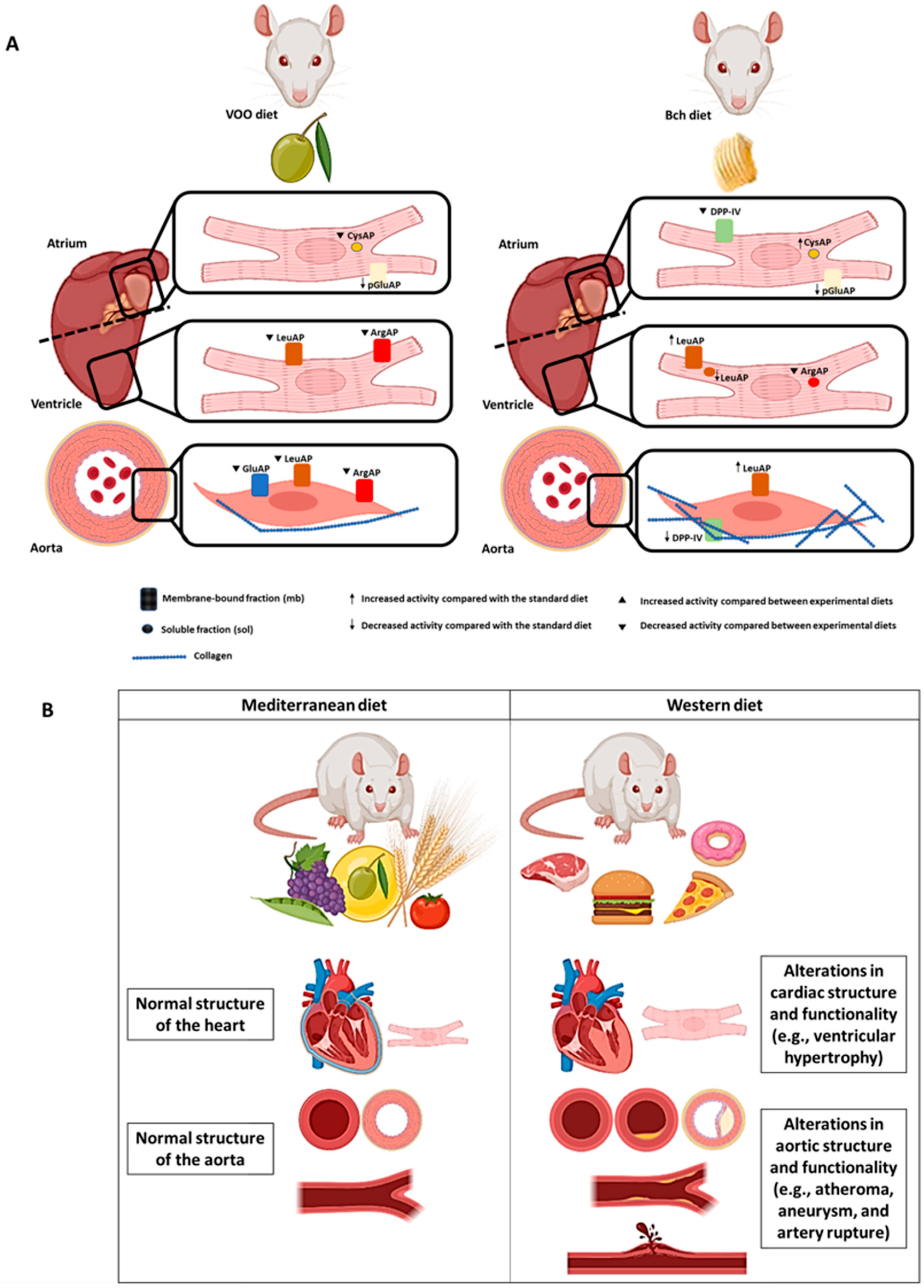
All the results predicted a differential effect of two high-fat diets on RAS regulation, functionality, and stability in the atrium, ventricle, and aorta (Figure 5). The inclusion of virgin olive oil in the diets moderated the normalization of RAS activities in the atrium (CysAP), ventricle (GluAP and ArgAP), and aorta (GluAP and ArgAP). However, the Bch diet had a significant increase in CysAP activity in the atrium, together with increases in the activity of functional marker LeuAP in the ventricle and aorta, while the olive oil normalized the LeuAP values. The VOO diet showed normal DPP-IV activity; however, the Bch diet reduced DPP-IV activity in the aorta. Interestingly, DPP-IV and angiotensinase activities were strongly correlated intragroup, especially with the VOO diet for AlaAP, ArgAP, AspAP, CysAP, and GluAP activities in the atrium ventricle. The replacement of saturated fats with monounsaturated fats would benefit by normalizing the activities that regulate homeostasis of the classical and non-classical pathways of RAS and DPP-IV for proper maintenance and functioning of the cardiovascular system.

Supplementary Materials

The following are available online at https://www.mdpi.com/article/10.3390/biomedicines9091149/s1, Figure S1: Group and intra-group correlations between significant glutamyl aminopeptidase, alanyl aminopeptidase, arginyl aminopeptidase, and cystinyl aminopeptidase activities analyzed in plasma and soluble and membrane-bound fractions of atrium, ventricle and aorta, Figure S2: Group and intra-group correlations between significant dipeptidyl peptidase IV activity analyzed in plasma and soluble fractions of atrium, Figure S3: Group and intra-group correlations between significant leucyl aminopeptidase activity analyzed in membrane-bound fractions of ventricle and aorta, Table S1: Angiotensinase activities in atrium, ventricle, aorta and plasma, Table S2: Dipeptidyl dipeptidase IV activity in atrium, ventricle, aorta and plasma, Table S3: Leucyl aminopeptidase, gamma-glutamyl transferase and pyroglutamyl aminopeptidase activities in atrium, ventricle, aorta and plasma.

Author Contributions

Conceptualization, I.P., M.R.-S., and G.D.-V.; methodology, I.P., A.B.S., and G.D.-V.; investigation, I.P., A.B.S., and G.D.-V.; data curation, A.B.S. and G.D.-V.; writing—original draft preparation, I.P. and G.D.-V.; writing—review and editing, I.P. and G.D.-V.; supervision, I.P., G.D.-V., and M.R.-S.; project administration, I.P.; funding acquisition, I.P. All authors have read and agreed to the published version of the manuscript.

Funding

This work was supported by grant code: ACCIÓN 1 PAIUJA 2019 2020: BIO221.

Institutional Review Board Statement

The study was conducted according to the guidelines of the Declaration of Helsinki, and the study was approved by the Institutional Animal Care and Use Committee of the University of Jaén (project number P.I.UJA_2005_acción 14), of the University of Jaén. Technical and human support provided by Recursos Científicos-Técnicos (CICT) Universidad de Jaén (UJA), Ministerio de Economía, Industria y Competitividad (MINECO), Junta de Andalucía, and Fondo Europeo de Desarrollo Regional (FEDER) is gratefully acknowledged.

Informed Consent Statement

Not applicable.

Data Availability Statement

Not applicable.

Acknowledgments

The authors would like to acknowledge the technical and human support provided by CICT and CEPA of the Universidad de Jaén (UJA, MINECO, Junta de Andalucía; FEDER).

Conflicts of Interest

The authors declare no conflict of interest.

Abbreviations

AlaAPalanyl-aminopeptidase
Angangiotensin
APaminopeptidase
ArgAParginyl-aminopeptidase
AspAPaspartyl-aminopeptidase
Bchbutter plus cholesterol
BPblood pressure
CysAPcystine aminopeptidase
CVDcardiovascular diseases
DPP-IVdipeptidyl-peptidase IV
GGTgamma-glutamyl transferase
GluAPglutamyl-aminopeptidase
HFDhigh-fat diet
IRAPinsulin-regulated aminopeptidase
LeuAPleucyl-aminopeptidase
MUFAmonounsaturated fatty acids
pGluAPpyroglutamyl-aminopeptidase
RASrenin–angiotensin system
Sstandard
SAFAsaturated fatty acids
VOOvirgin olive oil

References

  1. Vasilopoulou, D.; Markey, O.; Kliem, K.E.; Fagan, C.C.; Grandison, A.S.; Humphries, D.J.; Todd, S.; Jackson, K.G.; Givens, D.I.; Lovegrove, J.A. Reformulation initiative for partial replacement of saturated with unsaturated fats in dairy foods attenuates the increase in LDL cholesterol and improves flow-mediated dilatation compared with conventional dairy: The randomized, controlled REplacement of SaturatEd fat in dairy on Total cholesterol (RESET) study. Am. J. Clin. Nutr. 2020, 111, 739–748. [Google Scholar] [CrossRef]
  2. Cerf, M.E. High Fat Programming and Cardiovascular Disease. Medicina 2018, 54, 86. [Google Scholar] [CrossRef] [Green Version]
  3. Wali, J.A.; Jarzebska, N.; Raubenheimer, D.; Simpson, S.J.; Rodionov, R.N.; O’Sullivan, J.F. Cardio-Metabolic Effects of High-Fat Diets and Their Underlying Mechanisms—A Narrative Review. Nutrients 2020, 12, 1505. [Google Scholar] [CrossRef] [PubMed]
  4. Domínguez-Vías, G.; Segarra, A.B.; Ramírez-Sánchez, M.; Prieto, I. Effects of Virgin Olive Oil on Blood Pressure and Renal Aminopeptidase Activities in Male Wistar Rats. Int. J. Mol. Sci. 2021, 22, 5388. [Google Scholar] [CrossRef] [PubMed]
  5. Segarra, A.B.; Ramirez, M.; Banegas, I.; Alba, F.; Vives, F.; Gasparo, M.d.; Ortega, E.; Ruiz, E.; Prieto, I. Dietary fat influences testosterone, cholesterol, aminopeptidase A, and blood pressure in male rats. Horm. Metab. Res. 2008, 40, 289–291. [Google Scholar] [CrossRef]
  6. Villarejo, A.B.; Ramírez-Sánchez, M.; Segarra, A.B.; Martínez-Cañamero, M.; Prieto, I. Influence of extra virgin olive oil on blood pressure and kidney angiotensinase activities in spontaneously hypertensive rats. Planta Med. 2015, 81, 664–669. [Google Scholar] [CrossRef] [PubMed]
  7. Domínguez-Vías, G.; Segarra, A.B.; Martínez-Cañamero, M.; Ramírez-Sánchez, M.; Prieto, I. Influence of a Virgin Olive Oil versus Butter Plus Cholesterol-Enriched Diet on Testicular Enzymatic Activities in Adult Male Rats. Int. J. Mol. Sci. 2017, 18, 1701. [Google Scholar] [CrossRef] [Green Version]
  8. Martínez, N.; Prieto, I.; Hidalgo, M.; Segarra, A.B.; Martínez-Rodríguez, A.M.; Cobo, A.; Ramírez, M.; Gálvez, A.; Martínez-Cañamero, M. Refined versus Extra Virgin Olive Oil High-Fat Diet Impact on Intestinal Microbiota of Mice and Its Relation to Different Physiological Variables. Microorganisms 2019, 7, 61. [Google Scholar] [CrossRef] [Green Version]
  9. Domínguez-Vías, G.; Segarra, A.B.; Ramírez-Sánchez, M.; Prieto, I. The Role of High Fat Diets and Liver Peptidase Activity in the Development of Obesity and Insulin Resistance in Wistar Rats. Nutrients 2020, 12, 636. [Google Scholar] [CrossRef] [PubMed] [Green Version]
  10. Segarra, A.B.; Domínguez-Vías, G.; Redondo, J.; Martínez-Cañamero, M.; Ramírez-Sánchez, M.; Prieto, I. Hypothalamic Renin–Angiotensin System and Lipid Metabolism: Effects of Virgin Olive Oil versus Butter in the Diet. Nutrients 2021, 13, 480. [Google Scholar] [CrossRef]
  11. Sánchez-Aguilar, M.; Ibarra-Lara, L.; Del Valle-Mondragón, L.; Soria-Castro, E.; Torres-Narváez, J.C.; Carreón-Torres, E.; Sánchez-Mendoza, A.; Rubio-Ruíz, M.E. Nonclassical Axis of the Renin-Angiotensin System and Neprilysin: Key Mediators That Underlie the Cardioprotective Effect of PPAR-Alpha Activation during Myocardial Ischemia in a Metabolic Syndrome Model. PPAR Res. 2020, 2020, 8894525. [Google Scholar] [CrossRef] [PubMed]
  12. Ramírez, M.; Prieto, I.; Alba, F.; Vives, F.; Banegas, I.; de Gasparo, M. Role of central and peripheral aminopeptidase activities in the control of blood pressure: A working hypothesis. Heart Fail Rev. 2008, 13, 339–353. [Google Scholar] [CrossRef] [PubMed]
  13. Ramírez, M.; Prieto, I.; Martinez, J.M.; Vargas, F.; Alba, F. Renal aminopeptidase activities in animal models of hypertension. Regul. Pept. 1997, 72, 155–159. [Google Scholar] [CrossRef]
  14. Prieto, I.; Martinez, A.; Martinez, J.M.; Ramírez, M.J.; Vargas, F.; Alba, F.; Ramírez, M. Activities of aminopeptidases in a rat saline model of volume hypertension. Horm. Metab. Res. 1998, 30, 246–248. [Google Scholar] [CrossRef]
  15. Prieto, I.; Martínez, J.M.; Hermoso, F.; Ramírez, M.J.; de Gasparo, M.; Vargas, F.; Alba, F.; Ramírez, M. Effect of valsartan on angiotensin II- and vasopressin-degrading activities in the kidney of normotensive and hypertensive rats. Horm. Metab. Res. 2001, 33, 559–563. [Google Scholar] [CrossRef]
  16. Prieto, I.; Arechaga, G.; Segarra, A.B.; Alba, F.; de Gasparo, M.; Ramirez, M. Effects of dehydration on renal aminopeptidase activities in adult male and female rats. Regul. Pept. 2002, 106, 27–32. [Google Scholar] [CrossRef]
  17. Prieto, I.; Hermoso, F.; de Gasparo, M.; Vargas, F.; Alba, F.; Segarra, A.B.; Banegas, I.; Ramírez, M. Aminopeptidase activity in renovascular hypertension. Med. Sci. Monit. 2003, 9, 31–36. [Google Scholar]
  18. Prieto, I.; Hermoso, F.; Gasparo, M.d.; Vargas, F.; Alba, F.; Segarra, A.B.; Banegas, I.; Ramírez, M. Angiotensinase activities in the kidney of renovascular hypertensive rats. Peptides 2003, 24, 755–760. [Google Scholar] [CrossRef] [Green Version]
  19. Segarra, A.B.; Prieto, I.; Villarejo, A.B.; Banegas, I.; Wangensteen, R.; de Gasparo, M.; Vives, F.; Ramírez-Sánchez, M. Effects of antihypertensive drugs on angiotensinase activities in the testis of spontaneously hypertensive rats. Horm. Metab. Res. 2013, 45, 344–348. [Google Scholar] [CrossRef]
  20. Prieto, I.; Villarejo, A.B.; Segarra, A.B.; Banegas, I.; Wangensteen, R.; Martinez-Cañamero, M.; de Gasparo, M.; Vives, F.; Ramírez-Sánchez, M. Brain, heart and kidney correlate for the control of blood pressure and water balance: Role of angiotensinases. Neuroendocrinology 2014, 100, 198–208. [Google Scholar] [CrossRef] [PubMed]
  21. Villarejo, A.B.; Prieto, I.; Segarra, A.B.; Banegas, I.; Wangensteen, R.; Vives, F.; de Gasparo, M.; Ramírez-Sánchez, M. Relationship of angiotensinase and vasopressinase activities between hypothalamus, heart, and plasma in L-NAME-treated WKY and SHR. Horm. Metab. Res. 2014, 46, 561–567. [Google Scholar] [CrossRef] [PubMed]
  22. Prieto, I.; Villarejo, A.B.; Segarra, A.B.; Wangensteen, R.; Banegas, I.; de Gasparo, M.; Vanderheyden, P.; Zorad, S.; Vives, F.; Ramírez-Sánchez, M. Tissue distribution of CysAP activity and its relationship to blood pressure and water balance. Life Sci. 2015, 134, 73–78. [Google Scholar] [CrossRef] [PubMed]
  23. Prieto, I.; Segarra, A.B.; de Gasparo, M.; Martínez-Cañamero, M.; Ramírez-Sánchez, M. Divergent profile between hypothalamic and plasmatic aminopeptidase activities in WKY and SHR. Influence of beta-adrenergic blockade. Life Sci. 2018, 192, 9–17. [Google Scholar] [CrossRef] [PubMed]
  24. Keck, M.; Hmazzou, R.; Llorens-Cortes, C. Orally Active Aminopeptidase A Inhibitor Prodrugs: Current State and Future Directions. Curr. Hypertens. Rep. 2019, 21, 50. [Google Scholar] [CrossRef] [PubMed]
  25. Llorens-Cortes, C.; Touyz, R.M. Evolution of a New Class of Antihypertensive Drugs: Targeting the Brain Renin-Angiotensin System. Hypertension 2020, 75, 6–15. [Google Scholar] [CrossRef]
  26. Segarra, A.B.; Ruiz-Sanz, J.I.; Ruiz-Larrea, M.B.; Ramírez-Sánchez, M.; de Gasparo, M.; Banegas, I.; Martínez-Cañamero, M.; Vives, F.; Prieto, I. The profile of fatty acids in frontal cortex of rats depends on the type of fat used in the diet and correlates with neuropeptidase activities. Horm. Metab. Res. 2011, 43, 86–91. [Google Scholar] [CrossRef]
  27. Boitard, S.E.; Marc, Y.; Keck, M.; Mougenot, N.; Agbulut, O.; Balavoine, F.; Llorens-Cortes, C. Brain renin-angiotensin system blockade with orally active aminopeptidase A inhibitor prevents cardiac dysfunction after myocardial infarction in mice. J. Mol. Cell Cardiol. 2019, 127, 215–222. [Google Scholar] [CrossRef]
  28. Turgut, O.; Yilmaz, A.; Yalta, K.; Karadas, F.; Birhan-Yilmaz, M. Gamma-Glutamyltransferase is a promising biomarker for cardiovascular risk. Med. Hypotheses. 2006, 67, 1060–1064. [Google Scholar] [CrossRef]
  29. Danziger, R.S. Aminopeptidase N in arterial hypertension. Heart Fail. Rev. 2008, 13, 293–298. [Google Scholar] [CrossRef]
  30. Tsujimoto, M.; Goto, Y.; Maruyama, M.; Hattori, A. Biochemical and enzymatic properties of the M1 family of aminopeptidases involved in the regulation of blood pressure. Heart Fail Rev. 2008, 13, 285–291. [Google Scholar] [CrossRef]
  31. Mason, J.E.; Starke, R.D.; Van Kirk, J.E. Gamma-glutamyl transferase: A novel cardiovascular risk biomarker. Prev. Cardiol. 2010, 13, 36–41. [Google Scholar] [CrossRef] [PubMed]
  32. Quesada, A.; Vargas, F.; Montoro-Molina, S.; O’Valle, F.; Rodríguez-Martínez, M.D.; Osuna, A.; Prieto, I.; Ramírez, M.; Wangensteen, R. Urinary aminopeptidase activities as early and predictive biomarkers of renal dysfunction in cisplatin-treated rats. PLoS ONE 2012. [Google Scholar] [CrossRef] [PubMed]
  33. Chai, Y.; Gao, Y.; Xiong, H.; Lv, W.; Yang, G.; Lu, C.; Nie, J.; Ma, C.; Chen, Z.; Ren, J.; et al. A near-infrared fluorescent probe for monitoring leucine aminopeptidase in living cells. Analyst 2019, 144, 463–467. [Google Scholar] [CrossRef]
  34. Vargas, F.; Wangesteen, R.; Rodríguez-Gómez, I.; García-Estañ, J. Aminopeptidases in Cardiovascular and Renal Function. Role as Predictive Renal Injury Biomarkers. Int. J. Mol. Sci. 2020, 21, 5615. [Google Scholar] [CrossRef]
  35. Du, K.; Sheng, L.; Luo, X.; Fan, G.; Shen, D.; Wu, C.; Shen, R. A ratiometric fluorescent probe based on quinoline for monitoring and imaging of Leucine aminopeptidase in liver tumor cells. Spectrochim. Acta A Mol. Biomol. Spectrosc. 2021, 249, 119328. [Google Scholar] [CrossRef] [PubMed]
  36. Vauquelin, G.; Michotte, Y.; Smolders, I.; Sarre, S.; Ebinger, G.; Dupont, A.; Vanderheyden, P. Cellular targets for angiotensin II fragments: Pharmacological and molecular evidence. J. Renin Angiotensin Aldosterone Syst. 2002, 3, 195–204. [Google Scholar] [CrossRef] [Green Version]
  37. Wallis, M.G.; Lankford, M.F.; Keller, S.R. Vasopressin is a physiological substrate for the insulin-regulated aminopeptidase IRAP. Am. J. Physiol. Endocrinol. Metab. 2007, 293, 1092–1102. [Google Scholar] [CrossRef] [Green Version]
  38. Ramírez-Sánchez, M.; Prieto, I.; Wangensteen, R.; Banegas, I.; Segarra, A.B.; Villarejo, A.B.; Vives, F.; Cobo, J.; de Gasparo, M. The renin-angiotensin system: New insight into old therapies. Curr. Med. Chem. 2013, 20, 1313–1322. [Google Scholar] [CrossRef]
  39. Segarra, A.B.; Prieto, I.; Martinez-Canamero, M.; Vargas, F.; De Gasparo, M.; Vanderheyden, P.; Zorad, S.; Ramirez-Sanchez, M. Cystinyl and pyroglutamyl-beta-naphthylamide hydrolyzing activities are modified coordinately between hypothalamus, liver and plasma depending on the thyroid status of adult male rats. J. Physiol. Pharmacol. 2018, 69, 197–204. [Google Scholar] [CrossRef]
  40. Weber, K.T. Fibrosis and hypertensive heart disease. Curr. Opin. Cardiol. 2000, 15, 264–272. [Google Scholar] [CrossRef]
  41. Weber, K.T. Fibrosis in hypertensive heart disease: Focus on cardiac fibroblasts. J. Hypertens. 2004, 22, 47–50. [Google Scholar] [CrossRef]
  42. Aroor, A.R.; Habibi, J.; Kandikattu, H.K.; Garro-Kacher, M.; Barron, B.; Chen, D.; Hayden, M.R.; Whaley-Connell, A.; Bender, S.B.; Klein, T.; et al. Dipeptidyl peptidase-4 (DPP-4) inhibition with linagliptin reduces western diet-induced myocardial TRAF3IP2 expression, inflammation and fibrosis in female mice. Cardiovasc. Diabetol. 2017, 16, 61. [Google Scholar] [CrossRef] [PubMed] [Green Version]
  43. Sun, Y.; Zhang, J.; Lu, L.; Chen, S.S.; Quinn, M.T.; Weber, K.T. Aldosterone-induced inflammation in the rat heart: Role of oxidative stress. Am. J. Pathol. 2002, 161, 1773–1781. [Google Scholar] [CrossRef]
  44. Li, C.; Han, R.; Kang, L.; Wang, J.; Gao, Y.; Li, Y.; He, J.; Tian, J. Pirfenidone controls the feedback loop of the AT1R/p38 MAPK/renin-angiotensin system axis by regulating liver X receptor-α in myocardial infarction-induced cardiac fibrosis. Sci. Rep. 2017, 7, 40523. [Google Scholar] [CrossRef] [Green Version]
  45. Laurent, G.J. Dynamic state of collagen: Pathways of collagen degradation in vivo and their possible role in regulation of collagen mass. Am. J. Physiol. 1987, 252, 1–9. [Google Scholar] [CrossRef] [PubMed]
  46. Wang, Y.; Kou, J.; Zhang, H.; Wang, C.; Li, H.; Ren, Y.; Zhang, Y. The renin-angiotensin system in the synovium promotes periarticular osteopenia in a rat model of collagen-induced arthritis. Int. Immunopharmacol. 2018, 65, 550–558. [Google Scholar] [CrossRef]
  47. Lijnen, P.J.; Petrov, V.V.; Fagard, R.H. Collagen production in cardiac fibroblasts during inhibition of angiotensin-converting enzyme and aminopeptidases. J. Hypertens. 2004, 22, 209–216. [Google Scholar] [CrossRef]
  48. Mizutani, T.; Mizutani, H.; Kaneda, T.; Hagihara, M.; Nagatsu, T. Activity of dipeptidyl peptidase II and dipeptidyl peptidase IV in human gingiva with chronic marginal periodontitis. Arch. Oral Biol. 1990, 35, 891–894. [Google Scholar] [CrossRef]
  49. Kamori, M.; Hagihara, M.; Nagatsu, T.; Iwata, H.; Miura, T. Activities of dipeptidyl peptidase II, dipeptidyl peptidase IV, prolyl endopeptidase, and collagenase-like peptidase in synovial membrane from patients with rheumatoid arthritis and osteoarthritis. Biochem. Med. Metab. Biol. 1991, 45, 154–160. [Google Scholar] [CrossRef]
  50. Hatanaka, T.; Kawakami, K.; Uraji, M. Inhibitory effect of collagen-derived tripeptides on dipeptidylpeptidase-IV activity. J. Enzyme Inhib. Med. Chem. 2014, 29, 823–828. [Google Scholar] [CrossRef] [Green Version]
  51. Kenny, A.J.; Booth, A.G.; George, S.G.; Ingram, J.; Kershaw, D.; Wood, E.J.; Young, A.R. Dipeptidyl peptidase IV, a kidney brush-border serine peptidase. Biochem. J. 1976, 157, 169–182. [Google Scholar] [CrossRef] [PubMed] [Green Version]
  52. Nistala, R.; Savin, V. Diabetes, hypertension, and chronic kidney disease progression: Role of DPP4. Am. J. Physiol. Renal Physiol. 2017, 312, 661–670. [Google Scholar] [CrossRef]
  53. Zhong, J.; Rao, X.; Rajagopalan, S. An emerging role of dipeptidyl peptidase 4 (DPP4) beyond glucose control: Potential implications in cardiovascular disease. Atherosclerosis 2013, 226, 305–314. [Google Scholar] [CrossRef]
  54. Wolke, C.; Teumer, A.; Endlich, K.; Endlich, N.; Rettig, R.; Stracke, S.; Fiene, B.; Aymanns, S.; Felix, S.B.; Hannemann, A.; et al. Serum protease activity in chronic kidney disease patients: The GANI_MED renal cohort. Exp. Biol. Med. 2017, 242, 554–563. [Google Scholar] [CrossRef] [PubMed] [Green Version]
  55. Yamaguchi, T.; Watanabe, A.; Tanaka, M.; Shiota, M.; Osada-Oka, M.; Sano, S.; Yoshiyama, M.; Miura, K.; Kitajima, S.; Matsunaga, S.; et al. A dipeptidyl peptidase-4 (DPP-4) inhibitor, linagliptin, attenuates cardiac dysfunction after myocardial infarction independently of DPP-4. J. Pharmacol. Sci. 2019, 139, 112–119. [Google Scholar] [CrossRef]
  56. Shi, Z.X.; Xu, W.; Mergner, W.J.; Li, Q.L.; Cole, K.H.; Wilber, J.F. Localization of thyrotropin-releasing hormone mRNA expression in the rat heart by in situ hybridization histochemistry. Pathobiology 1996, 64, 314–319. [Google Scholar] [CrossRef] [PubMed]
  57. Arechaga, G.; Prieto, I.; Segarra, A.B.; Alba, F.; Ruiz-Larrea, M.B.; Ruiz-Sanz, J.I.; de Gasparo, M.; Ramirez, M. Dietary fatty acid composition affects aminopeptidase activities in the testes of mice. Int. J. Androl. 2002, 25, 113–118. [Google Scholar] [CrossRef] [PubMed]
  58. Zhou, Y.; Zhao, S.; Chen, K.; Hua, W.; Zhang, S. Predictive value of gamma-glutamyltransferase for ventricular arrhythmias and cardiovascular mortality in implantable cardioverter-defibrillator patients. BMC Cardiovasc. Disord. 2019, 19, 129. [Google Scholar] [CrossRef]
  59. Neuman, M.G.; Malnick, S.; Chertin, L. Gamma glutamyl transferase—An underestimated marker for cardiovascular disease and the metabolic syndrome. J. Pharm. Pharm. Sci. 2020, 23, 65–74. [Google Scholar] [CrossRef] [Green Version]
  60. Ruttmann, E.; Brant, L.J.; Concin, H.; Diem, G.; Rapp, K.; Ulmer, H.; Vorarlberg Health Monitoring and Promotion Program Study Group. Gamma-glutamyltransferase as a risk factor for cardiovascular disease mortality: An epidemiological investigation in a cohort of 163,944 Austrian adults. Circulation 2005, 112, 2130–2137. [Google Scholar] [CrossRef] [Green Version]
  61. Koenig, G.; Seneff, S. Gamma-Glutamyltransferase: A Predictive Biomarker of Cellular Antioxidant Inadequacy and Disease Risk. Dis. Markers 2015, 2015, 818570. [Google Scholar] [CrossRef] [Green Version]
  62. Kreutzer, F.; Krause, D.; Klaassen-Mielke, R.; Trampisch, H.J.; Diehm, C.; Rudolf, H. Gamma-glutamyl transferase as a risk factor for mortality and cardiovascular events in older adults—Results from a prospective cohort study in a primary care setting (getABI). Vasa 2019, 48, 313–319. [Google Scholar] [CrossRef]
  63. Lee, D.S.; Evans, J.C.; Robins, S.J.; Wilson, P.W.; Albano, I.; Fox, C.S.; Wang, T.J.; Benjamin, E.J.; D’Agostino, R.B.; Vasan, R.S. Gamma glutamyl transferase and metabolic syndrome, cardiovascular disease, and mortality risk: The Framingham Heart Study. Arterioscler. Thromb. Vasc. Biol. 2007, 27, 127–133. [Google Scholar] [CrossRef] [Green Version]
  64. Ndrepepa, G.; Colleran, R.; Kastrati, A. Gamma-glutamyl transferase and the risk of atherosclerosis and coronary heart disease. Clin. Chim. Acta 2018, 476, 130–138. [Google Scholar] [CrossRef]
  65. Greenberg, L.J. Fluorometric measurement of alkaline phosphatase and aminopeptidase activities in the order of 10-14 mole. Biochem. Biophys. Res. Commun. 1962, 9, 430–435. [Google Scholar] [CrossRef]
  66. Cheung, H.S.; Cushman, D.W. A soluble aspartate aminopeptidase from dog kidney. Biochim. Biophys. Acta 1971, 242, 190–193. [Google Scholar] [CrossRef]
  67. Tobe, H.; Kojima, F.; Aoyagi, T.; Umezawa, H. Purification by affinity chromatography using amastatin and properties of aminopeptidase A from pig kidney. Biochim. Biophys. Acta 1980, 613, 459–468. [Google Scholar] [CrossRef]
  68. Ramírez, M.; Prieto, I.; Banegas, I.; Segarra, A.B.; Alba, F. Neuropeptidases. Methods Mol. Biol. 2011, 789, 287–294. [Google Scholar] [CrossRef] [PubMed]
  69. Bradford, M.M. A rapid and sensitive method for the quantitation of microgram quantities of protein utilizing the principle of protein-dye binding. Anal. Biochem. 1976, 72, 248–254. [Google Scholar] [CrossRef]
  70. Sá, J.M.; Barbosa, R.M.; Menani, J.V.; De Luca, L.A., Jr.; Colombari, E.; Almeida-Colombari, D.S. Cardiovascular and hidroelectrolytic changes in rats fed with high-fat diet. Behav. Brain Res. 2019, 373, 112075. [Google Scholar] [CrossRef]
  71. Clifton, P.M.; Keogh, J.B. A systematic review of the effect of dietary saturated and polyunsaturated fat on heart disease. Nutr. Metab. Cardiovasc. Dis. 2017, 27, 1060–1080. [Google Scholar] [CrossRef] [PubMed]
  72. Arechaga, G.; Martínez, J.M.; Prieto, I.; Ramírez, M.J.; Sánchez, M.J.; Alba, F.; De Gasparo, M.; Ramírez, M. Serum aminopeptidase A activity of mice is related to dietary fat saturation. J. Nutr. 2001, 131, 1177–1179. [Google Scholar] [CrossRef] [PubMed] [Green Version]
  73. Summerhill, V.; Karagodin, V.; Grechko, A.; Myasoedova, V.; Orekhov, A. Vasculoprotective Role of Olive Oil Compounds via Modulation of Oxidative Stress in Atherosclerosis. Front. Cardiovasc. Med. 2018, 5, 188. [Google Scholar] [CrossRef] [PubMed] [Green Version]
  74. D’Agostino, R.; Barberio, L.; Gatto, M.; Muzzalupo, I.; Mandalà, M. Extra Virgin Olive Oil Phenols Dilate the Rat Mesenteric Artery by Activation of BKCa2+ Channels in Smooth Muscle Cells. Molecules 2020, 25, 2601. [Google Scholar] [CrossRef]
  75. Paknahad, Z.; Moosavian, S.P.; Mahdavi, R.; Rajabi, P. The effects of olive oil and cholesterol enriched diets on aortic fatty streak development and lipid peroxidation in rabbits. Nutr. Health 2021, 18, 2601060211022260. [Google Scholar] [CrossRef]
  76. Khan, S.A.; Sattar, M.Z.; Abdullah, N.A.; Rathore, H.A.; Abdulla, M.H.; Ahmad, A.; Johns, E.J. Obesity depresses baroreflex control of renal sympathetic nerve activity and heart rate in Sprague Dawley rats: Role of the renal innervation. Acta Physiol. 2015, 214, 390–401. [Google Scholar] [CrossRef]
  77. Segarra, A.B.; Prieto, I.; Martinez-Canamero, M.; Ruiz-Sanz, J.I.; Ruiz-Larrea, M.B.; De Gasparo, M.; Banegas, I.; Zorad, S.; Ramirez-Sanchez, M. Enkephalinase activity is modified and correlates with fatty acids in frontal cortex depending on fish, olive or coconut oil used in the diet. Endocr. Regul. 2019, 53, 59–64. [Google Scholar] [CrossRef] [Green Version]
  78. Brilla, C.G.; Maisch, B.; Weber, K.T. Renin-angiotensin system and myocardial collagen matrix remodeling in hypertensive heart disease: In vivo and in vitro studies on collagen matrix regulation. Clin. Investig. 1993, 71, 35–41. [Google Scholar] [CrossRef]
  79. Liu, T.; Wen, H.; Li, H.; Xu, H.; Xiao, N.; Liu, R.; Chen, L.; Sun, Y.; Song, L.; Bai, C.; et al. Oleic Acid Attenuates Ang II (Angiotensin II)-Induced Cardiac Remodeling by Inhibiting FGF23 (Fibroblast Growth Factor 23) Expression in Mice. Hypertension 2020, 75, 680–692. [Google Scholar] [CrossRef]
  80. Mascolo, A.; Sessa, M.; Scavone, C.; De Angelis, A.; Vitale, C.; Berrino, L.; Rossi, F.; Rosano, G.; Capuano, A. New and old roles of the peripheral and brain renin-angiotensin-aldosterone system (RAAS): Focus on cardiovascular and neurological diseases. Int. J. Cardiol. 2017, 227, 734–742. [Google Scholar] [CrossRef]
  81. Marc, Y.; Boitard, S.E.; Balavoine, F.; Azizi, M.; Llorens-Cortes, C. Targeting Brain Aminopeptidase A: A New Strategy for the Treatment of Hypertension and Heart Failure. Can. J. Cardiol. 2020, 36, 721–731. [Google Scholar] [CrossRef]
  82. Weber, K.T.; Sun, Y.; Tyagi, S.C.; Cleutjens, J.P. Collagen network of the myocardium: Function, structural remodeling and regulatory mechanisms. J. Mol. Cell. Cardiol. 1994, 26, 279–292. [Google Scholar] [CrossRef]
  83. Harikrishnan, V.; Titus, A.S.; Cowling, R.T. Collagen receptor cross-talk determines α-smooth muscle actin-dependent collagen gene expression in angiotensin II-stimulated cardiac fibroblasts. J. Biol. Chem. 2019, 294, 19723–19739. [Google Scholar] [CrossRef]
  84. Manrique, C.; Habibi, J.; Aroor, A.R.; Sowers, J.R.; Jia, G.; Hayden, M.R.; Garro, M.; Martinez-Lemus, L.A.; Ramirez-Perez, F.I.; Klein, T.; et al. Dipeptidyl peptidase-4 inhibition with linagliptin prevents western diet-induced vascular abnormalities in female mice. Cardiovasc. Diabetol. 2016, 15, 94. [Google Scholar] [CrossRef] [PubMed] [Green Version]
  85. Gutiérrez-Cuevas, J.; Sandoval-Rodriguez, A.; Meza-Rios, A.; Monroy-Ramírez, H.C.; Galicia-Moreno, M.; García-Bañuelos, J.; Santos, A.; Armendariz-Borunda, J. Molecular Mechanisms of Obesity-Linked Cardiac Dysfunction: An Up-Date on Current Knowledge. Cells 2021, 10, 629. [Google Scholar] [CrossRef]
  86. Santana, A.B.; de Souza Oliveira, T.C.; Bianconi, B.L.; Barauna, V.G.; Santos, E.W.; Alves, T.P.; Silva, J.C.; Fiorino, P.; Borelli, P.; Irigoyen, M.C.; et al. Effect of high-fat diet upon inflammatory markers and aortic stiffening in mice. BioMed Res. Int. 2014, 2014, 914102. [Google Scholar] [CrossRef] [Green Version]
  87. Arendse, L.B.; Danser, A.; Poglitsch, M.; Touyz, R.M.; Burnett, J.C., Jr.; Llorens-Cortes, C.; Ehlers, M.R.; Sturrock, E.D. Novel Therapeutic Approaches Targeting the Renin-Angiotensin System and Associated Peptides in Hypertension and Heart Failure. Pharmacol. Rev. 2019, 71, 539–570. [Google Scholar] [CrossRef] [PubMed] [Green Version]
  88. Cheng, H.C.; Abdel-Ghany, M.; Elble, R.C.; Pauli, B.U. Lung endothelial dipeptidyl peptidase IV promotes adhesion and metastasis of rat breast cancer cells via tumor cell surface-associated fibronectin. J. Biol. Chem. 1998, 273, 24207–24215. [Google Scholar] [CrossRef] [Green Version]
  89. Wang, X.M.; Holz, L.E.; Chowdhury, S.; Cordoba, S.P.; Evans, K.A.; Gall, M.G.; Vieira de Ribeiro, A.J.; Zheng, Y.Z.; Levy, M.T.; Yu, D.M.; et al. The pro-fibrotic role of dipeptidyl peptidase 4 in carbon tetrachloride-induced experimental liver injury. Immunol. Cell. Biol. 2017, 95, 443–453. [Google Scholar] [CrossRef]
  90. Oparil, S.; Schmieder, R.E. New approaches in the treatment of hypertension. Circ. Res. 2015, 116, 1074–1095. [Google Scholar] [CrossRef] [Green Version]
  91. Stewart, M.H.; Lavie, C.J.; Ventura, H.O. Future pharmacological therapy in hypertension. Curr. Opin. Cardiol. 2018, 33, 408–415. [Google Scholar] [CrossRef] [PubMed]
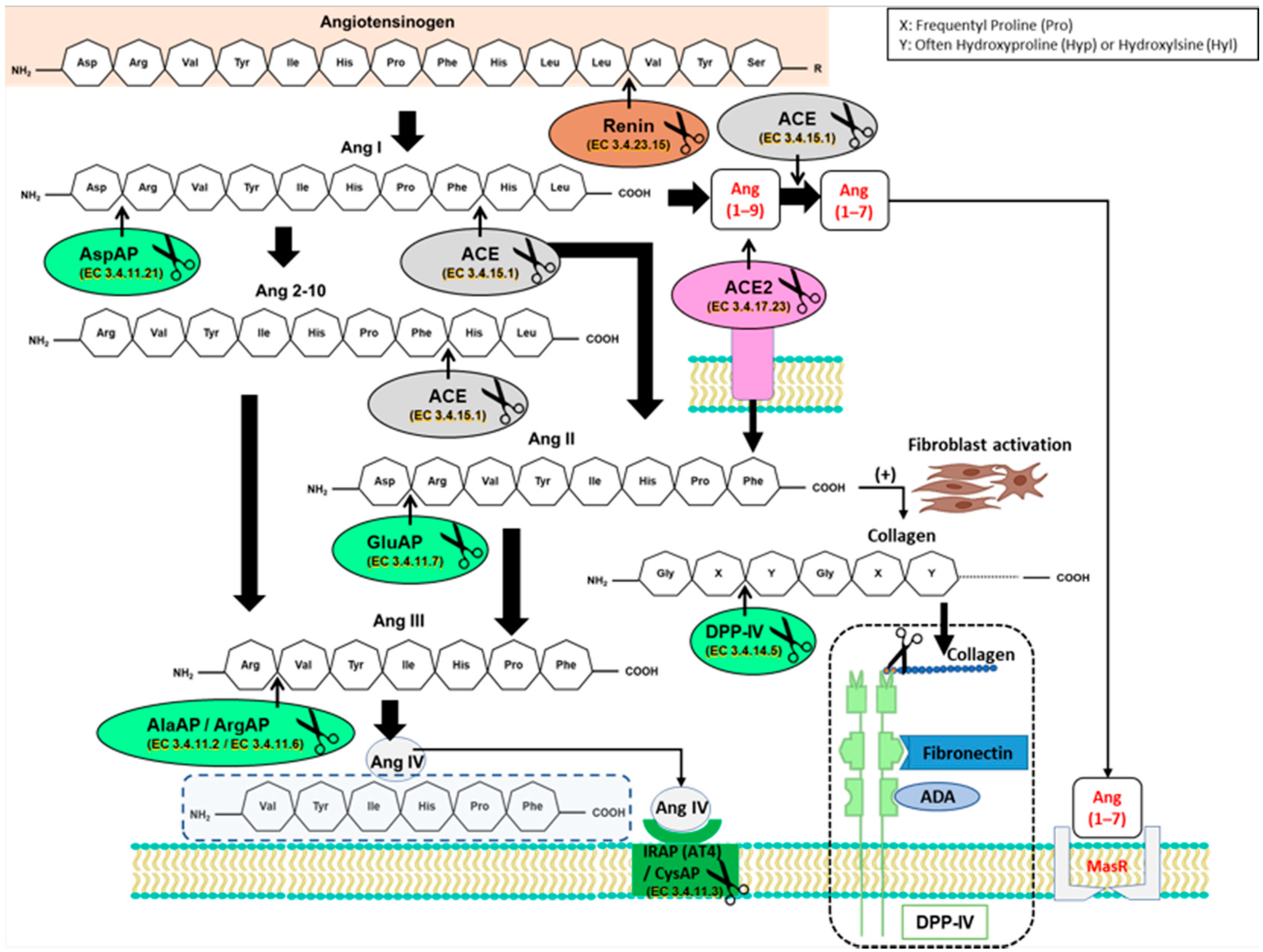
Figure 1. Partial diagram of angiotensinase activity downstream of the classical axis of the renin–angiotensin system (RAS) and the ACE2/Ang (1–7)/Mas receptor pathway, displaying the metabolic steps in which angiotensinase activities are involved. Ang II stimulates fibroblasts to produce collagen, this being a substrate for dipeptidyl peptidase IV (DPP-IV). ACE: angiotensin-converting enzyme; ACE2: angiotensin-converting enzyme 2; ADA: adenosine desaminase; AlaAP: alanyl aminopeptidase; Ang I: angiotensin I; Ang (1–7): angiotensin (1–7); Ang (1–9): angiotensin (1–9); Ang 2–10: angiotensin 2–10; Ang II: angiotensin II; Ang III: angiotensin III; Ang IV: angiotensin IV; ArgAP: arginyl aminopeptidase; AspAP: aspartyl aminopeptidase; AT4: angiotensin AT4 receptor; GluAP: glutamyl aminopeptidase; IRAP/CysAP: insulin-regulated aminopeptidase activity/cystinyl aminopeptidase; MasR: Mas receptor. The scissors symbol represents proteolytic activity of the enzyme, cleaving amino acids from the amino terminals.
Biomedicines 09 01149 g001
Figure 2. Mean values ± standard errors of significant (A) glutamyl aminopeptidase (GluAP), (BD) arginyl aminopeptidase (ArgAP), (E) cystinyl aminopeptidase (CysAP) activities in soluble (sol) and membrane-bound (mb) fractions of atrium, ventricle, and aorta. The small colored dots are data points per group. Values are expressed as pmol/min/mg prot in the rest of the tissues analyzed. * indicates significant differences between virgin olive oil diet (VOO) or butter plus cholesterol diet (Bch) vs. standard diet (S). * p < 0.05. # indicates significant differences between VOO and Bch, # p < 0.05.
Biomedicines 09 01149 g002
Figure 3. Mean values ± standard errors of significant (A,B) dipeptidyl peptidase IV (DPP-IV) activity in membrane-bound (mb) fraction of atrium and aorta. The small colored dots are data points per group. Values are expressed as pmol/min/mg prot in the rest of the tissues analyzed. * indicates significant differences between virgin olive oil diet (VOO) or butter plus cholesterol diet (Bch) vs. standard diet (S). * p < 0.05. # indicates significant differences between VOO and Bch, # p < 0.05.
Biomedicines 09 01149 g003
Figure 4. Mean values ± standard errors of significant (AC) leucyl aminopeptidase (LeuAP) and (D) pyroglutamyl aminopeptidase (pGluAP) activities in soluble and membrane-bound (mb) fractions of atrium, ventricle, and aorta. The small colored dots are data points per group. Values are expressed as pmol/min/mg prot in the rest of the tissues analyzed. * indicates significant differences between virgin olive oil diet (VOO) or butter plus cholesterol diet (Bch) vs. standard diet (S), * p < 0.05. # indicates significant differences between VOO and Bch, # p < 0.05.
Biomedicines 09 01149 g004
Figure 5. (A) Summary of the influence of the type of fat in the diet on the regulatory activities of the renin–angiotensin system, cardiac biomarker leucine aminopeptidase, and dipeptidyl peptidase IV. ArgAP: arginyl aminopeptidase; CysAP: cystinyl aminopeptidase; DPP-IV: dipeptidyl peptidase IV; GluAP: glutamyl aminopeptidase; LeuAP: leucyl aminopeptidase; pGluAP: pyroglutamyl aminopeptidase. VOO: diet enriched with virgin olive oil; Bch: diet enriched with butter plus cholesterol. (B) Differences in cardiovascular physiology with a long-term Mediterranean diet or Western diet at the structural or functional level in the heart and aorta.
Biomedicines 09 01149 g005
Table 1. Significant correlations of dipeptidyl peptidase IV (DPP-IV) activity with angiotensinase activities in atrium, ventricle, and aorta.
SampleDPP-IV (Fraction)Angiotensinase
(Fraction)
p-ValueRIntra-Group
Correlations
p-ValueR
AtriumDPP-IV (sol) vs.AlaAP (sol)0.0040.692VOO–VOO0.0070.969
ArgAP (sol)0.0020.734
CysAP (sol)<0.0010.777S–S
VOO–VOO
0.008
0.023
0.927
0.928
AtriumDPP-IV (mb) vs. AlaAP (mb)0.0120.609S–S
Bch-Bch
0.014
0.043
0.902
0.890
ArgAP (mb)0.0120.609S–S0.0110.912
CysAP (mb)0.0400.517
GluAP (mb)0.0370.526S-S0.0060.936
VentricleDPP-IV (sol) vs.AlaAP (sol)<0.0010.942VOO–VOO
Bch–Bch
0.002
0.018
0.984
0.940
ArgAP (sol)<0.0010.923VOO–VOO0.0040.979
AspAP (sol)<0.0010.813S–S
VOO–VOO
0.040
0.046
0.833
0.954
CysAP (sol)<0.0010.756VOO–VOO0.0230.929
GluAP (sol)<0.0010.904VOO–VOO0.0020.986
AortaDPP-IV (sol) vs.ArgAP (sol)0.0480.542
AlaAP (sol)0.0440.589
AortaDPP-IV (mb) vs.CysAP (mb)0.0190.638
Note: The values represent only those significant correlations between tissues. Values represent correlation of dipeptidyl peptidase IV vs. angiotensinases activities, in soluble (sol) and membrane-bound (mb) fractions of atrium, ventricle, and aorta. Intra-group correlations analyze the correlations between activities of the same dietary group. AlaAP: alanyl aminopeptidase; ArgAP: arginyl aminopeptidase; AspAP: aspartyl aminopeptidase; CysAP: cistinyl aminopeptidase; GluAP: glutamyl aminopeptidase; DPP-IV: dipeptidyl peptidase IV. p-value less than 0.05 was considered significant. R: linear correlation coefficient.
Publisher’s Note: MDPI stays neutral with regard to jurisdictional claims in published maps and institutional affiliations.