Article

Regulation of Oncogenic Targets by Tumor-Suppressive miR-150-3p in Lung Squamous Cell Carcinoma

1
Department of Pulmonary Medicine, Graduate School of Medical and Dental Sciences, Kagoshima University, Kagoshima 890-8520, Japan
2
Department of Functional Genomics, Chiba University Graduate School of Medicine, Chuo-ku, Chiba 260-8670, Japan
3
Department of Biochemistry and Genetics, Chiba University Graduate School of Medicine, Chuo-ku, Chiba 260-8670, Japan
*
Author to whom correspondence should be addressed.
These authors contributed equally to this work.
Biomedicines 2021, 9(12), 1883; https://doi.org/10.3390/biomedicines9121883
Submission received: 8 November 2021 / Revised: 3 December 2021 / Accepted: 9 December 2021 / Published: 11 December 2021
(This article belongs to the Special Issue MicroRNA in Solid Tumor and Hematological Diseases 2.0)

Abstract

:
Several recent studies have shown that both strands of certain miRNAs derived from miRNA duplexes are involved in cancer pathogenesis. Our own recent studies revealed that both strands of the miR-150 duplex act as tumor-suppressive miRNAs in lung adenocarcinoma (LUAD) through the targeting of several oncogenes. The aim of the study here was to further investigate the tumor-suppressive roles of miR-150-3p (the passenger strand) in lung squamous cell carcinoma (LUSQ) and its control of cancer-promoting genes in LUSQ cells. The downregulation of miR-150-3p in LUSQ tissues was confirmed by data in The Cancer Genome Atlas (TCGA). The ectopic expression of miR-150-3p attenuated cancer cell aggressive features, e.g., cell cycle arrest, migration and invasive abilities. Our target search strategy successfully identified a total of 49 putative targets that were listed as subjects of miR-150-3p regulation in LUSQ cells. Interestingly, among these targets, 17 genes were categorized as related to the “cell cycle” based on Gene Ontology (GO) classification, namely CENPA, CIT, CCNE1, CCNE2, TIMELESS, BUB1, MCM4, HELLS, SKA3, CDCA2, FANCD2, NUF2, E2F2, SUV39H2, CASC5, ZWILCH and CKAP2). Moreover, we show that the expression of HELLS (helicase, lymphoid specific) is directly controlled by miR-150-3p, and its expression promotes the malignant phenotype of LUSQ cells.

1. Introduction

Lung cancer is the deadliest cancer in the world, and approximately 1.8 million patients died from this disease in 2018 [1,2]. Lung cancer is histologically categorized into two types: non-small cell lung cancer (NSCLC) and small cell lung cancer. Approximately 85% of all lung cancers are NSCLC, which is further classified into three subtypes: adenocarcinoma (LUAD), squamous cell carcinoma (LUSQ) and large cell carcinoma [3].
The latest genomic analyses have revealed the presence of therapeutic target genes for LUAD, e.g., EGFR, MET and BRAF mutations and RET, ALK and ROS1 rearrangements. Treatment based on driver mutations was recently performed, resulting in the improved prognosis of patients with LUAD [4,5,6,7,8,9]. Even with intensive genomic analysis using next generation sequencing (NGS), the search for therapeutic targets for LUSQ has been difficult and is, thus, an urgent task.
MicroRNAs (miRNAs) belong to the class of noncoding RNAs and are found in all species [10,11]. They are involved in various biological processes, including fine tuning of gene expression. A single miRNA species is involved in controlling the expression of numerous genes [10,12]. Therefore, aberrant expression of a single miRNA can have a range of effects on orderly gene expression. A vast number of studies reveal that dysregulated miRNAs cause human diseases, including cancers [13,14,15,16,17]. Aberrant expression of miRNAs is deeply involved in human cancers, e.g., cancer cell progression, metastasis and drug resistance [18,19]. Therefore, the use of miRNAs as therapeutic agents (inhibitors and replacements) is very attractive. Recent developments in nanotechnology have made it possible to delivery miRNAs into diseased cells, e.g., lipid-based carriers, cationic polymer-based carriers and exosome-based carriers [20]. However, the fact that a single miRNA affects many target molecules has become a problem in clinical trial [21].
In general, the theory of miRNA biogenesis states that two types of miRNAs are derived from the pre-miRNA duplex. One is incorporated into the RNA-induced silencing complex (RISC) and controls the expression of target genes, defined as the “guide strand”. The miRNA complementary to the guide strand miRNA (called the passenger strand) is generally degraded in the cytoplasm [22].
An RNA-sequencing-based approach is suitable for creating cancer signatures based on miRNA expression. We recently established a number of miRNA expression signatures for pancreatic ductal adenocarcinoma (PDAC), head and neck squamous cell carcinoma (HNSCC), esophageal squamous cell carcinoma (ESCC) and breast cancer [23,24,25,26]. Our miRNA expression signatures revealed that both the guide and passenger strands of some miRNAs were significantly reduced in cancer tissues [27,28]. Moreover, our functional assays using ectopic expression of miRNAs showed that some miRNA passenger strands (e.g., miR-101-5p, miR-143-5p and miR-199a-3p) had tumor-suppressive functions via the direct control of oncogenic genes [27,29,30]. Therefore, elucidation of the passenger strands that control genes is a new challenge in understanding the molecular pathogenesis of cancer cells.
Our previous study demonstrated that miR-150-5p (the guide strand) acts as a tumor-suppressive miRNA in LUSQ cells. Matrix metalloproteinase 14 (MMP14) was found to be a direct target of miR-150-5p, and its overexpression promotes LUSQ cell migration and invasion [31]. Our present data indicate that miR-150-3p (the passenger strand) has a tumor-suppressive function, similarly to miR-150-5p. Interestingly, miR-150-3p-controlled genes were observed to be involved in cell cycle-related activities. Examining genes that are controlled by miRNAs (including passenger strand) should enhance our knowledge of the molecular pathology of LUSQ.

2. Materials and Methods

2.1. Human LUSQ Cell Lines and Cell Culture

LUSQ cell lines (EBC-1 and SK-MES-1) were obtained from the Japanese Cancer Research Resources Bank (JCRB) (Osaka, Japan) and the American Type Culture Collection (ATCC) (Manassas, VA, USA) respectively. EBC-1 cells were grown in RPMI-1640 medium (FUJIFILM Wako Pure Chemical Corporation, Osaka, Japan) supplemented with 10% fetal bovine serum (FBS). SK-MES-1 cells were grown in EMEM medium (ATCC) supplemented with 10% FBS. Both cell lines were incubated at 37 °C in a 5% CO2 atmosphere. Cell maintenance was performed as described in our previous publications [27,28,31,32].

2.2. RNA Extraction and Quantitative Reverse Transcription Polymerase Chain Reaction (qRT-PCR)

Isogen II (NIPPON GENE Co., Ltd., Tokyo, Japan) was used to extract total RNA from the cell lines. A NanoDrop 2000c spectrophotometer (Thermo Fisher Scientific Inc., Waltham, MA, USA) was used for assessment of total RNA quantification and quality. PrimeScriptTM RT Master Mix (catalog no.: RR036A, Takara Bio Inc., Shiga, Japan) and High Capacity cDNA Reverse Transcription Kit (Applied Biosystems, Foster City, CA, USA) were used to synthesize cDNA. TaqMan Real-Time PCR Assays were performed to analyze miRNA and gene expression according to our previous studies [27,28,31,32]. StepOnePlus Real-Time PCR System (Applied Biosystems) was used to run qRT-PCR reactions. The data were assessed in triplicate wells. Details of TaqMan probes and primers are shown in Table S1.

2.3. Transfection of miRNAs, siRNAs and Plasmid Vectors into LUSQ Cells

The miRNAs and siRNAs were transfected with Opti-MEM (Gibco, Carlsbad, CA, USA) and LipofectamineTM RNAiMax Transfection Reagent (Invitrogen, Carlsbad, CA, USA) into LUSQ cell lines. Plasmid vectors were transfected into cells using LipofectamineTM 2000 Transfection Reagent (Invitrogen). The procedures for miRNA, siRNA, and plasmid vector transfection into LUSQ cells were as previously described [27,28,31,32]. The miRNAs and siRNAs were transfected for 96 h in XTT assays and 72 h in the other experiments. The reagents using in this study are listed in Table S1.

2.4. Functional Assays for LUSQ Cells (Cell Proliferation, Migration, Invasion and Cell Cycle)

XTT assays using a Cell Proliferation Kit (Biological Industries, Beit-Haemek, Israel) were performed to assess cell proliferation. Cell migration was measured using wound healing assays. Cells were plated in six-well plates, and the cell monolayer was scraped using PIPETMAN DIAMOND Tips D200 (Gilson Incorporated Global Headquarters, Middleton, WI, USA) after transfection. The gap lengths 0 and 24 h after wounding were measured on photomicrographs. Cell invasion assessment was conducted using Corning BioCoatTM Matrigel Invasion Chambers (CORNING, Bedford, MA, USA). Flow cytometry (BD FACSCelestaTM Flow Cytometer, BD Biosciences, Franklin Lakes, NJ, USA) using BD CycletestTM Plus DNA Reagent Kits (BD Biosciences) was performed to evaluate the cell cycle. The results were analyzed using BD FACSDiva software (version 8.0.1.1, BD Biosciences). Cell proliferation, migration, invasion and cell cycle assays were performed as described in our previous publications [27,31]. All experiments were performed in triplicate.

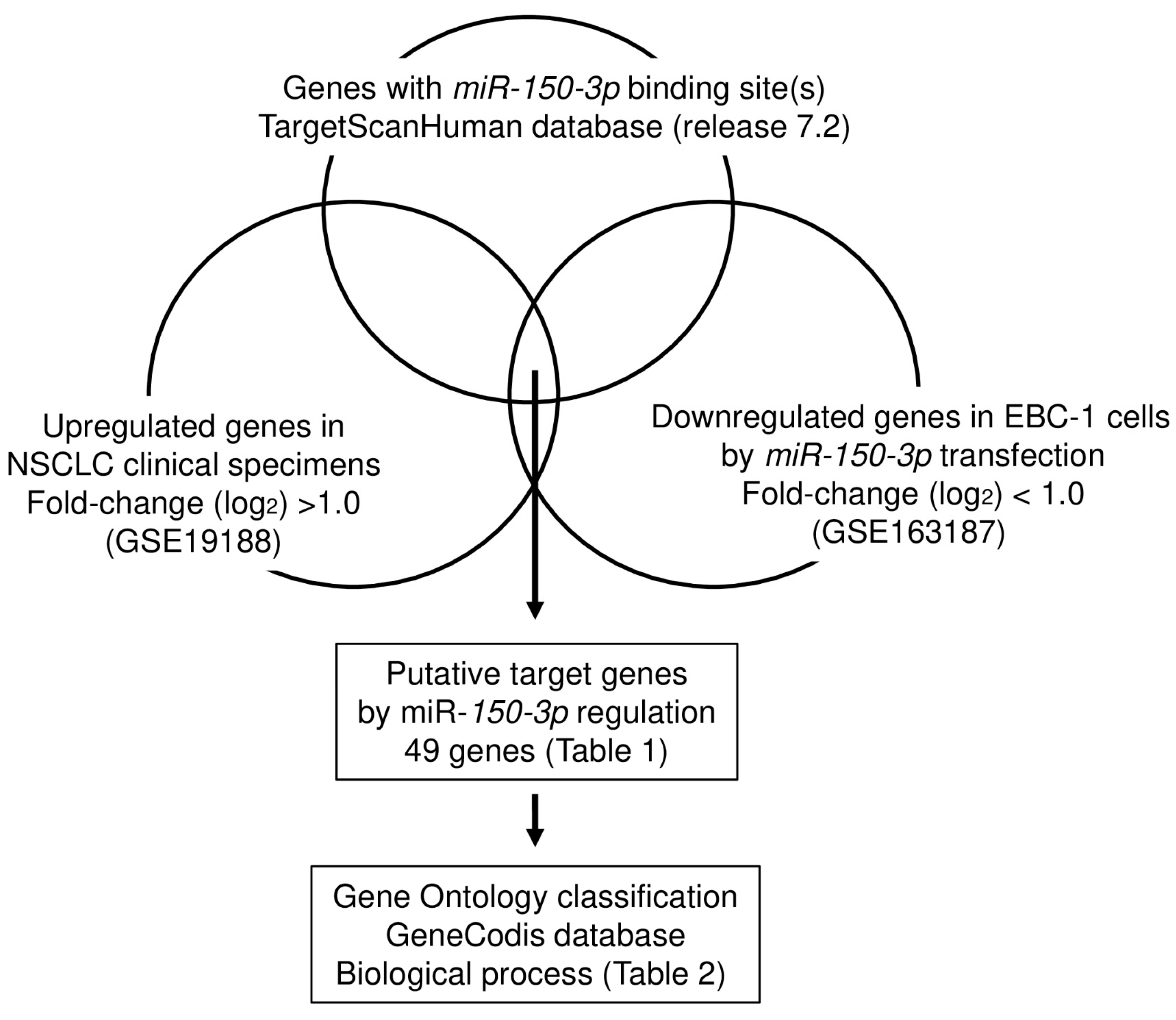
2.5. Identification of Oncogenic Targets by miR-150-3p Regulation in LUSQ Cells

The TargetScanHuman database (http://www.targetscan.org/vert_72/, accessed on 14 August 2019) was used to predict miRNA-binding sites. The expression profiles of miR-150-3p-transfected EBC-1 and SK-MES-1 cells were created, and expression data were deposited in the Gene Expression Omnibus (GEO) database (accession number: GSE163187).

2.6. In Silico Analysis of miRNA and Gene Expression Levels

The Cancer Genome Atlas (TCGA) datasets (https://www.cancer.gov/tcga) were used to assess the clinical significance of miRNAs and their target genes in lung squamous cell carcinoma. The gene expression levels and clinical data were obtained from cBioPortal (https://www.cbioportal.org/) and OncoLnc (http://www.oncolnc.org/) (data downloaded on 28 December 2019).

2.7. Plasmid Construction and Dual-Luciferase Reporter Assays

We prepared psiCHECK-2 plasmid vectors (catalog no.: C8021, Promega, Madison, WI, USA) which have a wild-type or a deletion-type of the miR-150-3p-binding site in the coding region of HELLS. miR-150-3p and the plasmid vectors were transfected into lung cancer cells for 36 hours. The Dual-Luciferase Reporter Assay System (Promega) was used to assess firefly and Renilla luciferase activities. Luciferase reporter assays were evaluated in triplicate wells. The dual-luciferase reporter assay procedure was described in our previous studies [27,31,32].

2.8. Western Blotting

The method for western blotting has been described in past reports [27,31,32]. In brief, RIPA Lysis Buffer System (catalog no.: sc-24948, Santa Cruz Biotechnology Inc., Dallas, TX, USA) was used to lyse cells. Protein concentrations were measured using the PierceTM BCA Protein Assay Kit (Thermo Fisher Scientific, Rockford, IL, USA). Protein (20 μg) was injected into each well of SuperSepTM Ace (6%, 13 well) (FUJIFILM Wako Pure Chemical Corporation, Osaka, Japan) and electrophoresis was performed. Precision Plus ProteinTM Dual Color Standards were used as molecular weight markers. Proteins were transferred onto polyvinylidene fluoride membranes (catalog no.: PPVH00010, Merck KgaA, Darmstadt, Germany). The membranes were incubated with blocking buffer (3% bovine serum albumin or 5% fat-free milk). After blocking, they were sequentially incubated, first with the primary antibody and subsequently in the blocking buffer containing the secondary antibody. Precision ProteinTM Strep Tactin-HRP Conjugate (catalog no.: #1610380, Bio-Rad Laboratories, Inc., Hercules, CA, USA) was used for chemiluminescent detection. The antibodies used in this study are shown in Table S1. Amersham ECL Prime Western Blotting Detection Reagent (Cytiva; Marlborough, MA, USA) was used to develop the signal. Visualization of the western blot was by chemiluminescence using FluorChem FC2 (Cell Biosciences, Santa Clara, CA, USA).

2.9. Immunohistochemical Staining

The procedure for immunohistochemical staining (IHC) was as previously described [27,28,31,32]. The tissue microarray slide (catalog no.: LC811a, US Biomax, Inc. Derwood, MD, USA) was de-waxed with xylene and ethanol. The slide was heated in 10× citrate buffer for heat epitope recovery, pH 6.0 (Diagnostic BioSystems, Pleasanton, CA, USA) for activation. After that, it was immersed in 0.3% H2O2 to block peroxidase activity. The antigen–antibody complex was detected by VECTASTAIN Universal Elite ABC Kit (Vector Laboratories, Burlingame, CA, USA). Primary antibody was diluted with Dako Real antibody diluent (Agilent, Santa Clara, CA, USA). Dako REALTM EnVisionTM Detection System Peroxidase/DAB+, rabbit/mouse (Agilent) was used in a chromogenic method. The primary antibody is described in Table S1. Two pathologists independently evaluated the immunostaining of lung cancer tissue. The IHC intensity was quantified on the basis of positive control.

2.10. Statistical Analysis

Statistical analyses were performed using GraphPad Prism 8 (GraphPad Software, La Jolla, CA, USA) and JMP Pro 14 (SAS Institute Inc., Cary, NC, USA). Multiple group comparison was achieved using one-way analysis of variance (ANOVA) and Tukey’s post hoc test. The significance of differences between two groups was assessed by Mann–Whitney U tests.

3. Results

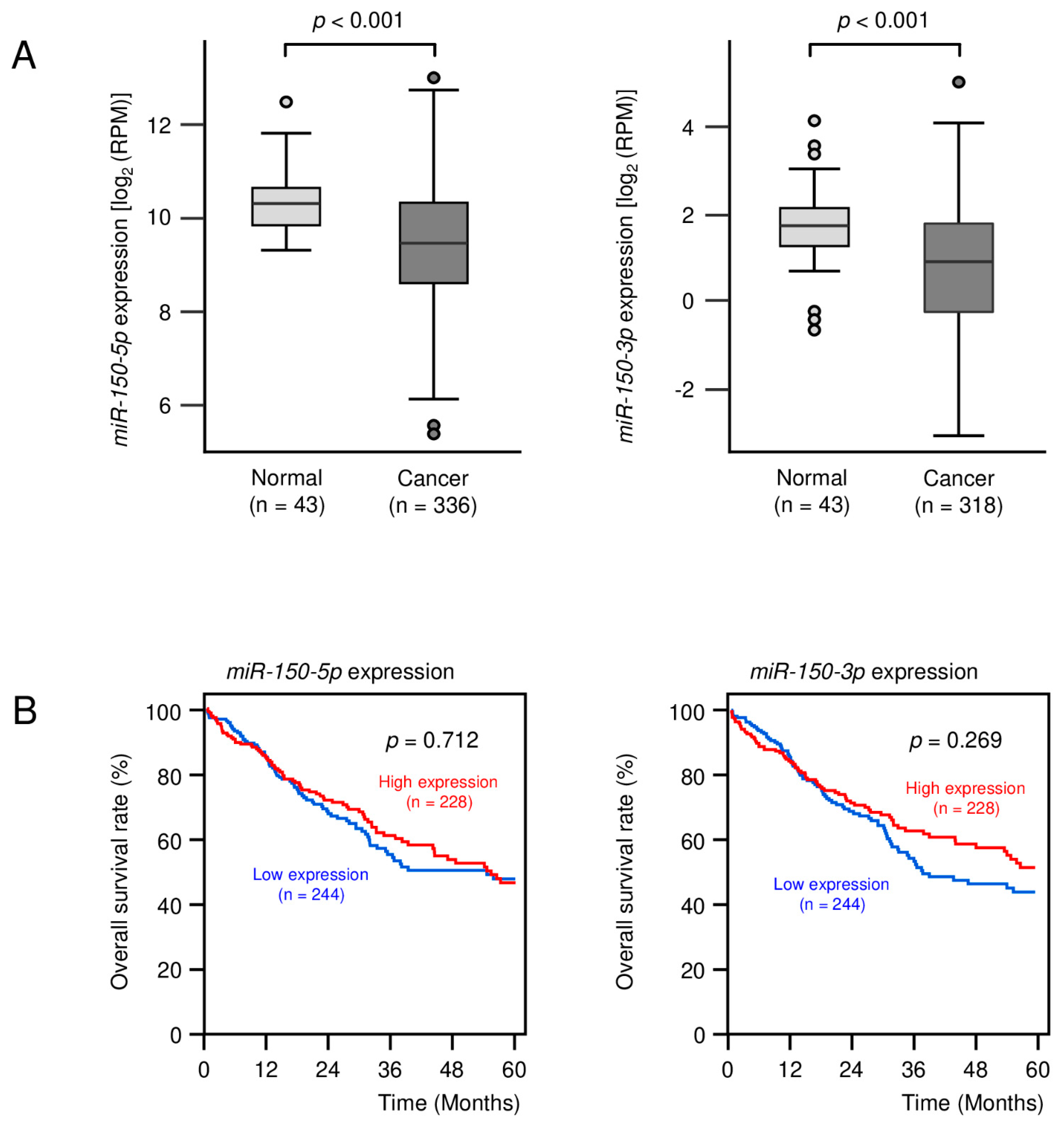
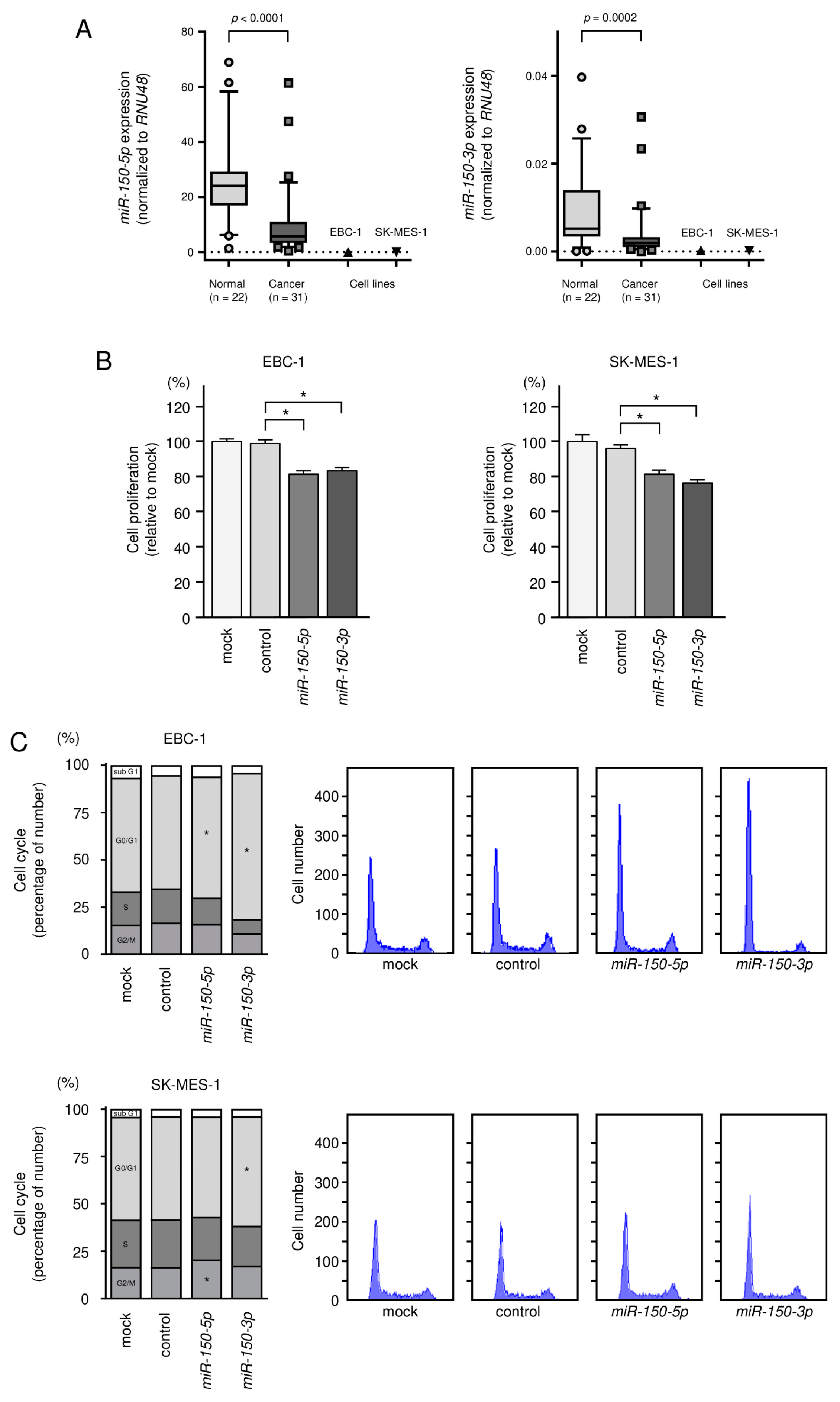
3.1. Expression Levels of miR-150-5p and miR-150-3p in Clinical LUSQ Tissues

The expression levels of miR-150-5p and miR-150-3p were evaluated by TCGA database analysis. The expression levels of both miR-150-5p and miR-150-3p were significantly downregulated in LUSQ tissues compared with normal tissues (both, p < 0.001; Figure 1A).
Cohort analyses using the TCGA-LUSQ datasets showed that the expression of miR-150-5p and miR-150-3p did not affect the 5-year survival rate of the patients (Figure 1B).

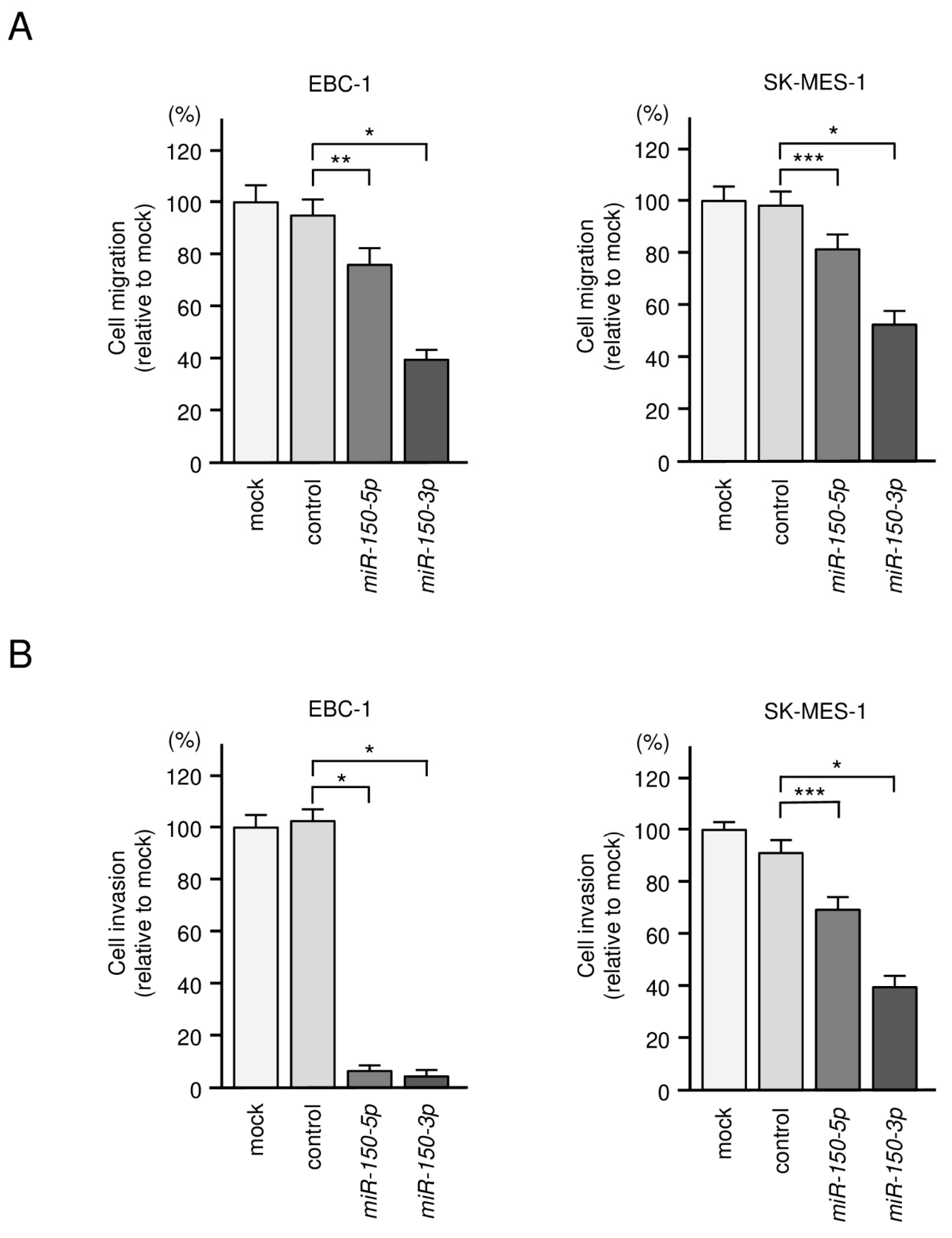
3.2. Tumor-Suppressive Roles of miR-150-5p and miR-150-3p in LUSQ Cells

For the validity of the gain-of-function assays, we examined the expression of miR-150-5p and miR-150-3p in the LUSQ cell lines EBC-1 and SK-MES-1. It was detected that the expression of miR-150-5p and miR-150-3p in the cell line was significantly lower than that of normal lung tissues (Figure 2A). Clinical information for the normal lung tissue is shown in Tables S2 and S3.
We assessed cell proliferation, cell cycle, migration and invasion based on transient transfection assays. Ectopic expression of the two miRNAs inhibited cancer cell proliferation in EBC-1 and SK-MES-1 cells (Figure 2B). Cell cycle assays showed that expression of miR-150-3p induced G0/G1 arrest in EBC-1 and SK-MES-1 cells (Figure 2C). Moreover, expression of miR-150-5p and miR-150-3p significantly blocked the migration and invasive abilities of LUSQ cells (Figure 3A,B and Figure S1).
Our data suggest that both miR-150-5p and miR-150-3p may have important role as tumor suppressors through downregulation of target oncogenic genes. We investigated the synergistic effect when both miRNAs were simultaneously transfected into cancer cells. However, we found no synergistic effect on cell viability inhibition based on miR-150-5p and miR-150-3p co-transfection (Figure S2). In the future, it will be necessary to conduct the analysis using an expression system that simultaneously expresses both miRNAs in cancer cells.
These data indicate that both strands of pre-miR-150 act as tumor-suppressive miRNAs in LUSQ cells. In contrast to miR-150-5p (the guide strand), there are not many reports of oncogenic genes in LUSQ cells that are controlled by miR-150-3p (the passenger strand). Therefore, we searched for oncogenic targets controlled by miR-150-3p in LUSQ cells.

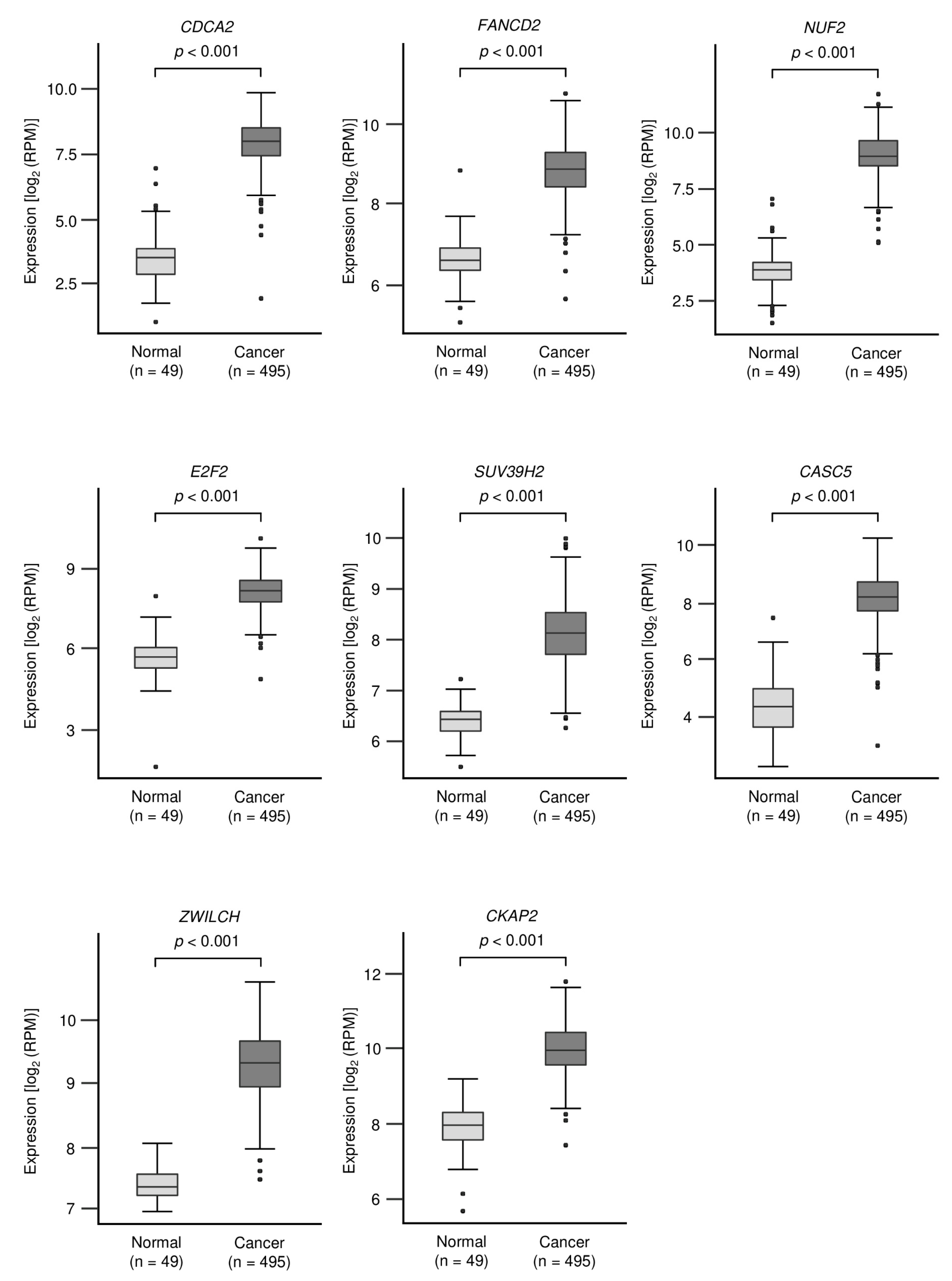
3.3. Identification of Oncogenic Targets Regulated by miR-150-3p in LUSQ Cells

In searching for oncogenic genes controlled by miR-150-3p in LUSQ cells, we made the following hypotheses: (1) target genes of miR-150-3p have one or more binding site(s), (2) target genes are upregulated in LUSQ tissues and (3) target genes will be downregulated following miR-150-3p transfection of LUSQ cells. We searched for genes that satisfied the three conditions by combining in silico analysis (TargetScanHuman database and GSE19188) and our gene expression data (GSE163187). A flowchart of the strategy used to narrow down the list of candidate genes is shown in Figure 4.
A total of 49 putative targets of miR-150-3p in LUSQ were identified (Table 1). These genes were additionally categorized according to Gene Ontology classification. Interestingly, the target molecules were significantly enriched for genes related to the “cell cycle” and “cell division” categories (Table 2).
The expression levels of genes involved in the “cell cycle” and “cell division” were evaluated by TCGA Database analysis. All genes were found to be significantly upregulated in LUSQ tissues (p < 0.001, Figure 5).

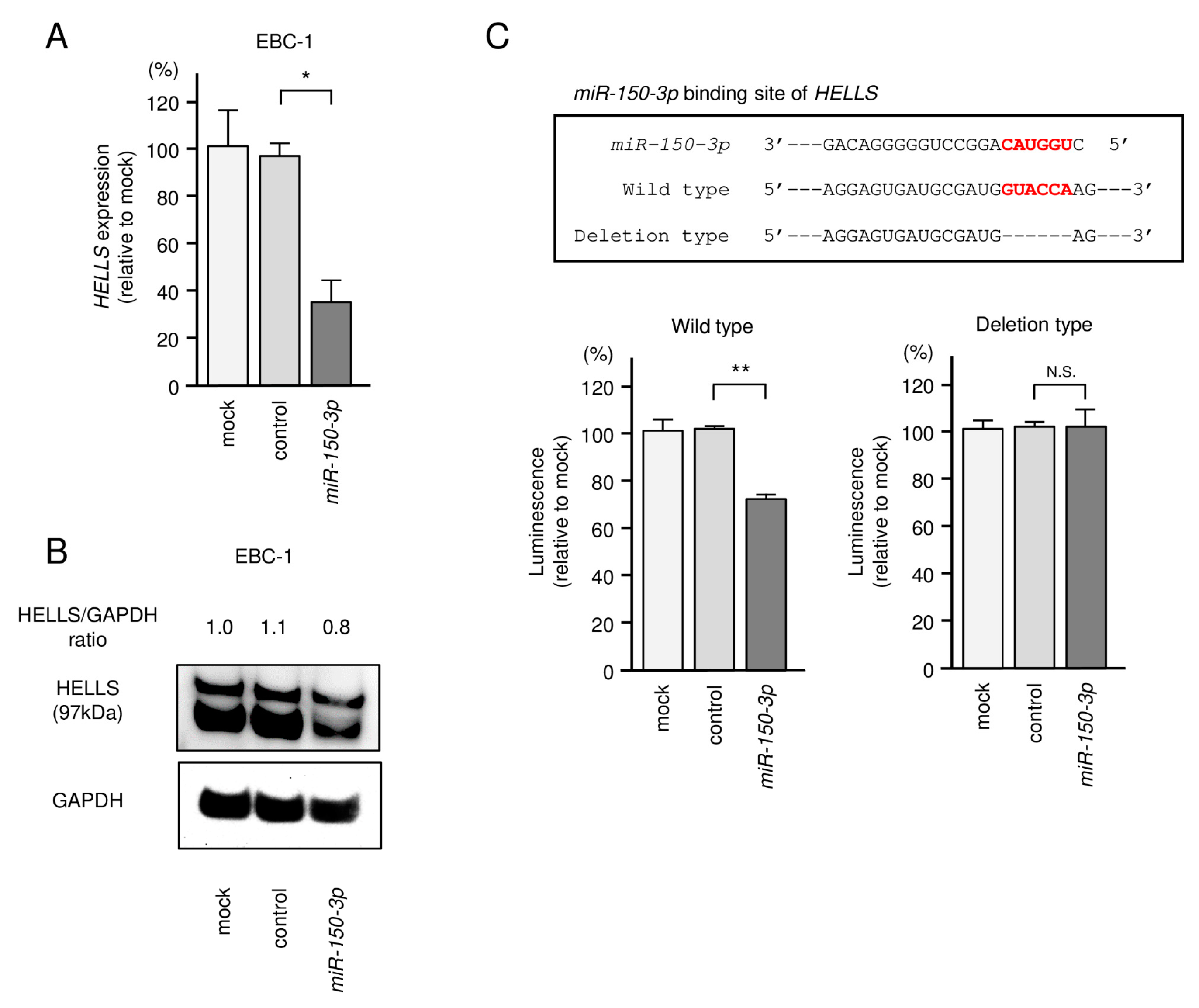
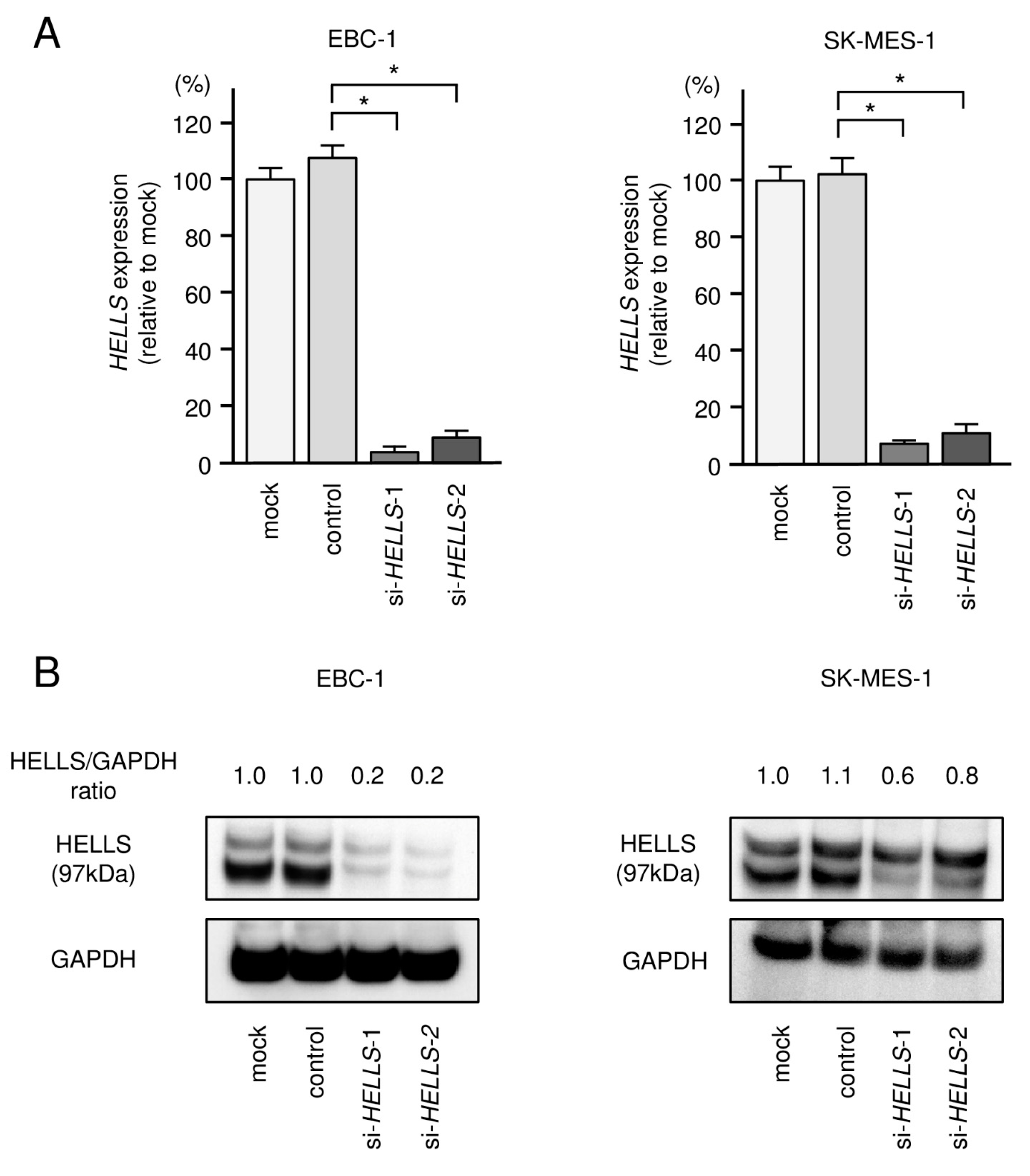
3.4. Direct Regulation of HELLS by miR-150-3p in LUSQ Cells

In this study, we focused on HELLS for the following reasons: (1) TargetScanHuman database analysis showed that a miR-150-3p binding site was present within the coding region of the HELLS gene. Generally, miRNA binding sites are located in the 3′UTR of the genes. (2) OncoLnc database analysis showed that the prognosis of patients with LUAD and patients with LUSQ differs depending on HELLS expression. (3) The functional analysis of HELLS has not been investigated in LUSQ cells.
In EBC-1 cells transfected with miR-150-3p, both HELLS mRNA and HELLS protein levels were significantly downregulated (Figure 6A,B). Full-sized images of western blot data are shown in Figure S3.
Analysis of the NCBI nucleotide and TargetScanHuman databases revealed that the miR-150-3p binding site (CAUGGU) was located within the coding region of HELLS gene (Figure S4). The presence of the miR-150-3p binding site within the coding region in the LUSQ and LUAD cell lines (EBC-1, SK-MES-1, A549 and H1299) was confirmed through sequencing (Figure S5).
Next, we performed a luciferase reporter assay to determine whether miR-150-3p directly binds to HELLS. Luciferase activity was significantly reduced in cells transfected with the wild-type vector (with miR-150-3p binding site) but not in cells transfected with the deletion construct (without miR-150-3p binding site) (Figure 6C). Our present data demonstrate that miR-150-3p directly bound to its target sequence HELLS and negatively controlled its expression in LUSQ cells. The sequence used for luciferase reporter assays is shown in Figure S5.

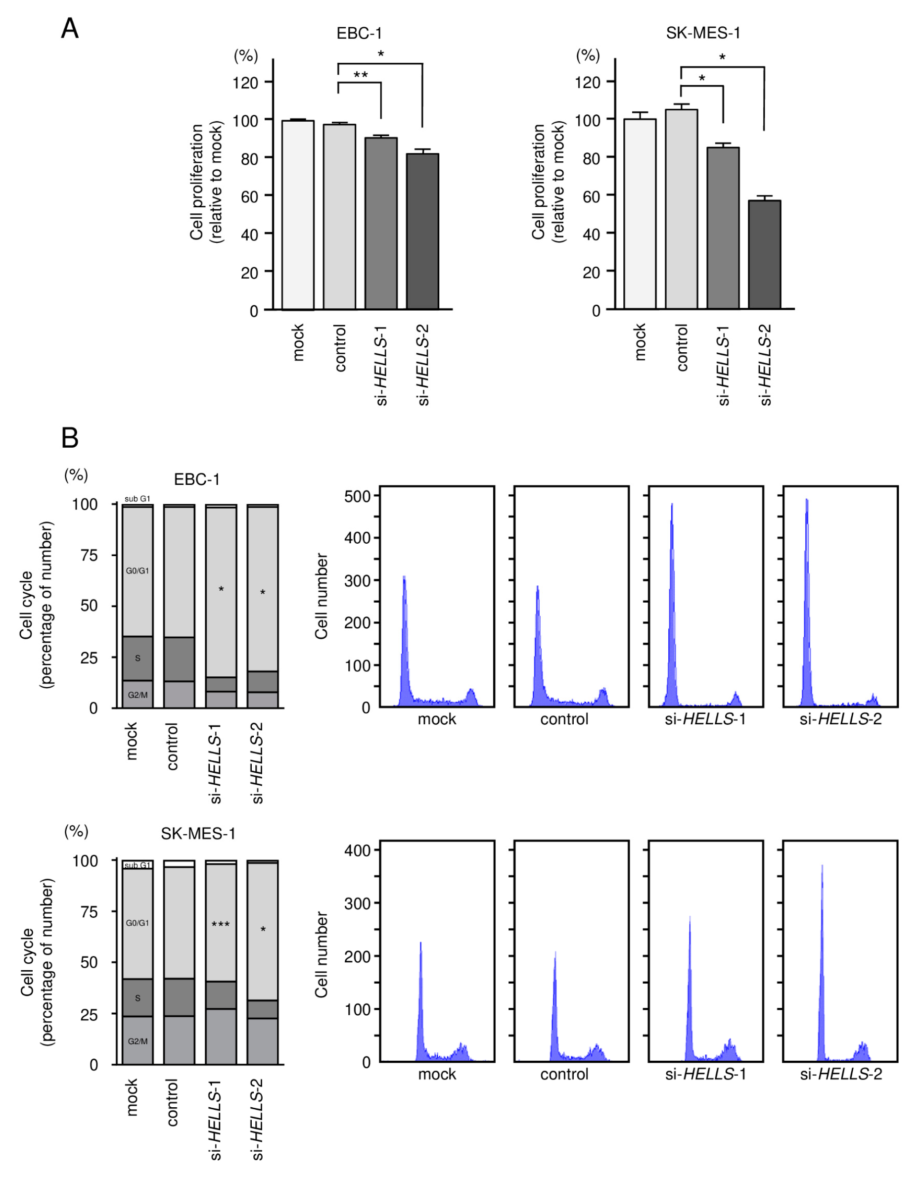
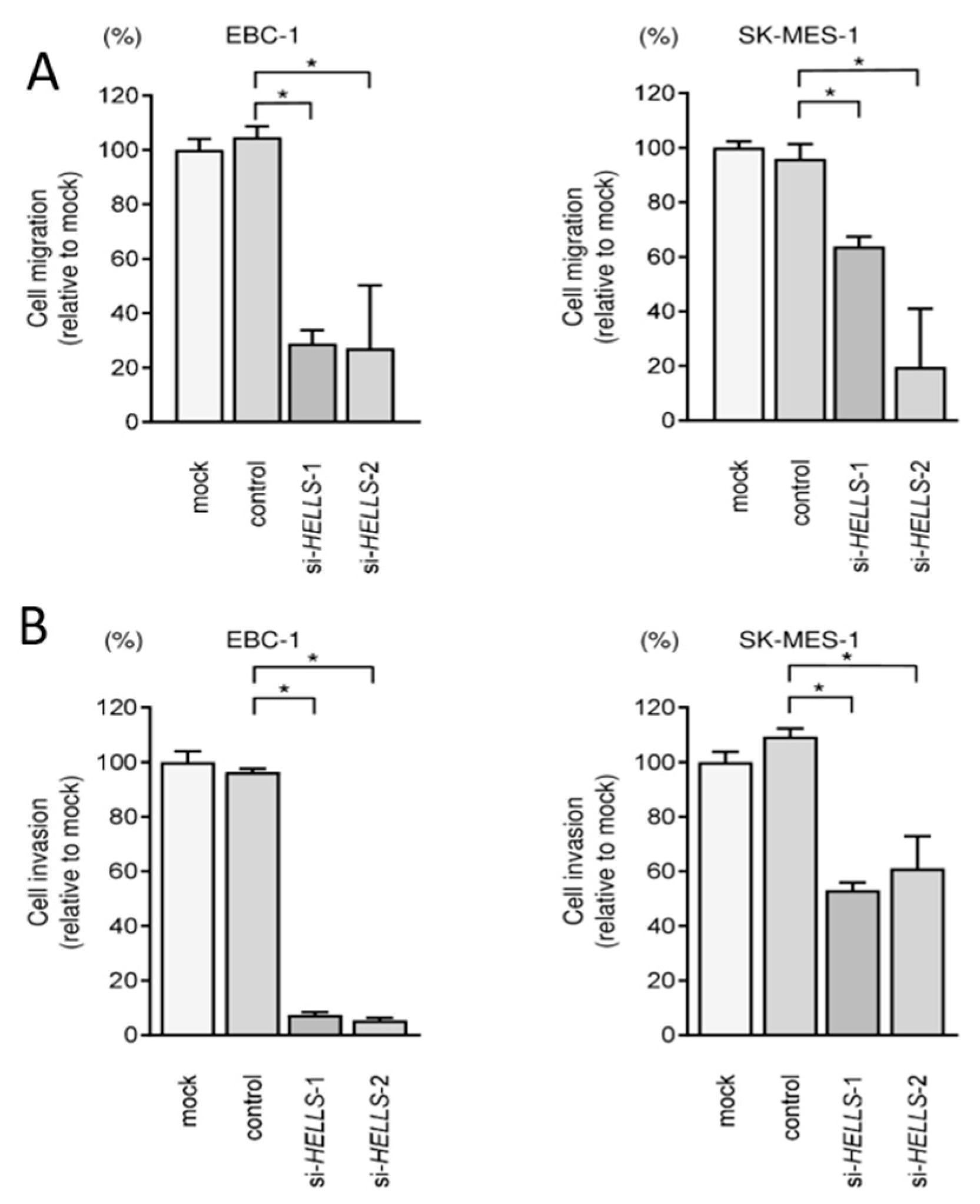
3.5. Effects of Knockdown of HELLS in LUSQ Cells

To investigate the oncogenic roles of HELLS in LUSQ cells, we used knockdown assays based on two types of small interfering RNAs (siRNAs) targeting HELLS. First, to validate the knockdown efficiencies of siRNAs in LUSQ cells, the mRNA and protein levels of HELLS were shown to be successfully downregulated by either siRNAs in EBC-1 and SK-MES-1 cells (Figure 7A,B). Full-sized images of western blot data are shown in Figure S6.
Transient transfection of the two siRNAs into EBC-1 and SK-MES-1 cells resulted in the inhibition of cancer cell proliferation (Figure 8A). Cell cycle assays showed that expression of the two miRNAs induced G0/G1 arrest in EBC-1 and SK-MES-1 cells (Figure 8B). These results indicate that the expression of HELLS enhances LUSQ cell proliferation, and therefore, acts as an oncogene. Knockdown of HELLS in LUSQ cells also blocked migration and invasion (Figure 9). The images of migration and invasion assays are shown in Figure S7.

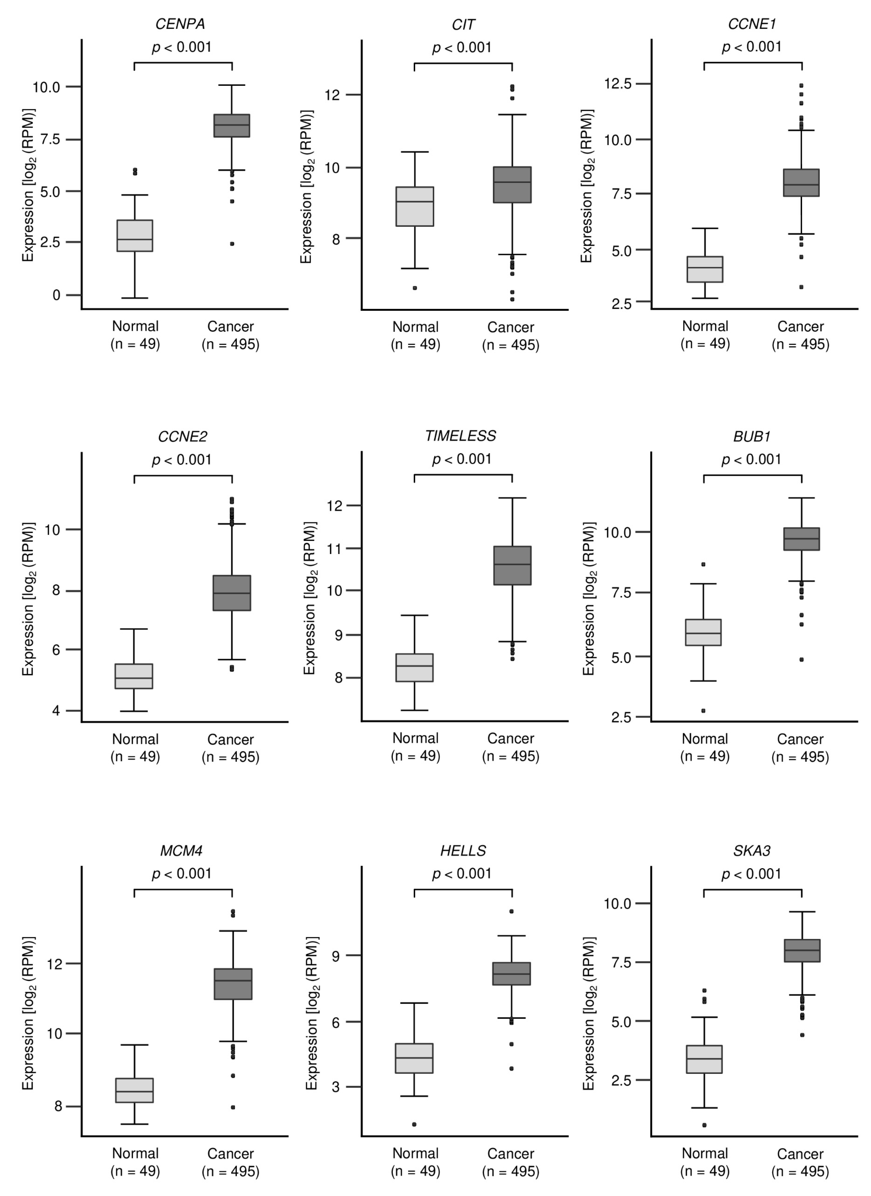
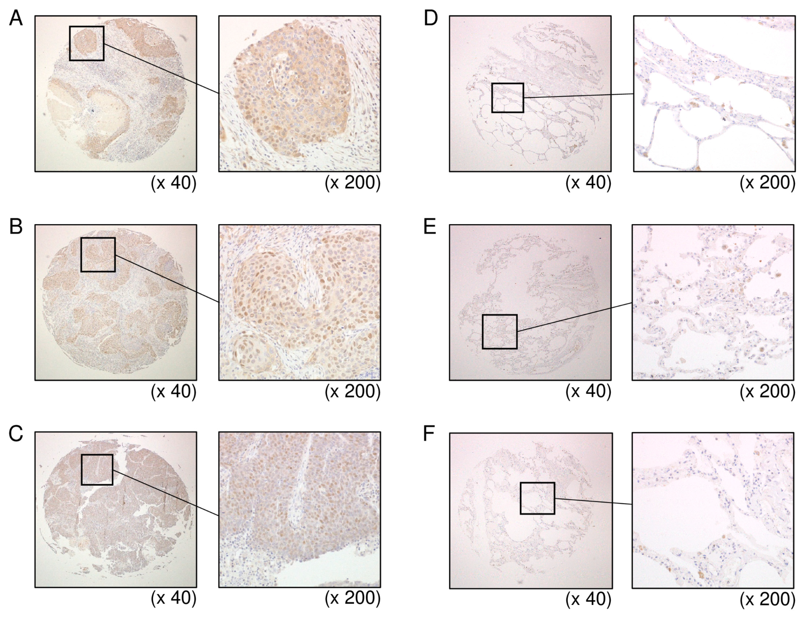
3.6. Overexpression of HELLS in LUSQ Clinical Specimens

Expression of HELLS protein was investigated using LUSQ tissues. It was found that HELLS was overexpressed in cancer lesions of LUSQ tissue (Figure 10A–C). In contrast to cancer lesions, HELLS expression was weak in normal lung tissues (Figure 10D–F). Table S4 provides information on the clinical specimens used for immunostaining.
Furthermore, we examined the relationship between the clinical outcome of lung cancer and HELLS expression using TCGA data (TCGA-LUSQ and TCGA-LUAD). In LUAD, patients with high HELLS expression had a significantly worse prognosis compared to those with low expression. Notably, in LUSQ, patients with low HELLS expression had a significantly worse prognosis compared with those with high expression (Figure S8). Forest plot presenting the results of a multivariate Cox regression analysis of the prognostic value of HELLS identified in an LUSQ and LUAD dataset from TCGA. In Figure S8, the expression level of HELLS was determined to be independent prognostic factors in terms of the 5-year overall survival rate after adjustments for age, sex, disease stage and pathological T and N stage (p < 0.05). Therefore, the expression of HELLS predicts opposite outcomes in patients with LUAD and LUSQ when used as a prognostic marker. At present, there is no clear explanation for this phenomenon.

4. Discussion

The analysis of miRNA expression signatures based on RNA sequencing revealed that some passenger strands of miRNAs (e.g., miR-99a-3p, miR-101-5p, miR-143-5p and miR-145-3p) are significantly downregulated in several types of cancer tissues and that they act as tumor suppressors in different types of cancer [26,28,29,33]. Recently, a large number of in silico analyses revealed that in some miRNAs, both strands (5p and 3p) coordinately modulate oncogenic pathways, e.g., miR-28, miR-30a, miR-139, miR-143 and miR-145 [27,28,34,35,36]. These findings show that several miRNA passenger strands are deeply associated with the molecular pathogenesis of human cancers. Simultaneous investigation of 5p and 3p strands of miRNA duplexes will elucidate novel molecular mechanism in human cancer cells.
In the last 10 years, several studies have revealed that aberrant expression of miR-150-5p (the guide strand) significantly contributes to human oncogenesis [31,37,38,39]. Initially, miR-150-5p was considered to be a hematopoietic-specific miRNA, and associations with hematological malignancies were investigated [40]. Previous studies in solid cancers showed that miR-150-5p behaves differently (as an oncogene or as a tumor suppressor) depending on the type of cancer [31,41]. In NSCLC, it was shown that miR-150-5p is overexpressed in NSCLC tissues, where it negatively regulates the tumor-suppressor TP53 [41]. Another study showed that miR-150-5p was present in NSCLC tissues and that its overexpression promotes cellular proliferation and migration [42]. In contrast to the oncogenic function of miR-150-5p in NSCLC cells, miR-150-5p was significantly downregulated in cancer stem cells in NSCLC [39], and the expression of miR-150-5p was demonstrated to inhibit the HMGA2 and β-catenin signaling pathways [39]. Our previous study supports the function of miR-150-5p in acting as a tumor-suppressive miRNA in LUSQ cells [31].
Few studies have simultaneously analyzed both the 5p and 3p strands of miR-150 because passenger strands have not received much attention to date. Previous reports have demonstrated that the 5p and 3p strands of the miR-150 duplex have tumor suppressive functions in prostate cancer, ESCC and HNSCC [37,38]. In this study, we demonstrated that ectopic expression of miR-150-3p (the passenger strand) attenuated the malignant phenotypes of LUSQ cells, suggesting that it acts as a tumor-suppressive miRNA. The results from both our present investigation plus our previous study show that both strands of the miR-150 duplex act as tumor-suppressive miRNAs in LUSQ cells.
We explored the molecular networks that were controlled by miR-150-3p in LUSQ cells. A total of 49 genes were identified as putative targets of miR-150-3p regulation in LUSQ cells. Among them, 17 genes were classified as related to the “cell cycle”. For example, aberrant expression of MCM4 was detected in LUAD tissues and MCM4 knockdown suppressed cell growth, migration and invasion of LUAD cells [27]. CENPA downregulation was found to induce G0/G1 cell cycle arrest and apoptosis. Downregulation of CENPA inhibited migration and invasion in vitro and suppressed the growth of xenografted A549 cells [43]. SKA3 induces the expression of matrix metalloproteinase (MMP)-2, -7 and -9. It also binds and activates epidermal growth factor receptor (EGFR). These processes activate the PI3K–AKT pathway and promote lung adenocarcinoma metastasis [44]. E2F transcription factors regulate cell cycle, apoptosis and DNA damage responses. E2F2 knockdown suppresses cell viability and colony formation in non-small cell lung cancer [45].
In this study, we focused on HELLS (helicase, lymphoid-specific), because its expression level was reduced by ectopic expression of miR-150-3p in LUSQ cells. Notably, miR-150-3p binding site was detected in coding region of HELLS gene. It is established concept of miRNA biogenesis that miRNAs are binding 3′-untranslated regions of their target genes, and induce mRNAs degradation [46,47]. In contrast to this theory, the molecular mechanisms by which miRNAs bind to coding regions, and regulate gene expression, is under discussion [46,47]. Several studies have indicated that miRNAs can bind to coding region of mRNAs and control of their expressions [46,47]. It has suggested that the molecular mechanisms of miRNAs expression regulation were differs depending on the site to which miRNAs bound. Previous reports suggested that when the miRNAs binding site are coding region, the major regulatory effect of miRNAs is translational inhibition [46,47]. In this study, we provided the possibility that miR-150-3p can bind coding region of HELLS, and reduced expression of HELLS in LUSQ cells. Additional analysis is needed on the regulatory mechanisms of gene expression for miR-150-3p that bind to the coding region of the genes. Other miRNAs and lncRNAs analyses involved in epigenetic regulation of HELLS are also required.
In this study, we focused on HELLS (helicase, lymphoid specific), which belongs to the SNF2 family of chromatin-remodeling ATPases [48]. HELLS is recruited to specific DNA sites to control the transcription of targeted genes [49]. Importantly, HELLS expression is directly controlled by the E2F1 transcription factor, and HELLS acts as an E2F3 transcriptional co-activator, resulting in the expression of cell cycle-related genes [50]. Overexpression of HELLS was reported in several types of cancers, e.g., nasopharyngeal carcinoma, hepatocellular carcinoma, colorectal cancer and pancreatic cancer [49,51,52,53]. In pancreatic cancer patients, the expression of HELLS is correlated with clinical stage and the prediction of short survival. Moreover, HELLS knockdown leads to cell cycle arrest and enhanced cisplatin sensitivity of pancreatic cancer cells [51]. Our present study of LUSQ cells confirmed that depletion of HELLS attenuates cancer cell aggressive phenotypes. Consequently, aberrant HELLS expression is closely linked to the malignant progression of several cancers, suggesting that HELLS is a candidate therapeutic target for these cancers.
In this study, a total of 49 putative target was identified as miR-150-3p regulation in LUSQ cells. Large-scale cohort studies of these genes may reveal new diagnostic markers for LUSQ. Furthermore, it is also an important issue to investigate the relationship between genomic mutations or treatment response in patients with LUSQ and the expression of these genes. Further analysis of miRNA-controlled molecular networks will lead to the search for diagnostic markers and therapeutic targets for this disease.

5. Conclusions

Analysis of the TCGA database showed that miR-150-3p expression was significantly downregulated in LUSQ clinical specimens. Ectopic expression assays demonstrated that miR-150-3p inhibited the aggressiveness of LUSQ cells, suggesting that miR-150-3p acts as a tumor suppressor. Interestingly, miR-150-3p was found to controlled cell cycle-related genes. The expression of HELLS, among the various cell cycle regulators, was demonstrated to be directly controlled by miR-150-3p, and its aberrant expression enhanced the malignant phenotypes of LUSQ cells. Results here and from our previous studies demonstrate that both strands of pre-miR-150 (the miR-150-5p guide strand and miR-150-3p passenger strand) have extremely important tumor suppressor functions in LUSQ cells. Involvement of the passenger strand of miRNA in LUSQ molecular pathogenesis is a new concept in cancer research.

Supplementary Materials

The following are available online at https://www.mdpi.com/article/10.3390/biomedicines9121883/s1, Figure S1: Cell migration and invasion assays in LUSQ cells with ectopic expression of miR-150-5p or miR-150-3p. Figure S2: Cell proliferation was assessed with XTT assays after transfection of miR-150-5p, -3p or 5p + 3p. Figure S3: Downregulation of HELLS protein expression by miR-150-3p. Figure S4: The nucleotide sequence of HELLS gene (NM_001289067.2). Figure S5: The sequence of the region containing the miR-150-3p binding site of each cell line. Figure S6: HELLS protein expression downregulated by si-HELLS. Figure S7: Cell migration and invasion assays in EBC-1 and SK-MES-1 after knockdown of HELLS by si-HELLS. Figure S8: Kaplan–Meier curves of 5-year overall survival due to difference in HELLS expression in LUSQ and LUAD patients from TCGA database. Table S1: Reagents used in this study. Table S2: Characteristics of the patients used for normal lung tissue near the tumor in Figure 2A. Table S3: Characteristics of the patients used for lung cancer tissue in Figure 2A. Table S4: Characteristics of the patients used for immunostaining.

Author Contributions

Conceptualization, H.I., N.S. and K.M. (Keiko Mizuno); methodology, K.M. (Keiko Mizuno), K.M. (Kentaro Machida) and N.S.; validation, H.S. and T.S.; investigation, K.T., H.S., S.M. (Shunsuke Misono), K.M. (Keiko Mizuno), K.M. (Kentaro Machida), S.A. and S.M. (Shogo Moriya); resources, T.S. and H.I.; writing (original draft preparation), M.K., K.T., A.U. and N.S.; writing (review and editing), K.M. (Kentaro Machida) and H.I.; visualization, M.K., K.T., S.M. (Shunsuke Misono) and T.S.; supervision, N.S.; funding acquisition, S.M. (Shunsuke Misono), H.S., A.U. and K.M. (Keiko Mizuno) All authors have read and agreed to the published version of the manuscript.

Funding

The present study was supported by KAKENHI grants 19K08605, 19K08627, 19K08656 and 19K17640.

Institutional Review Board Statement

The study was conducted according to the guidelines of the Declaration of Helsinki, and approved by Ethics Committee of Kagoshima University (protocol code: 190118 eki, date of approval: 20 September 2019).

Informed Consent Statement

Not applicable.

Data Availability Statement

Data is contained within the article or supplementary material.

Conflicts of Interest

The authors declare no conflict of interest.

References

  1. Fitzmaurice, C.; Allen, C.; Barber, R.M.; Barregard, L.; Bhutta, Z.A.; Brenner, H.; Dicker, D.J.; Chimed-Orchir, O.; Dandona, R.; Dandona, L.; et al. Global, regional, and national cancer incidence, mortality, years of life lost, years lived with disability, and disability-adjusted life-years for 32 cancer groups, 1990 to 2015: A systematic analysis for the global burden of disease study. JAMA Oncol. 2017, 3, 524–548. [Google Scholar] [CrossRef]
  2. Bray, F.; Ferlay, J.; Soerjomataram, I.; Siegel, R.L.; Torre, L.A.; Jemal, A. Global cancer statistics 2018: GLOBOCAN estimates of incidence and mortality worldwide for 36 cancers in 185 countries. CA Cancer J. Clin. 2018, 68, 394–424. [Google Scholar] [CrossRef] [PubMed] [Green Version]
  3. Travis, W.D.; Brambilla, E.; Nicholson, A.G.; Yatabe, Y.; Austin, J.H.M.; Beasley, M.B.; Chirieac, L.R.; Dacic, S.; Duhig, E.; Flieder, D.B.; et al. The 2015 World Health Organization classification of lung tumors: Impact of genetic, clinical and radiologic advances since the 2004 classification. J. Thorac. Oncol. 2015, 10, 1243–1260. [Google Scholar] [CrossRef] [PubMed] [Green Version]
  4. Hida, T.; Nokihara, H.; Kondo, M.; Kim, Y.H.; Azuma, K.; Seto, T.; Takiguchi, Y.; Nishio, M.; Yoshioka, H.; Imamura, F.; et al. Alectinib versus crizotinib in patients with ALK-positive non-small-cell lung cancer (J-ALEX): An open-label, randomised phase 3 trial. Lancet 2017, 390, 29–39. [Google Scholar] [CrossRef]
  5. Ramalingam, S.S.; Vansteenkiste, J.; Planchard, D.; Cho, B.C.; Gray, J.E.; Ohe, Y.; Zhou, C.; Reungwetwattana, T.; Cheng, Y.; Chewaskulyong, B.; et al. Overall survival with osimertinib in untreated, EGFR-mutated advanced NSCLC. N. Engl. J. Med. 2020, 382, 41–50. [Google Scholar] [CrossRef]
  6. Paik, P.K.; Felip, E.; Veillon, R.; Sakai, H.; Cortot, A.B.; Garassino, M.C.; Mazieres, J.; Viteri, S.; Senellart, H.; Van Meerbeeck, J.; et al. Tepotinib in non-small-cell lung cancer with MET exon 14 skipping mutations. N. Engl. J. Med. 2020, 383, 931–943. [Google Scholar] [CrossRef]
  7. Drilon, A.; Oxnard, G.R.; Tan, D.S.W.; Loong, H.H.F.; Johnson, M.; Gainor, J.; McCoach, C.E.; Gautschi, O.; Besse, B.; Cho, B.C.; et al. Efficacy of selpercatinib in RET fusion-positive non-small-cell lung cancer. N. Engl. J. Med. 2020, 383, 813–824. [Google Scholar] [CrossRef]
  8. Planchard, D.; Smit, E.F.; Groen, H.J.M.; Mazieres, J.; Besse, B.; Helland, Å.; Giannone, V.; D’Amelio, A.M., Jr.; Zhang, P.; Mookerjee, B.; et al. Dabrafenib plus trametinib in patients with previously untreated BRAF(V600E)-mutant metastatic non-small-cell lung cancer: An open-label, phase 2 trial. Lancet Oncol. 2017, 18, 1307–1316. [Google Scholar] [CrossRef]
  9. Drilon, A.; Siena, S.; Dziadziuszko, R.; Barlesi, F.; Krebs, M.G.; Shaw, A.T.; de Braud, F.; Rolfo, C.; Ahn, M.J.; Wolf, J.; et al. Entrectinib in ROS1 fusion-positive non-small-cell lung cancer: Integrated analysis of three phase 1-2 trials. Lancet Oncol. 2020, 21, 261–270. [Google Scholar] [CrossRef]
  10. Bartel, D.P. MicroRNAs: Target recognition and regulatory functions. Cell 2009, 136, 215–233. [Google Scholar] [CrossRef] [PubMed] [Green Version]
  11. Bartel, D.P. MicroRNAs: Genomics, biogenesis, mechanism, and function. Cell 2004, 116, 281–297. [Google Scholar] [CrossRef] [Green Version]
  12. Catalanotto, C.; Cogoni, C.; Zardo, G. MicroRNA in control of gene expression: An overview of nuclear functions. Int. J. Mol. Sci. 2016, 17, 1712. [Google Scholar] [CrossRef] [PubMed] [Green Version]
  13. Lin, S.; Gregory, R.I. MicroRNA biogenesis pathways in cancer. Nat. Rev. Cancer. 2015, 15, 321–333. [Google Scholar] [CrossRef] [PubMed]
  14. Baer, C.; Claus, R.; Plass, C. Genome-wide epigenetic regulation of miRNAs in cancer. Cancer Res. 2013, 73, 473–477. [Google Scholar] [CrossRef] [PubMed] [Green Version]
  15. Di Leva, G.; Garofalo, M.; Croce, C.M. MicroRNAs in cancer. Ann. Rev. Pathol. 2014, 9, 287–314. [Google Scholar] [CrossRef] [Green Version]
  16. Gulyaeva, L.F.; Kushlinskiy, N.E. Regulatory mechanisms of microRNA expression. J. Transl. Med. 2016, 14, 143. [Google Scholar] [CrossRef] [Green Version]
  17. Ramassone, A.; Pagotto, S.; Veronese, A.; Visone, R. Epigenetics and MicroRNAs in Cancer. Int. J. Mol. Sci. 2018, 19, 459. [Google Scholar] [CrossRef] [Green Version]
  18. Ma, L. MicroRNA and metastasis. Adv. Cancer Res. 2016, 132, 165–207. [Google Scholar] [CrossRef]
  19. Bayraktar, R.; Van Roosbroeck, K. miR-155 in cancer drug resistance and as target for miRNA-based therapeutics. Cancer Metastasis Rev. 2018, 37, 33–44. [Google Scholar] [CrossRef]
  20. Desantis, V.; Saltarella, I.; Lamanuzzi, A.; Melaccio, A.; Solimando, A.G.; Mariggiò, M.A.; Racanelli, V.; Paradiso, A.; Vacca, A.; Frassanito, M.A. MicroRNAs-based nano-strategies as new therapeutic approach in multiple myeloma to overcome disease progression and drug resistance. Int. J. Mol. Sci. 2020, 21, 3084. [Google Scholar] [CrossRef]
  21. Zhang, S.; Cheng, Z.; Wang, Y.; Han, T. The risks of miRNA therapeutics: In a drug target perspective. Drug Des. Devel. Ther. 2021, 15, 721–733. [Google Scholar] [CrossRef]
  22. Matranga, C.; Tomari, Y.; Shin, C.; Bartel, D.P.; Zamore, P.D. Passenger-strand cleavage facilitates assembly of siRNA into Ago2-containing RNAi enzyme complexes. Cell 2005, 123, 607–620. [Google Scholar] [CrossRef] [PubMed] [Green Version]
  23. Koshizuka, K.; Nohata, N.; Hanazawa, T.; Kikkawa, N.; Arai, T.; Okato, A.; Fukumoto, I.; Katada, K.; Okamoto, Y.; Seki, N. Deep sequencing-based microRNA expression signatures in head and neck squamous cell carcinoma: Dual strands of pre-miR-150 as antitumor miRNAs. Oncotarget 2017, 8, 30288–30304. [Google Scholar] [CrossRef] [PubMed] [Green Version]
  24. Yonemori, K.; Seki, N.; Idichi, T.; Kurahara, H.; Osako, Y.; Koshizuka, K.; Arai, T.; Okato, A.; Kita, Y.; Arigami, T.; et al. The microRNA expression signature of pancreatic ductal adenocarcinoma by RNA sequencing: Anti-tumour functions of the microRNA-216 cluster. Oncotarget 2017, 8, 70097–70115. [Google Scholar] [CrossRef] [Green Version]
  25. Toda, H.; Kurozumi, S.; Kijima, Y.; Idichi, T.; Shinden, Y.; Yamada, Y.; Arai, T.; Maemura, K.; Fujii, T.; Horiguchi, J.; et al. Molecular pathogenesis of triple-negative breast cancer based on microRNA expression signatures: Antitumor miR-204-5p targets AP1S3. J. Hum. Genet. 2018, 63, 1197–1210. [Google Scholar] [CrossRef]
  26. Wada, M.; Goto, Y.; Tanaka, T.; Okada, R.; Moriya, S.; Idichi, T.; Noda, M.; Sasaki, K.; Kita, Y.; Kurahara, H.; et al. RNA sequencing-based microRNA expression signature in esophageal squamous cell carcinoma: Oncogenic targets by antitumor miR-143-5p and miR-143-3p regulation. J. Hum. Genet. 2020, 65, 1019–1034. [Google Scholar] [CrossRef] [PubMed]
  27. Sanada, H.; Seki, N.; Mizuno, K.; Misono, S.; Uchida, A.; Yamada, Y.; Moriya, S.; Kikkawa, N.; Machida, K.; Kumamoto, T.; et al. Involvement of dual strands of miR-143 (miR-143-5p and miR-143-3p) and their target oncogenes in the molecular pathogenesis of lung adenocarcinoma. Int. J. Mol. Sci. 2019, 20, 4482. [Google Scholar] [CrossRef] [Green Version]
  28. Misono, S.; Seki, N.; Mizuno, K.; Yamada, Y.; Uchida, A.; Arai, T.; Kumamoto, T.; Sanada, H.; Suetsugu, T.; Inoue, H. Dual strands of the miR-145 duplex (miR-145-5p and miR-145-3p) regulate oncogenes in lung adenocarcinoma pathogenesis. J. Hum. Genet. 2018, 63, 1015–1028. [Google Scholar] [CrossRef]
  29. Toda, H.; Seki, N.; Kurozumi, S.; Shinden, Y.; Yamada, Y.; Nohata, N.; Moriya, S.; Idichi, T.; Maemura, K.; Fujii, T.; et al. RNA-sequence-based microRNA expression signature in breast cancer: Tumor-suppressive miR-101-5p regulates molecular pathogenesis. Mol. Oncol. 2020, 14, 426–446. [Google Scholar] [CrossRef] [PubMed] [Green Version]
  30. Koshizuka, K.; Hanazawa, T.; Kikkawa, N.; Arai, T.; Okato, A.; Kurozumi, A.; Kato, M.; Katada, K.; Okamoto, Y.; Seki, N. Regulation of ITGA3 by the anti-tumor miR-199 family inhibits cancer cell migration and invasion in head and neck cancer. Cancer Sci. 2017, 108, 1681–1692. [Google Scholar] [CrossRef] [Green Version]
  31. Suetsugu, T.; Koshizuka, K.; Seki, N.; Mizuno, K.; Okato, A.; Arai, T.; Misono, S.; Uchida, A.; Kumamoto, T.; Inoue, H. Downregulation of matrix metalloproteinase 14 by the antitumor miRNA, miR-150-5p, inhibits the aggressiveness of lung squamous cell carcinoma cells. Int. J. Oncol. 2018, 52, 913–924. [Google Scholar] [CrossRef] [PubMed]
  32. Misono, S.; Seki, N.; Mizuno, K.; Yamada, Y.; Uchida, A.; Sanada, H.; Moriya, S.; Kikkawa, N.; Kumamoto, T.; Suetsugu, T.; et al. Molecular pathogenesis of gene regulation by the miR-150 duplex: miR-150-3p regulates TNS4 in lung adenocarcinoma. Cancers 2019, 11, 601. [Google Scholar] [CrossRef] [PubMed] [Green Version]
  33. Arai, T.; Okato, A.; Yamada, Y.; Sugawara, S.; Kurozumi, A.; Kojima, S.; Yamazaki, K.; Naya, Y.; Ichikawa, T.; Seki, N. Regulation of NCAPG by miR-99a-3p (passenger strand) inhibits cancer cell aggressiveness and is involved in CRPC. Cancer Med. 2018, 7, 1988–2002. [Google Scholar] [CrossRef]
  34. Lv, Y.; Yang, H.; Ma, X.; Wu, G. Strand-specific miR-28-3p and miR-28-5p have differential effects on nasopharyngeal cancer cells proliferation, apoptosis, migration and invasion. Cancer Cell Int. 2019, 19, 187. [Google Scholar] [CrossRef]
  35. Liu, X.; Ji, Q.; Zhang, C.; Liu, X.; Liu, Y.; Liu, N.; Sui, H.; Zhou, L.; Wang, S.; Li, Q. miR-30a acts as a tumor suppressor by double-targeting COX-2 and BCL9 in H. pylori gastric cancer models. Sci. Rep. 2017, 7, 7113. [Google Scholar] [CrossRef] [Green Version]
  36. Okada, R.; Goto, Y.; Yamada, Y.; Kato, M.; Asai, S.; Moriya, S.; Ichikawa, T.; Seki, N. Regulation of oncogenic targets by the tumor-suppressive miR-139 duplex (miR-139-5p and miR-139-3p) in renal cell carcinoma. Biomedicines 2020, 8, 599. [Google Scholar] [CrossRef] [PubMed]
  37. Koshizuka, K.; Hanazawa, T.; Kikkawa, N.; Katada, K.; Okato, A.; Arai, T.; Idichi, T.; Osako, Y.; Okamoto, Y.; Seki, N. Antitumor miR-150-5p and miR-150-3p inhibit cancer cell aggressiveness by targeting SPOCK1 in head and neck squamous cell carcinoma. Auris Nasus Larynx 2018, 45, 854–865. [Google Scholar] [CrossRef]
  38. Osako, Y.; Seki, N.; Koshizuka, K.; Okato, A.; Idichi, T.; Arai, T.; Omoto, I.; Sasaki, K.; Uchikado, Y.; Kita, Y.; et al. Regulation of SPOCK1 by dual strands of pre-miR-150 inhibit cancer cell migration and invasion in esophageal squamous cell carcinoma. J. Hum. Genet. 2017, 62, 935–944. [Google Scholar] [CrossRef]
  39. Dai, F.Q.; Li, C.R.; Fan, X.Q.; Tan, L.; Wang, R.T.; Jin, H. miR-150-5p inhibits non-small-cell lung cancer metastasis and recurrence by targeting HMGA2 and β-catenin signaling. Mol. Ther. Nucleic Acids. 2019, 16, 675–685. [Google Scholar] [CrossRef] [PubMed] [Green Version]
  40. Bellon, M.; Lepelletier, Y.; Hermine, O.; Nicot, C. Deregulation of microRNA involved in hematopoiesis and the immune response in HTLV-I adult T-cell leukemia. Blood 2009, 113, 4914–4917. [Google Scholar] [CrossRef] [Green Version]
  41. Wang, P.Y.; Li, Y.J.; Zhang, S.; Li, Z.L.; Yue, Z.; Xie, N.; Xie, S.Y. Regulating A549 cells growth by ASO inhibiting miRNA expression. Mol. Cell. Biochem. 2010, 339, 163–171. [Google Scholar] [CrossRef] [PubMed]
  42. Wu, Z.; Li, W.; Li, J.; Zhang, Y.; Zhang, X.; Xu, Y.; Hu, Y.; Li, Q.; Sun, Q.; Ma, Z. Higher expression of miR-150-5p promotes tumorigenesis by suppressing LKB1 in non-small cell lung cancer. Pathol Res. Pract. 2020, 216, 153145. [Google Scholar] [CrossRef]
  43. Wu, Q.; Chen, Y.F.; Fu, J.; You, Q.H.; Wang, S.M.; Huang, X.; Feng, X.J.; Zhang, S.H. Short hairpin RNA-mediated down-regulation of CENP-A attenuates the aggressive phenotype of lung adenocarcinoma cells. Cell. Oncol. 2014, 37, 399–407. [Google Scholar] [CrossRef]
  44. Hu, D.D.; Chen, H.L.; Lou, L.M.; Zhang, H.; Yang, G.L. SKA3 promotes lung adenocarcinoma metastasis through the EGFR-PI3K-Akt axis. Biosci Rep. 2020, 40. [Google Scholar] [CrossRef] [Green Version]
  45. Chen, L.; Yu, J.H.; Lu, Z.H.; Zhang, W. E2F2 induction in related to cell proliferation and poor prognosis in non-small cell lung carcinoma. Int. J. Clin. Exp. Pathol. 2015, 8, 10545–10554. [Google Scholar]
  46. Zhang, K.; Zhang, X.; Cai, Z.; Zhou, J.; Cao, R.; Zhao, Y.; Chen, Z.; Wang, D.; Ruan, W.; Zhao, Q.; et al. A novel class of microRNA-recognition elements that function only within open reading frames. Nat. Struct. Mol. Biol. 2018, 25, 1019–1027. [Google Scholar] [CrossRef] [PubMed]
  47. Brümmer, A.; Hausser, J. MicroRNA binding sites in the coding region of mRNAs: Extending the repertoire of post-transcriptional gene regulation. Bioessays 2014, 36, 617–626. [Google Scholar] [CrossRef]
  48. Lungu, C.; Muegge, K.; Jeltsch, A.; Jurkowska, R.Z. An ATPase-deficient variant of the SNF2 family member HELLS shows altered dynamics at pericentromeric heterochromatin. J. Mol. Biol. 2015, 427, 1903–1915. [Google Scholar] [CrossRef] [PubMed]
  49. Law, C.T.; Wei, L.; Tsang, F.H.; Chan, C.Y.; Xu, I.M.; Lai, R.K.; Ho, D.W.; Lee, J.M.; Wong, C.C.; Ng, I.O.; et al. HELLS regulates chromatin remodeling and epigenetic silencing of multiple tumor suppressor genes in human hepatocellular carcinoma. Hepatology 2019, 69, 2013–2030. [Google Scholar] [CrossRef]
  50. Von Eyss, B.; Maaskola, J.; Memczak, S.; Möllmann, K.; Schuetz, A.; Loddenkemper, C.; Tanh, M.D.; Otto, A.; Muegge, K.; Heinemann, U.; et al. The SNF2-like helicase HELLS mediates E2F3-dependent transcription and cellular transformation. EMBO J. 2012, 31, 972–985. [Google Scholar] [CrossRef] [Green Version]
  51. Hou, X.; Yang, L.; Wang, K.; Zhou, Y.; Li, Q.; Kong, F.; Liu, X.; He, J. HELLS, a chromatin remodeler is highly expressed in pancreatic cancer and downregulation of it impairs tumor growth and sensitizes to cisplatin by reexpressing the tumor suppressor TGFBR3. Cancer Med. 2021, 10, 350–364. [Google Scholar] [CrossRef] [PubMed]
  52. Liu, X.; Hou, X.; Zhou, Y.; Li, Q.; Kong, F.; Yan, S.; Lei, S.; Xiong, L.; He, J. Downregulation of the helicase lymphoid-specific (HELLS) gene impairs cell proliferation and induces cell cycle arrest in colorectal cancer cells. OncoTargets Ther. 2019, 12, 10153–10163. [Google Scholar] [CrossRef] [PubMed] [Green Version]
  53. Waseem, A.; Ali, M.; Odell, E.W.; Fortune, F.; Teh, M.T. Downstream targets of FOXM1: CEP55 and HELLS are cancer progression markers of head and neck squamous cell carcinoma. Oral Oncol. 2010, 46, 536–542. [Google Scholar] [CrossRef] [PubMed] [Green Version]
Figure 1. Expression levels of miR-150-5p and -3p and prognosis due to the difference in expression levels in LUSQ. (A) Expression levels of miR-150-5p and miR-150-3p in normal and cancer specimens were assessed using The Cancer Genome Atlas (TCGA). (B) Relationships between the expression levels of miR-150-5p and miR-150-3p and Kaplan–Meier curves of 5-year overall survival in LUSQ patients were evaluated using TCGA database.
Biomedicines 09 01883 g001
Figure 2. Expression levels of miR-150-5p and miR-150-3p in LUSQ clinical specimens and LUSQ cell lines and effects of ectopic expression of miR-150-5p and miR-150-3p in LUSQ cells. (A) Expression levels of miR-150-5p and miR-150-3p were assessed in clinical specimens and cell lines (EBC-1 and SK-MES-1). Data were normalized to RNU48. (B) XTT assays were performed after transfection of miR-150-5p or miR-150-3p to assess cell proliferation. (C) Cell cycle status after transfection of miR-150-5p or miR-150-3p was tested by flow cytometry. * p ≤ 0.0001.
Biomedicines 09 01883 g002
Figure 3. Effects of ectopic expression of miR-150-5p and miR-150-3p in LUSQ cells. (A) Wound healing assays were performed after transfection with miR-150-5p or miR-150-3p to measure cell migration. (B) Matrigel invasion assay was performed after transfection with miR-150-5p or miR-150-3p to determine cell invasion. * p < 0.0001, ** p < 0.001, *** p < 0.05.
Biomedicines 09 01883 g003
Figure 4. Flowchart for searching for oncogenic targets subject to miR-150-3p regulation in LUSQ cells. To identify genes controlled by miR-150-3p in LUSQ cells, we used the TargetScanHuman database and two gene expression profile datasets (GES19188 and GSE163187). A total of 49 genes were identified as possibly controlled by miR-150-3p.
Biomedicines 09 01883 g004
Figure 5. Expression levels of miR-150-3p target genes in LUSQ tissues. Analysis of the expression levels of miR-150-3p target genes involved in the “cell cycle” and “cell division” pathways. A total of 17 genes were evaluated in LUSQ clinical specimens using TCGA database.
Biomedicines 09 01883 g005aBiomedicines 09 01883 g005b
Figure 6. Direct regulation of HELLS by miR-150-3p in LUSQ cells. (A) qRT-PCR was performed after transfection of miR-150-3p to validate the mRNA expression of HELLS. GUSB was used as an internal control. (B) Western blotting of HELLS and GAPDH was performed after transfection of miR-150-3p. GAPDH was used as a loading control. ((C), upper) The miR-150-3p binding site of HELLS was predicted using TargetScanHuman database ver.7.2. ((C), lower) The luciferase reporter assay confirms the presence of an miR-150-3p binding site in HELLS. Normalized data were calculated as Renilla/firefly luciferase activity ratios. * p < 0.001, ** p < 0.01, N.S.: not significant.
Biomedicines 09 01883 g006
Figure 7. (A) qRT-PCR was performed to assess the expression of HELLS after transfection with si-HELLS. GUSB was used as the internal control. (B) Western blotting was carried out using anti-HELLS antibody after transfection with si-HELLS. GAPDH was used as the loading control. * p < 0.0001.
Biomedicines 09 01883 g007
Figure 8. Effects of HELLS knockdown in LUSQ cells. (A) Cell proliferation was assessed using the XTT assay. (B) The cell cycle was analyzed by flow cytometry. G0/G1 arrest was observed after si-HELLS transfection. * p < 0.0001, ** p = 0.0002, *** p < 0.01.
Biomedicines 09 01883 g008
Figure 9. Evaluation of migration and invasion after knockdown of HELLS in LUSQ cells. (A) Wound healing assays were performed after transfection with si-HELLS to measure cell migration. (B) Matrigel invasion assays were performed after transfection with si-HELLS to determine cell invasion. * p < 0.0001.
Biomedicines 09 01883 g009
Figure 10. Expression of HELLS in LUSQ clinical specimens. (AC) Overexpression of HELLS in the nuclei was observed in LUSQ tissues through immunostaining. (DF) Weakly staining of HELLS in normal lung tissue.
Biomedicines 09 01883 g010
Table 1. Candidate target genes regulated by miR-150-3p in LUSQ cells.
Entrez Gene IDGene SymbolGene NameTotal SitesGSE19188 Log2 FCEBC-1 miR-150-3p Transfectant Log2 FCTCGA OncoLnc
p-Value
1058CENPAcentromere protein A 13.49−1.560.097
83540NUF2NUF2 component of NDC80 kinetochore complex 23.44−2.300.366
699BUB1BUB1 mitotic checkpoint serine/threonine kinase 13.21−1.860.172
3070HELLShelicase, lymphoid specific 13.15−1.750.002
4173MCM4minichromosome maintenance complex component 4 13.13−2.070.315
150468CKAP2Lcytoskeleton associated protein 2 like 23.03−1.890.700
9837GINS1GINS complex subunit 1 12.99−2.290.509
84951TNS4tensin 4 12.56−1.050.908
157313CDCA2cell division cycle associated 2 12.39−1.530.538
29028ATAD2ATPase family AAA domain containing 2 12.25−1.960.329
898CCNE1cyclin E1 12.23−1.510.255
57082CASC5cancer susceptibility candidate 5 12.23−1.910.377
55771PRR11proline rich 11 22.20−1.830.548
2146EZH2enhancer of zeste 2 polycomb repressive complex 2 subunit 12.06−1.020.095
9134CCNE2cyclin E2 12.04−2.730.175
221150SKA3spindle and kinetochore associated complex subunit 3 12.01−2.530.616
84733CBX2chromobox 2 11.99−1.160.422
1870E2F2E2F transcription factor 2 11.70−2.870.584
8357HIST1H3Hhistone cluster 1 H3 family member h 11.70−2.140.251
84798C19orf48chromosome 19 open reading frame 48 11.68−1.350.159
8970HIST1H2BJhistone cluster 1 H2B family member j 11.66−1.080.451
8914TIMELESStimeless circadian regulator 11.65−2.160.433
79172CENPOcentromere protein O 11.62−1.480.259
26586CKAP2cytoskeleton associated protein 2 11.54−1.200.454
94032CAMK2N2calcium/calmodulin dependent protein kinase II inhibitor 2 11.54−2.370.994
55055ZWILCHzwilch kinetochore protein 11.53−2.410.223
25886POC1APOC1 centriolar protein A 11.43−2.100.084
79723SUV39H2suppressor of variegation 3–9 homolog 2 11.39−1.580.261
81831NETO2neuropilin and tolloid like 2 21.38−1.850.379
8358HIST1H3Bhistone cluster 1 H3 family member b 11.36−2.240.127
5563PRKAA2protein kinase AMP-activated catalytic subunit alpha 211.32−1.050.0382
54830NUP62CLnucleoporin 62 C-terminal like 11.32−1.200.794
146956EME1essential meiotic structure-specific endonuclease 1 11.30−1.690.234
11113CITcitron rho-interacting serine/threonine kinase 11.28−2.490.553
201725C4orf46chromosome 4 open reading frame 46 11.28−2.390.249
126433FBXO27F-box protein 27 11.27−2.290.950
5738PTGFRNprostaglandin F2 receptor inhibitor 21.24−2.510.645
22824HSPA4Lheat shock protein family A (Hsp70) member 4 like 11.24−1.020.889
140707BRI3BPBRI3 binding protein 11.23−1.870.829
80179MYO19myosin XIX 11.19−1.500.333
2177FANCD2FA complementation group D2 11.15−1.540.403
9802DAZAP2DAZ associated protein 2 21.14−1.780.479
79071ELOVL6ELOVL fatty acid elongase 6 11.13−1.110.693
374655ZNF710zinc finger protein 710 11.11−1.200.0206
145282MIPOL1mirror-image polydactyly 1 11.11−1.030.254
10849CD3EAPCD3e molecule associated protein 11.10−1.280.267
150223YDJCYdjC chitooligosaccharide deacetylase homolog 11.09−1.490.352
80010RMI1RecQ mediated genome instability 1 11.01−1.260.621
26355FAM162Afamily with sequence similarity 162 member A 11.01−1.870.0475
FC: fold change; TCGA: The Cancer Genome Atlas.
Table 2. Significantly enriched annotations of target genes regulated by miR-150-3p.
DescriptionAnnotationNumber of Genesp-ValueGenes
cell cycleGO:0007049179.64 × 10−12CENPA, CIT, CCNE1, CCNE2, TIMELESS, BUB1, MCM4, HELLS, SKA3, CDCA2, FANCD2, NUF2, E2F2, SUV39H2, CASC5, ZWILCH, CKAP2
cell divisionGO:0051301121.36 × 10−8CENPA, CIT, CCNE1, CCNE2, TIMELESS, BUB1, HELLS, SKA3, CDCA2, NUF2, CASC5, ZWILCH
chromosome segregationGO:000705950.000241047BUB1, SKA3, CDCA2, NUF2, CASC5
chromatin organizationGO:000632570.00128717HIST1H3B, HIST1H3H, PRKAA2, EZH2, CBX2, SUV39H2, ATAD2
DNA replication initiationGO:000627030.00275067CCNE1, CCNE2, MCM4
homologous chromosome pairing at meiosisGO:000712930.0027733CCNE1, CCNE2, FANCD2
CENP-A containing nucleosome assemblyGO:003408030.00566854CENPA, CENPO, CASC5
negative regulation of gene expression, epigeneticGO:004581430.00814561HIST1H3B, HIST1H3H, EZH2
mitotic cytokinesisGO:000028130.00864655CENPA, CIT, CKAP2
regulation of gene silencingGO:006096820.00873016HIST1H3B, HIST1H3H
GO: Gene Ontology.
Publisher’s Note: MDPI stays neutral with regard to jurisdictional claims in published maps and institutional affiliations.

