
Using Deep Convolutional Neural Networks for Enhanced Ultrasonographic Image Diagnosis of Differentiated Thyroid Cancer

 , , ,
, , ,
Abstract
:1. Introduction
2. Materials and Methods
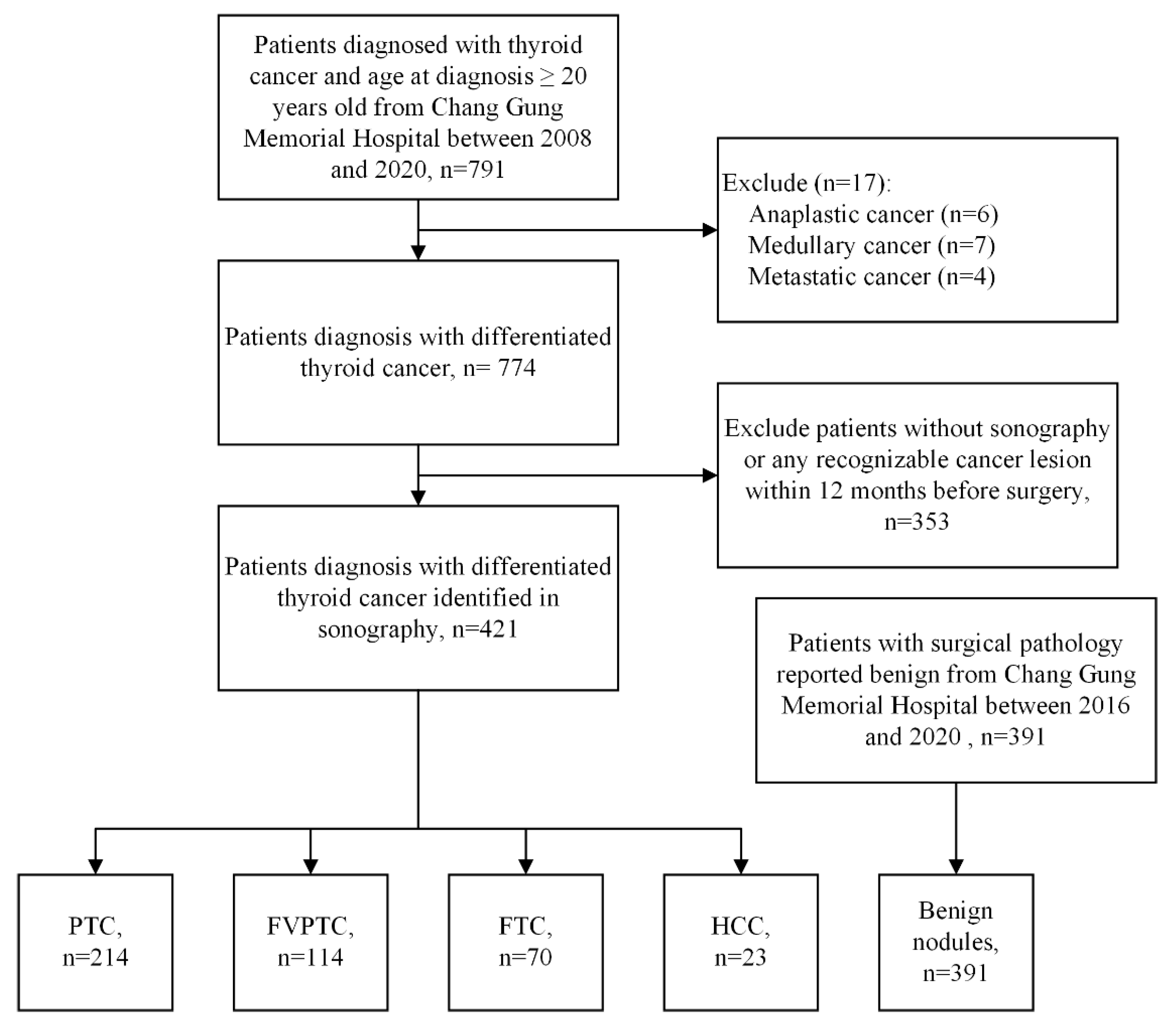
2.1. Data Sources
2.2. Data Collection
2.3. Study Design
2.4. Statistical Analysis
3. Results
3.1. Study Population
3.2. Demographics
3.3. Performance Assessment on CNNs and Physician
4. Discussion
5. Conclusions
Supplementary Materials
Author Contributions
Funding
Institutional Review Board Statement
Informed Consent Statement
Data Availability Statement
Acknowledgments
Conflicts of Interest
References
- Dean, D.S.; Gharib, H. Epidemiology of thyroid nodules. Best Pract. Res. Clin. Endocrinol. Metab. 2008, 22, 901–911. [Google Scholar] [CrossRef]
- Uppal, A.; White, M.G.; Nagar, S.; Aschebrook-Kilfoy, B.; Chang, P.J.; Angelos, P.; Kaplan, E.L.; Grogan, R.H. Benign and Malignant Thyroid Incidentalomas Are Rare in Routine Clinical Practice: A Review of 97,908 Imaging Studies. Cancer Epidemiol. Prev. Biomark. 2015, 24, 1327–1331. [Google Scholar] [CrossRef] [PubMed] [Green Version]
- Zevallos, J.P.; Hartman, C.M.; Kramer, J.R.; Sturgis, E.M.; Chiao, E.Y. Increased thyroid cancer incidence corresponds to increased use of thyroid ultrasound and fine-needle aspiration: A study of the Veterans Affairs health care system. Cancer 2015, 121, 741–746. [Google Scholar] [CrossRef]
- Ganly, I.; Nixon, I.J.; Wang, L.Y.; Palmer, F.L.; Migliacci, J.C.; Aniss, A.; Sywak, M.; Eskander, A.E.; Freeman, J.L.; Campbell, M.J.; et al. Survival from Differentiated Thyroid Cancer: What Has Age Got to Do with It? Thyroid 2015, 25, 1106–1114. [Google Scholar] [CrossRef] [Green Version]
- Carling, T.; Udelsman, R. Thyroid cancer. Annu. Rev. Med. 2014, 65, 125–137. [Google Scholar] [CrossRef] [PubMed]
- Bible, K.C.; Kebebew, E.; Brierley, J.; Brito, J.P.; Cabanillas, M.E.; Clark, T.J., Jr.; Di Cristofano, A.; Foote, R.; Giordano, T.; Kasperbauer, J.; et al. 2021 American Thyroid Association Guidelines for Management of Patients with Anaplastic Thyroid Cancer. Thyroid 2021, 31, 337–386. [Google Scholar] [CrossRef]
- Wells, S.A., Jr.; Asa, S.L.; Dralle, H.; Elisei, R.; Evans, D.B.; Gagel, R.F.; Lee, N.; Machens, A.; Moley, J.F.; Pacini, F.; et al. Revised American Thyroid Association guidelines for the management of medullary thyroid carcinoma. Thyroid 2015, 25, 567–610. [Google Scholar] [CrossRef]
- Kushchayeva, Y.; Duh, Q.Y.; Kebebew, E.; D’Avanzo, A.; Clark, O.H. Comparison of clinical characteristics at diagnosis and during follow-up in 118 patients with Hurthle cell or follicular thyroid cancer. Am. J. Surg. 2008, 195, 457–462. [Google Scholar] [CrossRef] [PubMed]
- Wong, K.T.; Ahuja, A.T. Ultrasound of thyroid cancer. Cancer Imaging 2005, 5, 157–166. [Google Scholar] [CrossRef] [PubMed] [Green Version]
- Han, K.; Ha, H.J.; Kong, J.S.; Kim, J.S.; Myung, J.K.; Koh, J.S.; Park, S.; Shin, M.S.; Song, W.T.; Seol, H.S.; et al. Cytological Features That Differentiate Follicular Neoplasm from Mimicking Lesions. J. Pathol. Transl. Med. 2018, 52, 110–120. [Google Scholar] [CrossRef]
- Ferrari, S.M.; Fallahi, P.; Ruffilli, I.; Elia, G.; Ragusa, F.; Paparo, S.R.; Ulisse, S.; Baldini, E.; Giannini, R.; Miccoli, P.; et al. Molecular testing in the diagnosis of differentiated thyroid carcinomas. Gland. Surg. 2018, 7, S19–S29. [Google Scholar] [CrossRef] [PubMed]
- Polyzos, S.A.; Anastasilakis, A.D. Clinical complications following thyroid fine-needle biopsy: A systematic review. Clin. Endocrinol. 2009, 71, 157–165. [Google Scholar] [CrossRef]
- Mesko, B. The role of artificial intelligence in precision medicine. Expert Rev. Precis. Med. Drug Dev. 2017, 2, 239–241. [Google Scholar] [CrossRef]
- Chi, J.; Walia, E.; Babyn, P.; Wang, J.; Groot, G.; Eramian, M. Thyroid Nodule Classification in Ultrasound Images by Fine-Tuning Deep Convolutional Neural Network. J. Digit. Imaging 2017, 30, 477–486. [Google Scholar] [CrossRef]
- Song, J.; Chai, Y.J.; Masuoka, H.; Park, S.W.; Kim, S.J.; Choi, J.Y.; Kong, H.J.; Lee, K.E.; Lee, J.; Kwak, N.; et al. Ultrasound image analysis using deep learning algorithm for the diagnosis of thyroid nodules. Medicine 2019, 98, e15133. [Google Scholar] [CrossRef] [PubMed]
- Abdolali, F.; Kapur, J.; Jaremko, J.L.; Noga, M.; Hareendranathan, A.R.; Punithakumar, K. Automated thyroid nodule detection from ultrasound imaging using deep convolutional neural networks. Comput. Biol. Med. 2020, 122, 103871. [Google Scholar] [CrossRef]
- Guan, Q.; Wang, Y.; Du, J.; Qin, Y.; Lu, H.; Xiang, J.; Wang, F. Deep learning based classification of ultrasound images for thyroid nodules: A large scale of pilot study. Ann. Transl. Med. 2019, 7, 137. [Google Scholar] [CrossRef]
- Liu, C.; Xie, L.; Kong, W.; Lu, X.; Zhang, D.; Wu, M.; Zhang, L.; Yang, B. Prediction of suspicious thyroid nodule using artificial neural network based on radiofrequency ultrasound and conventional ultrasound: A preliminary study. Ultrasonics 2019, 99, 105951. [Google Scholar] [CrossRef] [PubMed]
- Liu, R.; Zhou, S.; Guo, Y.; Wang, Y.; Chang, C. Nodule Localization in Thyroid Ultrasound Images with a Joint-Training Convolutional Neural Network. J. Digit. Imaging 2020, 33, 1266–1279. [Google Scholar] [CrossRef]
- Liu, Z.; Zhong, S.; Liu, Q.; Xie, C.; Dai, Y.; Peng, C.; Chen, X.; Zou, R. Thyroid nodule recognition using a joint convolutional neural network with information fusion of ultrasound images and radiofrequency data. Eur. Radiol. 2021, 31, 5001–5011. [Google Scholar] [CrossRef] [PubMed]
- Park, V.Y.; Han, K.; Seong, Y.K.; Park, M.H.; Kim, E.K.; Moon, H.J.; Yoon, J.H.; Kwak, J.Y. Diagnosis of Thyroid Nodules: Performance of a Deep Learning Convolutional Neural Network Model vs. Radiologists. Sci. Rep. 2019, 9, 17843. [Google Scholar] [CrossRef]
- Wang, L.; Yang, S.; Yang, S.; Zhao, C.; Tian, G.; Gao, Y.; Chen, Y.; Lu, Y. Automatic thyroid nodule recognition and diagnosis in ultrasound imaging with the YOLOv2 neural network. World J. Surg. Oncol. 2019, 17, 12. [Google Scholar] [CrossRef] [Green Version]
- Akkus, Z.; Cai, J.; Boonrod, A.; Zeinoddini, A.; Weston, A.D.; Philbrick, K.A.; Erickson, B.J. A Survey of Deep-Learning Applications in Ultrasound: Artificial Intelligence-Powered Ultrasound for Improving Clinical Workflow. J. Am. Coll. Radiol. 2019, 16, 1318–1328. [Google Scholar] [CrossRef]
- Shin, H.C.; Roth, H.R.; Gao, M.; Lu, L.; Xu, Z.; Nogues, I.; Yao, J.; Mollura, D.; Summers, R.M. Deep Convolutional Neural Networks for Computer-Aided Detection: CNN Architectures, Dataset Characteristics and Transfer Learning. IEEE Trans. Med. Imaging 2016, 35, 1285–1298. [Google Scholar] [CrossRef] [PubMed] [Green Version]
- Haugen, B.R.; Alexander, E.K.; Bible, K.C.; Doherty, G.M.; Mandel, S.J.; Nikiforov, Y.E.; Pacini, F.; Randolph, G.W.; Sawka, A.M.; Schlumberger, M.; et al. 2015 American Thyroid Association Management Guidelines for Adult Patients with Thyroid Nodules and Differentiated Thyroid Cancer: The American Thyroid Association Guidelines Task Force on Thyroid Nodules and Differentiated Thyroid Cancer. Thyroid 2016, 26, 1–133. [Google Scholar] [CrossRef] [PubMed] [Green Version]
- McHenry, C.R.; Phitayakorn, R. Follicular adenoma and carcinoma of the thyroid gland. Oncologist 2011, 16, 585–593. [Google Scholar] [CrossRef] [PubMed] [Green Version]
- Brito, J.P.; Gionfriddo, M.R.; Al Nofal, A.; Boehmer, K.R.; Leppin, A.L.; Reading, C.; Callstrom, M.; Elraiyah, T.A.; Prokop, L.J.; Stan, M.N.; et al. The accuracy of thyroid nodule ultrasound to predict thyroid cancer: Systematic review and meta-analysis. J. Clin. Endocrinol. Metab. 2014, 99, 1253–1263. [Google Scholar] [CrossRef]
- Liu, F.H.; Liou, M.J.; Hsueh, C.; Chao, T.C.; Lin, J.D. Thyroid follicular neoplasm: Analysis by fine needle aspiration cytology, frozen section, and histopathology. Diagn. Cytopathol. 2010, 38, 801–805. [Google Scholar] [CrossRef]
- Wu, M.-H.; Chen, K.-Y.; Hsieh, M.-S.; Chen, A.; Chen, C.-N. Risk Stratification in Patients With Follicular Neoplasm on Cytology: Use of Quantitative Characteristics and Sonographic Patterns. Front. Endocrinol. 2021, 12, 350. [Google Scholar] [CrossRef]
- Russakovsky, O.; Deng, J.; Su, H.; Krause, J.; Satheesh, S.; Ma, S.; Huang, Z.; Karpathy, A.; Khosla, A.; Bernstein, M.; et al. Imagenet Large Scale Visual Recognition Challenge. Int. J. Comput. Vis. 2015, 115, 211–252. [Google Scholar] [CrossRef] [Green Version]
- Yoon, J.H.; Kim, E.K.; Hong, S.W.; Kwak, J.Y.; Kim, M.J. Sonographic features of the follicular variant of papillary thyroid carcinoma. J. Ultrasound Med. 2008, 27, 1431–1437. [Google Scholar] [CrossRef] [PubMed]
- Li, P.; Liu, P.; Zhang, H. Ultrasonic diagnosis for thyroid Hürthle cell tumor. Cancer Biomark. 2017, 20, 235–240. [Google Scholar] [CrossRef] [PubMed]
- Sillery, J.C.; Reading, C.C.; Charboneau, J.W.; Henrichsen, T.L.; Hay, I.D.; Mandrekar, J.N. Thyroid follicular carcinoma: Sonographic features of 50 cases. AJR Am. J. Roentgenol. 2010, 194, 44–54. [Google Scholar] [CrossRef] [PubMed]
- Fionda, B.; Boldrini, L.; D’Aviero, A.; Lancellotta, V.; Gambacorta, M.A.; Kovacs, G.; Patarnello, S.; Valentini, V.; Tagliaferri, L. Artificial intelligence (AI) and interventional radiotherapy (brachytherapy): State of art and future perspectives. J. Contemp. Brachyther. 2020, 12, 497–500. [Google Scholar] [CrossRef]


| Pathological Types | ||||||
|---|---|---|---|---|---|---|
| Malignant Group (n = 421) | Benign Group (n = 391) | |||||
| PTC | FVPTC | FTC | HCC | Benign | p-Value | |
| Number of patients | 214 | 114 | 70 | 23 | 391 | |
| Age (years), mean (SD) | 47.36 ± 13.70 | 44.90 ± 15.12 | 46.61 ± 17.10 | 51.17 ± 15.62 | 54.18 ± 13.15 | <0.0001 |
| Sex (n, %) | ||||||
| Male | 53 (25) | 28 (25) | 15 (21) | 4 (17) | 80 (20) | 0.6985 |
| Female | 161 (75) | 86 (75) | 55 (79) | 19 (83) | 311 (80) | |
| Number of US images | 470 | 215 | 131 | 38 | 937 | |
| Number of cropped images | 533 | 272 | 175 | 53 | 1275 | |
| Location (n, %) | ||||||
| Left | 88 (41.12) | 52 (45.61) | 39 (55.71) | 13 (56.52) | 143 (36.57) | 0.0017 |
| Right | 109 (50.93) | 57 (50.00) | 27 (38.57) | 10 (43.48) | 192 (49.10) | |
| Both (Left + Right) | 11 (5.15) | 4 (3.51) | 1 (1.43) | 0 (0.00) | 47 (12.03) | |
| Isthmus | 6 (2.80) | 1 (0.88) | 3 (4.29) | 0 (0.00) | 9 (2.30) | |
| Ultrasound brands (%) | ||||||
| Aloka | 3.93 | 2.46 | 6.67 | 0.00 | 7.57 | <0.0001 |
| GE Healthcare | 37.12 | 62.30 | 64.01 | 78.26 | 45.87 | |
| Hitachi | 7.42 | 2.46 | 1.33 | 0.00 | 7.34 | |
| Philips | 4.37 | 0.82 | 8.00 | 8.70 | 2.06 | |
| Siemens | 28.82 | 10.66 | 5.33 | 8.70 | 18.81 | |
| Toshiba | 17.90 | 16.39 | 13.33 | 4.34 | 18.12 | |
| Others | 0.44 | 4.91 | 1.33 | 0.00 | 0.23 | |
| Aloka | GE Healthcare | Hitachi | Philips | Siemens | Toshiba | Others | p-Value | |
|---|---|---|---|---|---|---|---|---|
| PTC | 18.00 | 19.90 | 32.07 | 35.72 | 39.52 | 27.15 | 11.11 | <0.0001 |
| FVPTC | 6.00 | 17.80 | 5.66 | 3.57 | 7.78 | 13.25 | 66.67 | <0.0001 |
| FTC | 10.00 | 11.24 | 1.89 | 21.43 | 2.40 | 6.62 | 11.11 | <0.0001 |
| HCC | 0.00 | 4.22 | 0.00 | 7.14 | 1.20 | 0.66 | 0.00 | <0.0001 |
| Benign | 66.00 | 46.84 | 60.38 | 32.14 | 49.10 | 52.32 | 11.11 | <0.0001 |
| Histopathology | Number (%) | ||
|---|---|---|---|
| Malignant group (n = 421) | PTC features (n = 214) | Classic PTC | 208 (97.19) |
| Diffuse sclerosing variant | 3 (1.40) | ||
| Tall cell variant | 1 (0.47) | ||
| Cribriform morular variant | 1 (0.47) | ||
| Encapsulated variant | 1 (0.47) | ||
| FTC features (n = 207) | Follicular variant of PTC | 106 (51.21) | |
| Follicular carcinoma, minimally invasive | 70 (33.82) | ||
| Hürthle cell carcinoma | 23 (11.11) | ||
| Encapsulated follicular variant of PTC | 8 (3.86) | ||
| Benign group (n = 391) | Nodular hyperplasia | 289 (73.91) | |
| Follicular adenoma | 48 (12.28) | ||
| Cyst | 47 (12.02) | ||
| Hürthle cell adenoma | 7 (1.79) | ||
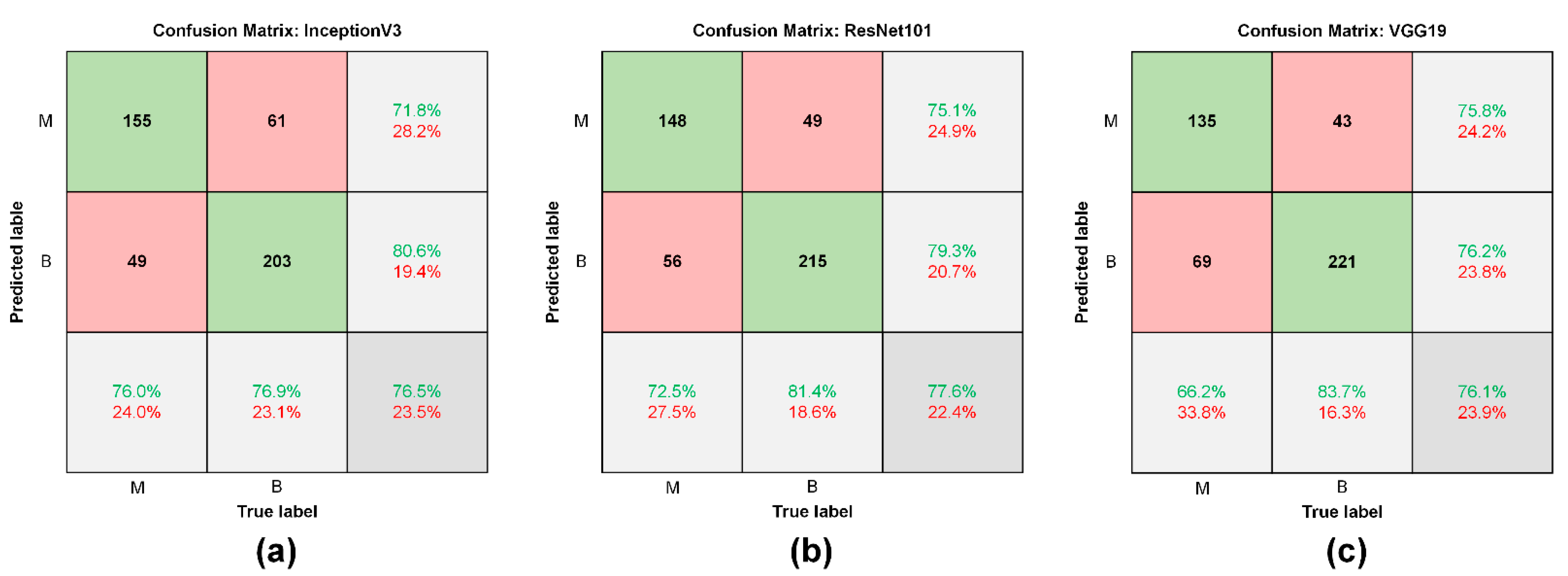
| Sensitivity | Specificity | PPV | NPV | Accuracy | AUC | |
|---|---|---|---|---|---|---|
| InceptionV3 | 76.0 | 76.9 | 71.8 | 80.6 | 76.5 | 0.82 |
| ResNet101 | 72.5 | 81.4 | 75.1 | 79.3 | 77.6 | 0.83 |
| VGG19 | 66.2 | 83.7 | 75.8 | 76.2 | 76.1 | 0.83 |
| Endocrinologist 1 | 38.7 | 74.2 | 53.7 | 61.1 | 58.8 | - |
| Endocrinologist 2 | 35.3 | 82.6 | 61.0 | 62.3 | 62.0 | - |
| Malignant Group | Benign Group | ||||||
|---|---|---|---|---|---|---|---|
| PTC | FVPTC | FTC | HCC | NH | FA | C | |
| InceptionV3 | 81.4 | 72.9 | 72.7 | 66.7 | 75.0 | 65.0 | 92.5 |
| ResNet101 | 73.2 | 74.6 | 69.7 | 66.7 | 79.4 | 80.0 | 90.0 |
| VGG19 | 64.9 | 71.2 | 63.6 | 60.0 | 82.4 | 75.0 | 95.0 |
| Endocrinologist 1 | 58.8 | 20.3 | 27.3 | 13.3 | 73.0 | 80.0 | 80.0 |
| Endocrinologist 2 | 53.6 | 17.0 | 30.3 | 6.7 | 81.9 | 75.0 | 90.0 |
Publisher’s Note: MDPI stays neutral with regard to jurisdictional claims in published maps and institutional affiliations. |
© 2021 by the authors. Licensee MDPI, Basel, Switzerland. This article is an open access article distributed under the terms and conditions of the Creative Commons Attribution (CC BY) license (https://creativecommons.org/licenses/by/4.0/).
Share and Cite
Chan, W.-K.; Sun, J.-H.; Liou, M.-J.; Li, Y.-R.; Chou, W.-Y.; Liu, F.-H.; Chen, S.-T.; Peng, S.-J. Using Deep Convolutional Neural Networks for Enhanced Ultrasonographic Image Diagnosis of Differentiated Thyroid Cancer. Biomedicines 2021, 9, 1771. https://doi.org/10.3390/biomedicines9121771
Chan W-K, Sun J-H, Liou M-J, Li Y-R, Chou W-Y, Liu F-H, Chen S-T, Peng S-J. Using Deep Convolutional Neural Networks for Enhanced Ultrasonographic Image Diagnosis of Differentiated Thyroid Cancer. Biomedicines. 2021; 9(12):1771. https://doi.org/10.3390/biomedicines9121771
Chicago/Turabian StyleChan, Wai-Kin, Jui-Hung Sun, Miaw-Jene Liou, Yan-Rong Li, Wei-Yu Chou, Feng-Hsuan Liu, Szu-Tah Chen, and Syu-Jyun Peng. 2021. "Using Deep Convolutional Neural Networks for Enhanced Ultrasonographic Image Diagnosis of Differentiated Thyroid Cancer" Biomedicines 9, no. 12: 1771. https://doi.org/10.3390/biomedicines9121771
APA StyleChan, W.-K., Sun, J.-H., Liou, M.-J., Li, Y.-R., Chou, W.-Y., Liu, F.-H., Chen, S.-T., & Peng, S.-J. (2021). Using Deep Convolutional Neural Networks for Enhanced Ultrasonographic Image Diagnosis of Differentiated Thyroid Cancer. Biomedicines, 9(12), 1771. https://doi.org/10.3390/biomedicines9121771